Tumor cell xenograft model in zebrafish, and methods of constructing and using the same

a tumor cell and zebrafish technology, applied in the field of biomedicine, can solve the problems of affecting the survival rate of patients, the limited treatment options of gastric cancer, and the major public health problem of tumor diseases, and achieve the effect of accurate screening out, accurate guidance for clinical medication, and accurate screening out patients

Inactive Publication Date: 2019-11-21
NANJING EMORY BIOTECH CO LTD
View PDF0 Cites 4 Cited by
  • Summary
  • Abstract
  • Description
  • Claims
  • Application Information

AI Technical Summary

Benefits of technology

[0028]The use of the tumor cell xenograft model in zebrafish of the present invention in screening drugs for gastric cancer can accurately screen out the patients that 5-FU (5-fluorouracil) is effective and patients that 5-FU (5-fluorouracil) is ineffective, providing accurate guidance for clinical medication. The use of the tumor cell xenograft model in zebrafish of the present invention in screening drugs for lung cancer can accurately screen out the patients that gefitinib, cisplatin or docetaxel is effective and patients that gefitinib, cisplatin or docetaxel is ineffective, providing accurate guidance for clinical medication.
[0029]The CM-Dil of the present invention is a dye which labels the cells by binding to a lipid molecule on the membrane structure, and has strong and stable red fluorescence (excitation peak 553 nm / emission peak 570 nm). Unlike Dil, CM-Dil has good water solubility, so it is more convenient and efficient for cell staining. The CM group (i.e., chloromethyl substituent) can react with the sulfhydryl group on the polypeptide and protein to keep the molecule stable in the aldehydes, so the CM-Dil-labeled cells can be fixed, cracked and paraffin-embedded without affecting the fluorescence. Therefore, it is an ideal fluorescent cell labeling dye for immunofluorescence, immunohistochemistry and in situ hybridization. In addition, CM-Dil is non-toxic to cells, is stable and long-lasting, and can well trace cells for a long time. Studies have confirmed that after labeling with CM-Dil, the fluorescence is stably expressed in the cells, and the positive labeling rate is over 98%. The labeled cells have good morphology, and the differentiation of cells in vitro can be effectively observed; or the labeled cells can be injected into the body, to effectively show the migration and differentiation of transplanted cells in living tissues. CM-Dil has a chemical name below: 3H-Indolium,5-[[[4-(chloromethyl)benzoyl]amino]methyl]-2-[3-(1,3-dihydro-3,3-dimethyl-1-octadecyl-2H-indol-2-ylidene)-1-propenyl]-3,3-dimethyl-1-octadecyl-, chloride.
[0030]The zebrafish used in the present invention has the characteristics of small volume, fast growth, and transparent body during the early development. The zebrafish-based PDX model has the advantages of low cost, high throughput, simple operation, and easy observation in vivo. More importantly, the experimental period with the zebrafish-based PDX model is short and is only one week. Therefore, the zebrafish-based PDX model is currently the only animal model that is expected to satisfy the clinical demand to guide individualized medications for solid tumors such as gastric cancer and lung cancer in real time.
[0032]In the method of the present invention, a fluorescence method is used to effectively evaluate a tumor treatment drug, and the method is simple and effective, and can meet clinical needs. The zebrafish model provided in the present invention provides a simple and effective method for evaluating the therapeutic effects of 5-FU (5-fluorouracil) on patients with gastric cancer or gefitinib, cisplatin or docetaxel on patients with lung cancer alone or in combination.

Problems solved by technology

Tumor diseases have become a major public health problem in the world.
However, due to the non-obvious early symptoms of gastric cancer and the lack of popularization of routine gastroscopy, about 80% of patients with gastric cancer in China have reached the advanced stage upon clinical consultation.
The existing treatments for gastric cancer are limited, and the overall survival rate of surgery alone is only about 20%.
Because gastric cancer is a highly heterogeneous tumor, many existing clinical evidences have shown that chemotherapy can prolong the survival time of patients with gastric cancer, but no “gold standard” therapeutic regimen with recognized obvious advantages and individualized medications has been found.
Many patients lose their original treatment window because they fail to receive the drug that best matches the individual.
In addition, lung cancer is the leading cause of malignant tumor-related death today.
Although the treatment of lung cancer has developed rapidly in recent years, the overall prognosis has not improved significantly.
There are many types of lung cancer, the most common of which is non-small cell lung cancer (NSCLC), and because of the lack of specific symptoms in early stage of lung cancer, the condition has progressed to the middle or late stage when most patients are diagnosed, so the difficulty of treatment is increased.
Although there are many drugs for treating lung cancer, the survival rate of patients has not been improved significantly, and chemotherapy is palliative, mainly aiming at prolonging the survival of patients and improving their quality of life.
At present, the commonly used anti-tumor chemotherapeutic drugs are less than 70% effective in the treatment of patients, and because of the lack of genetic analysis in individualized treatment with chemotherapy drugs, 20% to 40% of patients may even receive the incorrect drug treatment.
Many patients lose their original treatment window because they fail to receive the drug that best matches the individual.
However, because the duration from tumor transplantation, tumor formation to efficacy evaluation in mice is usually 3 months, and many patients have a survival period of less than 3 months, the existing PDX model cannot meet the significant demands for clinical real-time guidance for individualized medication.
More importantly, the experimental period with the zebrafish-based PDX model is short and is only one week.

Method used


Image

  • Tumor cell xenograft model in zebrafish, and methods of constructing and using the same
  • Tumor cell xenograft model in zebrafish, and methods of constructing and using the same
  • Tumor cell xenograft model in zebrafish, and methods of constructing and using the same

Examples


example 1

ion of a Patient-Derived Gastric Cancer Cell Xenograft Model in Zebrafish of the Present Invention

[0047]1. Isolation of Primary Cells from Gastric Cancer Tissue

[0048]The patient-derived clinical tissue biopsy that was a surgical specimen of gastric cancer was placed in physiological saline. The blood clot, necrotic tissue, fat and connective tissues on the surface of the tumor tissue were removed under aseptic conditions. The tissue was cut by an ophthalmic scissor after sterilization, and washed 2 times with sterile phosphate buffer (pH 7.4). A small amount of phosphate buffer was added, and the tissue was repeatedly cut with an ophthalmic scissor until the tissue became a paste and was about 1 mm3. 0.25% trypsin was added and the tissue was digested at 37° C. for 10 minutes. After the tissue mass was observed to be completely dissociated, centrifugation was performed to remove trypsin. The cells were re-suspended in RPMI-1640 medium containing 10% FBS (fetal calf serum).

[0049]2. S...

example 2

n of the Clinical Anticancer Effect of 5-FU with 4 Patient-Derived Xenograft Zebrafish Models

[0056]1. Determination of Safe Dose

[0057]Two days after fertilization, the zebrafish embryos were treated (soaked) with various concentrations of 5-FU for three days, and the highest 5-FU concentration within the safety range for embryos was determined to be 4000 nM.

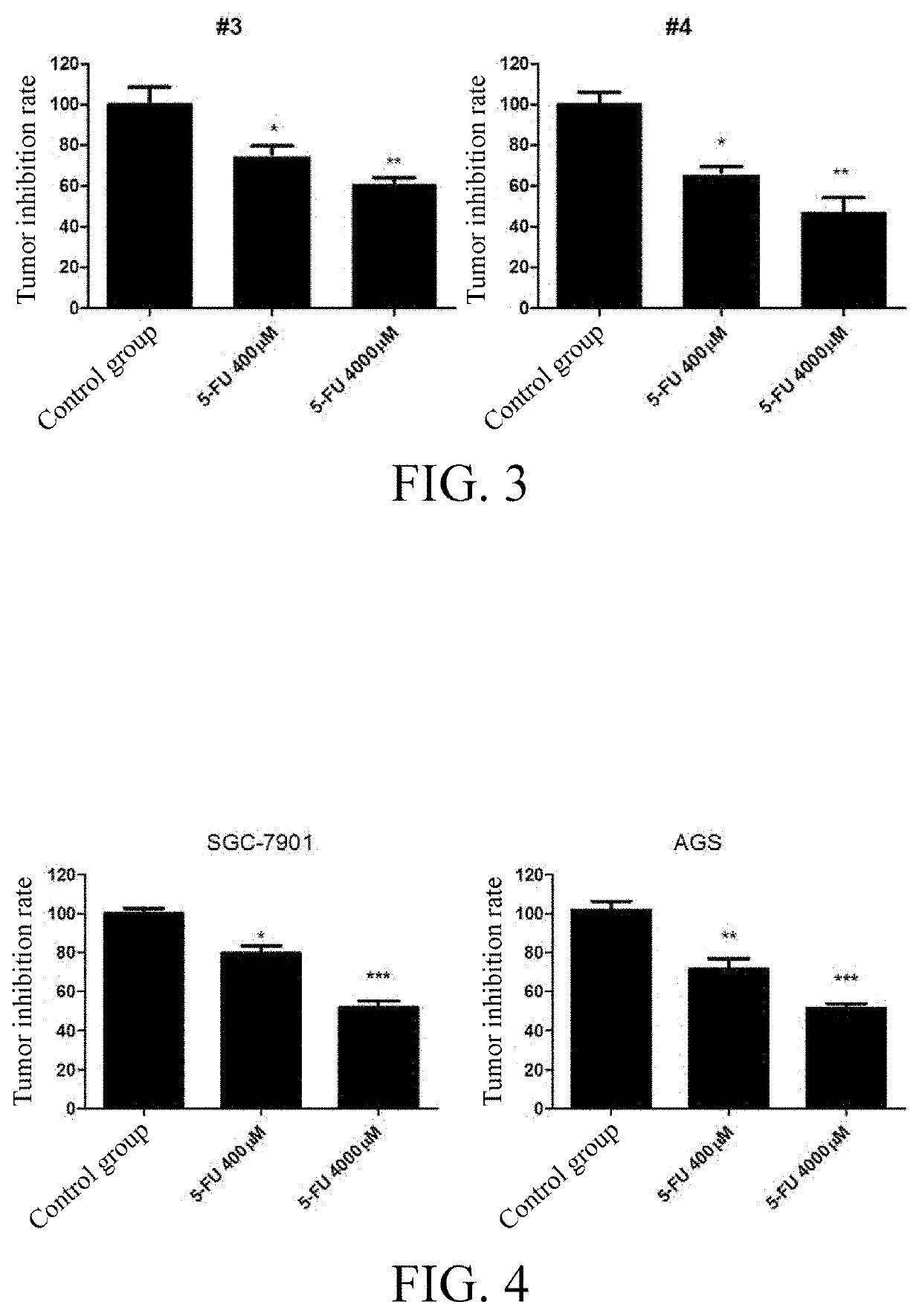
[0058]2. Drug Treatment of Zebrafish Embryos

[0059]The 4000-μM and 400 μM 5-FU were used to soak the zebrafish embryo model injected with the primary gastric cancer cells derived from different patients prepared by the method of Example 1 for three days, and 0.1% DMSO was used as a solvent control.

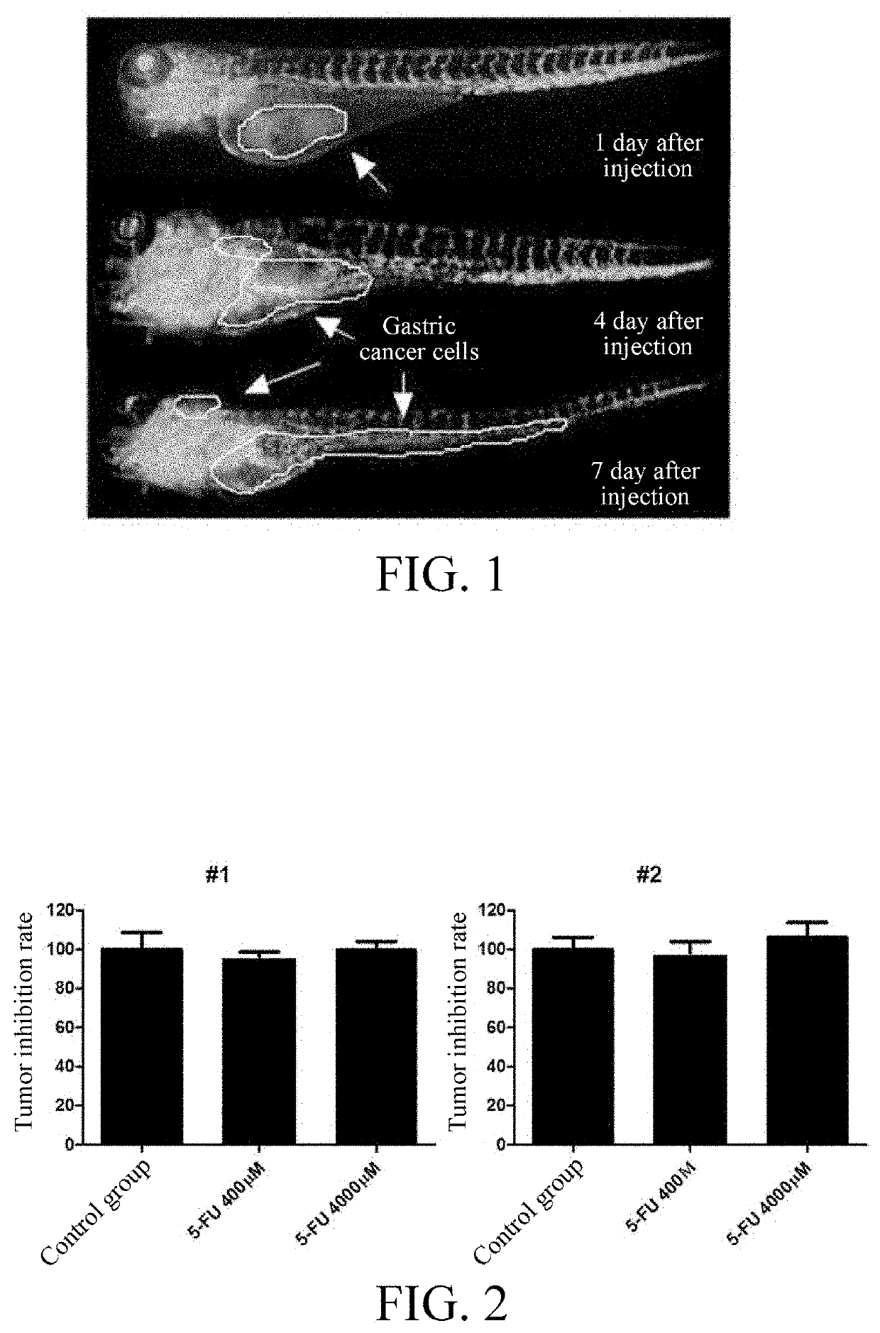
[0060]3. Observation of Antitumor Effect Under Fluorescence Microscope

[0061]The proliferation and spread of red patient-derived cells in the zebrafish embryos were observed after treatment. The red cells were photographed under a fluorescence microscope, and the red fluorescence intensity was quantified by Image Pro Plus software to calc...

example 3

n of the Anticancer Effects of 5-FU with Two Human Gastric Cancer Cell Line (SGC-7901 and AGS) Xenograft Models in Zebrafish

[0064]1. Drug Treatment of Zebrafish Embryos

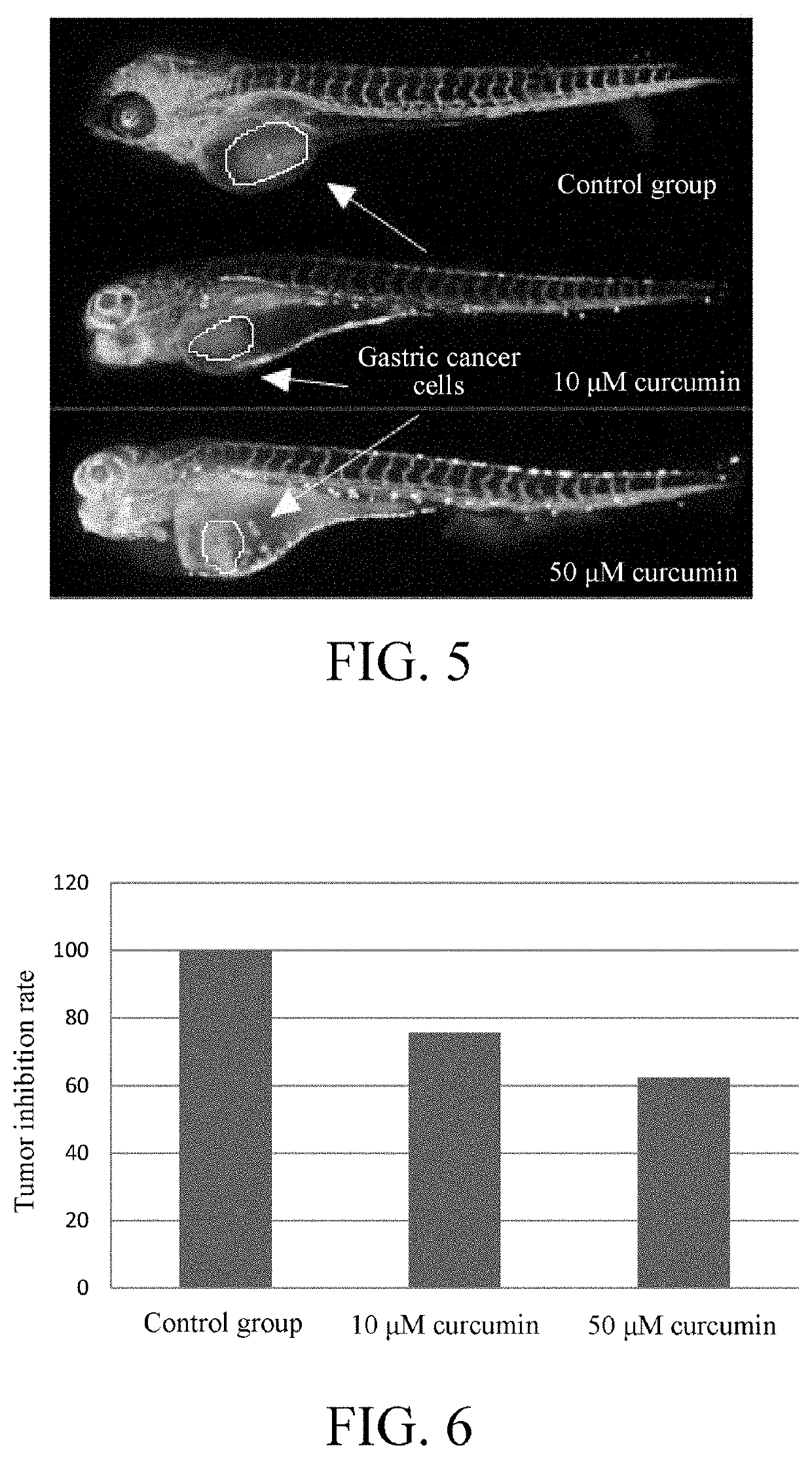
[0065]4000 μM and 400 μM5-FU were used to soak the zebrafish embryos injected with gastric cancer cell lines (SGC-7901 and AGS) (constructed as in Example 1) for three consecutive days, and 0.1% DMSO was used as a solvent control.

[0066]2. Observation of Antitumor Effect Under Fluorescence Microscope

[0067]The proliferation and spread of red patient-derived cells in the zebrafish embryos were observed after treatment. The red cells were photographed under a fluorescence microscope, and the red fluorescence intensity was quantified by Image Pro Plus software to calculate the anti-tumor effect of 5-FU by a formula: Tumor inhibition rate of the drug=(the fluorescence intensity of the treatment group / the fluorescence intensity of the control group)*100% (see FIG. 4).

[0068]The results with human gastric cancer cell line xeno...


PUM


Abstract

A tumor cell xenograft model in zebrafish, and methods of constructing and using the same. Primary cells dissociated from the tumor tissue of a patient are transplanted into zebrafish, so as to obtain a patient-derived tumor xenograft model. The tumor xenograft model retains the pathological features of human gastric cancer tissues in clinic, has higher clinical relevance, and can be used in the systematic research into the mechanisms underlying the proliferation, metastasis, spread and drug resistance of tumors, and to screen effective drugs for tumor treatments.

Description

BACKGROUNDTechnical Field[0001]The present invention relates to the technical field of biomedicine, and in particular to a patient-derived gastric cancer xenograft model in zebrafish, and methods of constructing and using the same.Related Art[0002]Tumor diseases have become a major public health problem in the world. The most common tumors include lung cancer, gastric cancer, and breast cancer. Among them, gastric cancer is one of the most common malignant tumors of the digestive system found in the world, and has high incidence in East Asia. According to the “World Cancer Report 2014” published by the World Health Organization (WHO), the new cases of gastric cancer and deaths in China accounted for more than 40% of the total number in the whole world in 2012. In 2016, the Cancer Statistics in China, 2015 published in an authoritative journal “CA Cancer J Clin” sponsored by the American Cancer Society (ACS) showed that there were 679,100 new cases of gastric cancer in China in 2015,...

Claims


Application Information

Patent Timeline
Patent Type & Authority Applications(United States)
IPC IPC(8): A61K49/00A01K67/027
CPCA01K2267/0331A01K2207/12A61K49/0008A01K67/0271A01K2227/40A61K35/38A01K61/10
Inventor HE, MINGFANGWANG, RUIXUELI, JIANYING
Owner NANJING EMORY BIOTECH CO LTD
Why Eureka
Try Eureka