Supercharge Your Innovation With Domain-Expert AI Agents!

Xenotransplantation products and methods

Active Publication Date: 2020-04-09
XENOTHERAPEUTICS INC
View PDF0 Cites 2 Cited by
  • Summary
  • Abstract
  • Description
  • Claims
  • Application Information

AI Technical Summary

Benefits of technology

The present invention provides a method for delaying, reducing, or preventing rejection, separation, or adverse reactions in a human recipient of xenotransplanted tissues or organs from a pig. This is achieved by selectively replacing a portion of the pig's major histocompatibility complex (MHC) gene with a corresponding portion of the human recipient's MHC gene. The invention also includes identifying and replacing sections of the MHC gene that are critical to avoid in order to minimize immunological rejection. The invention provides biologically reprogrammed swine that have a well-functioning immune system and are substantially free of critical pathogens. The invention also includes reprogramming small sections of genetic code rather than inserting a transgenic animal's genome into the recipient's genome. The invention also includes removing or neutralizing structures that cause rejection from the transplantation product to minimize or eliminate immunological reaction and rejection.

Problems solved by technology

Sadly, each day, 30 people either die or are removed from the waiting list (because they are too sick for the requisite surgery) due to this inadequate supply of organs.
While the rate of divergence between available donors and unmet need of recipients has been improved marginally, this disparity has continued to present day and remains considerable; the supply remains disastrously inadequate.
Beyond the shortage of organs, other significant issues related to allotransplantation exist and present a multifaceted problem, involving safety, logistical, ethical, legal, institutional, and cultural complications.
Allogeneic tissues from human donors carry significant infectious disease risks.
Due to technical and timing constraints surrounding organ viability and post-mortem screening, absolute testing is hindered, and this risk cannot be eliminated.
Benichou, et al., describes the response to an allogeneic skin graft as a “potent” immune response involving engagement of both the innate and adaptive immune systems Abbas A K, Lichtman A H H, Pillai S (2017) Cellular and Molecular Immunology can blunt the natural immunological processes; unfortunately, these medications are often a lifelong requirement after organ transplantation and increase recipient susceptibility to otherwise routine pathogens.
While these drugs allow transplant recipients to tolerate the presence of foreign organs, they also increase the risk of infectious disease and symptoms associated with a compromised immune system, as a “broad array of organisms may be transmitted with human allografts.” Fishman J A, Greenwald M A, Grossi P A (2012) Transmission of infection With Human Allografts: Essential Considerations in Donor Screening.
One, often overlooked logistical constraint involves the intricate and timely preservation methods of donated organs necessary to maintain viability between the time of procurement and transplantation.
Limitations on organ preservation are considerable and directly impact surgical capacity.
The fundamental reason for these restrictive, limiting timeframes is due to the inability to preserve the fundamental vital capacity of the numerous and diverse cells comprising the larger tissue or organ.
Interruption or complete termination of necessary and highly complex, interdependent cellular activity thereby erodes the potential clinical utility of the transplant.
Despite these drawbacks, organ transplantation is unquestionably the preferred therapy for most patients with end stage organ failure, in large part due to a lack of viable alternatives.
However, the advent of organ transplantation as a successful life-saving therapeutic intervention, juxtaposed against the paucity of organs available to transplant, unfortunately places medical professionals in an ideologically vexing position of having to decide who lives and who dies.
During this critical period, patients with severe burns are at risk of deteriorating clinical condition due to infection from opportunistic pathogens, disrupted skin barrier, and impairment of immune response, as well as hypovolemia through fluid loss at the burn site.
This is frequently followed by electrolyte, temperature and pH imbalances that contribute to organ failure, and possibly death.
While it would be ideal to utilize autograft to treat such injuries, the supply of the patient's own skin is often limited during the acute phase of burn care (within 96 hours of injury) especially in cases where burns cover 20% or more of total body surface area.
More importantly, autologous graft harvest is often clinically contraindicated as it results in further insult to the patient's already compromised homeostasis and worsens morbidity.
Unfortunately, given the inherent logistical and supply constraints, as well as infectious agent concerns, the availability of HCA is severely limited.
Many of these wound cover and closure products have limited indications and other limitations as described in the literature.
Such products do not vascularize and are not intended for treatment of severe and extensive, deep partial and full thickness burn wounds and allow for vascularization of the wound bed.
Prior to the present invention, no skin substitute is able to approximate the biologic properties of viable human skin and there is no satisfactory replacement for human allograft skin.
Xenotransplantation from swine to humans, however, has significant roadblocks, not the least of which is hyperacute rejection where natural human antibodies target epitopes on the animal cells, causing rejection and failure of the transplanted organs, cells or tissue.
Other roadblocks include delayed xenograft rejection, acute cellular rejection, chronic rejection, risks of cross-species transmission of disease or parasites.
Thus, similar to the alpha-1,3-galactosyltransferase epitope, Neu5Gc is expressed on swine cells, but is recognized as foreign by human recipients of xenotransplant products, leading to eventual rejection.
While the previously used methods require immunosuppressive drugs, use of immunosuppressants often lead to life-threatening malignancies and infections.
In other cases, the prior art methods resolved to knockout animal genes and / or to insert human genes to create transgenic animals, but doing so resulted in animals that were unable to survive and thrive due to compromised immune systems and other phenotypic complications.
In such cases, disruption of swine glycan and / or SLA genes made the cells vulnerable to lysis by natural killer cells.
Prior xenotransplantation studies have reported problems including natural killer cell-mediated lysis after transplantation, as well as difficulties maintaining antiviral immunity, and autoimmune disorders in the donor animals.
Despite such publications, complex issues concerning xenotransplantation products in humans remain unsolved and guidance for providing viable xenotransplantation products remains vague, contradictory, and generic where specificity is required for safety and efficacy.
There is no xenotransplantation product comprising live animal cells, tissues, or organs approved for marketing and very few clinical trials.
The industry has faced safety concerns, e.g., potential transmission of pathogens, coupled with the lack of meaningful regulatory guidance on approved manufacturing procedures and product characteristics.
While various publications have reported advances, e.g., in academic settings, none has resulted in an approved xenotransplantation product comprising live animal cells, tissues, or organs, and good manufacturing practice (GMP) regulations have not been promulgated for such products or manufacturing processes.

Method used


Image

  • Xenotransplantation products and methods
  • Xenotransplantation products and methods
  • Xenotransplantation products and methods

Examples


example 1

DPF Closed Colony Skin Graft (Monkey Studies)

[0543]It has been discovered that skin grafts derived from a DPF Closed Colony, α-1,3-galactosyltransferase [Gal-T] knockout pigs produced in accordance with the present invention exhibit significantly longer rejection times than skin grafts derived from α-1,3-galactosyltransferase [Gal-T] knockout pigs but that were not derived from DPF Closed Colony pigs.

[0544]Numerous prior studies evaluating rejection time of α-1,3-galactosyltransferase [Gal-T] knockout pigs (not derived from a DPF Closed Colony) on monkeys show rejection times in the range of 11-13 days. See, e.g., Albritton et al., Lack of Cross-Sensitization Between alpha-1, 3-Galactosyltransferase Knockout Porcine and Allogeneic Skin Grafts Permits Serial Grafting, Transplantation & Volume 97, Number 12, Jun. 27, 2014, (Gal-T-KO skin grafts on recipient baboons fully rejected by 12 or 13 days); Barone et al., “Genetically modified porcine split-thickness skin grafts as an alternat...

example 2

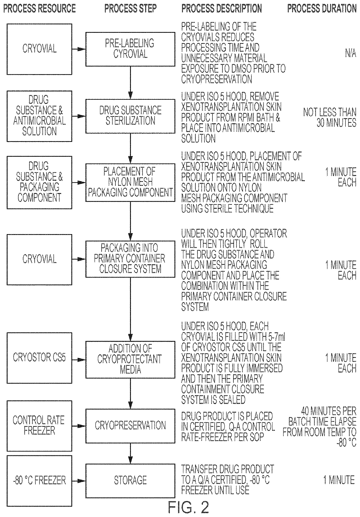
[0564]The following example provides a description of a process of harvesting and processing skin from a designated pathogen free α-1,3-galactosyltransferase [Gal-T] knockout swine produced in accordance with the present invention, with the skin to be used as a xenogeneic skin product for human transplantation. In some of these aspects, the xenotransplantation product consists of split thickness grafts consisting of dermal and epidermal tissue layers containing vital, non-terminally sterilized porcine cells derived from specialized, genetically engineered, Designated Pathogen Free (DPF), source animals (alpha 1,3 galactosyltransferase knockout [Gal-T-KO] miniature swine).

[0565]The genetically engineered source animals in this example do not contain any foreign, introduced DNA into the genome; the gene modification employed is exclusively a knock-out of a single gene that was responsible for encoding for an enzyme that causes ubiquitous expression of a cell-surface antigen. The xenot...

example 3

Dermis Epidermus Combo Product

[0675]The following example provides a description of a skin product derived from a designated pathogen free α-1,3-galactosyltransferase [Gal-T] knockout swine for use in human transplantation produced in accordance with the present invention. The product, process and techniques disclosed herein are but examples, and do not limit the scope of the invention.

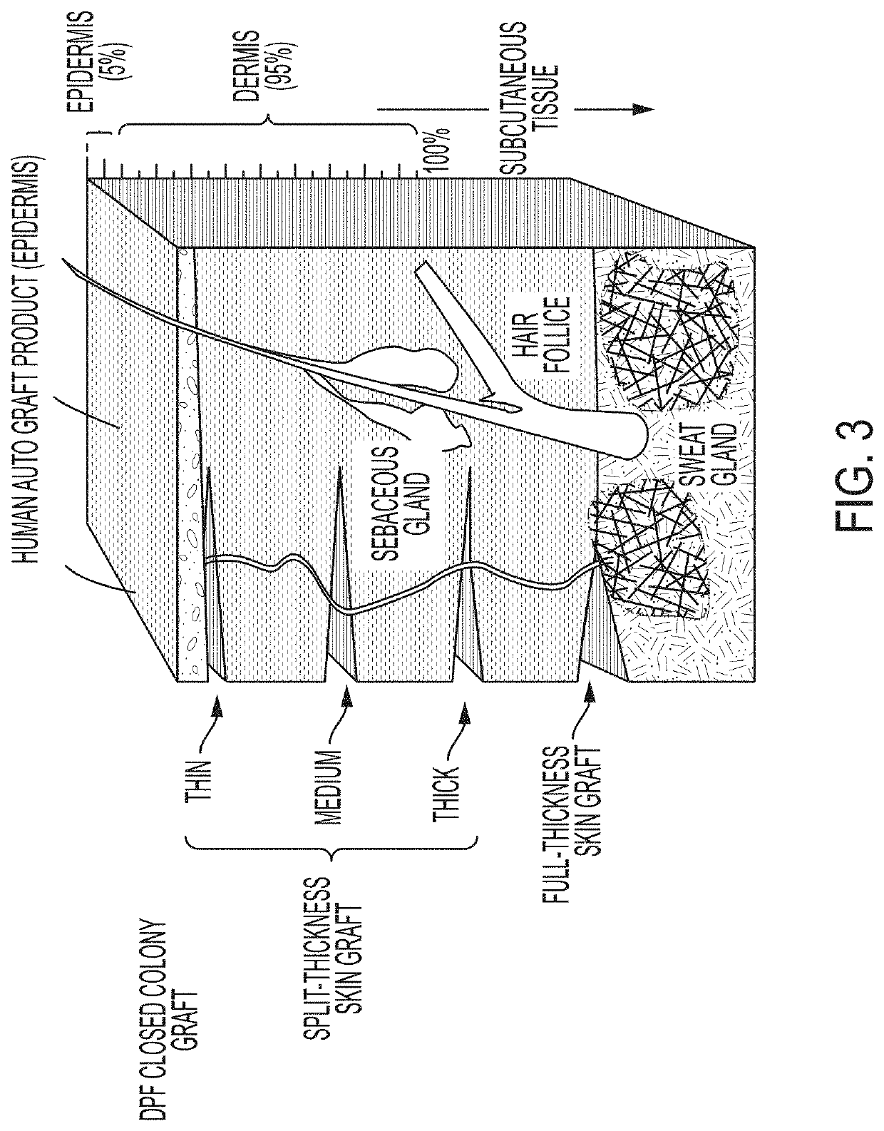
[0676]Some skin transplantation products for the treatment of burns and other ailments utilize cultured epidermal autografts (see, e.g., products produced by Vericel Corporation under the Epicel® brand name). Such epidermal autografts can be utilized for patients with burns (including severe burns) and result in reduced or no rejection in the transplanted epidermal material since the material is derived from the patient's own skin.

[0677]However, such products are limited to the epidermis only, and do not include the dermis portion of the skin. Referring to FIG. 3, it will be understood that the dermis...


PUM

PropertyMeasurementUnit
Fractionaaaaaaaaaa
Fractionaaaaaaaaaa
Fractionaaaaaaaaaa

Abstract

A biological product for clinical xenotransplantation into a human and a method of preparing biological product for clinical xenotransplantation into a human involving producing a non-wild type, biologically engineered swine having a biologically engineered genome such that the swine does not express one or more extracellular surface glycan epitopes, is free of certain pathogens, is reared according to a bioburden-reducing procedure in a closed designated pathogen free herd, wherein the biological product is harvested following the swine being euthanized and the product is aseptically removed from the swine, the biological product is processed involving sterilization and storing the product in a sterile container, and the product does not contain one or more extracellular surface glycans, is free of certain designated pathogens, is biologically active and comprises live cells and tissues capable of vascularizing after xenotransplantation.

Description

CROSS-REFERENCE TO RELATED APPLICATIONS[0001]This application claims priority benefit of U.S. provisional application Nos. 62 / 742,188, filed Oct. 5, 2018; 62 / 756,925, filed Nov. 7, 2018; U.S. 62 / 756,955 filed Nov. 7, 2018; U.S. 62 / 756,977, filed Nov. 7, 2018; U.S. 62 / 756,993, filed Nov. 7, 2018; U.S. 62 / 792,282, filed Jan. 14, 2019; U.S. 62 / 795,527, filed Jan. 22, 2019; U.S. 62 / 823,455, filed Mar. 25, 2019; and U.S. 62 / 848,272, filed May 15, 2019, the disclosures of all of which are incorporated herein by reference in their entireties.FIELD OF THE INVENTION[0002]The subject matter disclosed herein relates to biological products derived from genetically engineered organisms, and related methods, for use in xenotransplantation.BACKGROUND OF THE INVENTION[0003]According to the United Network for Organ Sharing (“UNOS”), every ten minutes, someone is added to the national transplant waiting list, and 20 people die each day waiting for a transplant. As of August 2019, there were about 114...

Claims


Application Information

Patent Timeline
IPC IPC(8): A61L27/36
CPCA61L27/3687A61L27/3691A61L27/3604A61L27/362A61L27/3683A61L27/38A61K35/36A01K2227/108A01K2267/025A01K67/0276C12N15/907A01K2217/075A61K35/12A61K2300/00C12Y114/18002A61L2430/34A61L27/60C12Y204/01092C12N2310/20
Inventor HOLZER, PAUL W.ADKINS, JONMONROY, RODNEY L.CHANG, ELIZABETH J.
Owner XENOTHERAPEUTICS INC
Features
  • R&D
  • Intellectual Property
  • Life Sciences
  • Materials
  • Tech Scout
  • Unparalleled Data Quality
  • Higher Quality Content
  • 60% Fewer Hallucinations
Learn More