
Apparatus, systems and methods for transvascular access to the brain

a brain and transvascular technology, applied in the field of brain access apparatus, can solve the problems of increased hospital stay, neurologic deficit, and even death, and the conventional methods of accessing brain tissue are prone to surgery complications, and the hospital admissions are associated with high costs and significant complications

Active Publication Date: 2022-06-30
VONOVA INC
View PDF5 Cites 3 Cited by
  • Summary
  • Abstract
  • Description
  • Claims
  • Application Information

AI Technical Summary

Benefits of technology

The endovascular catheter described in this patent is made from a special blend of polymers and reinforcement materials, which results in a thin-walled catheter with strong flexibility, high-tensile strength, torque, and durability. The catheter is designed to be biologically inert, preventing inflammation and thrombogenicity, and resistant to biofilm formation. It is also coated with hydrophilic or lubricous materials to prevent cytotoxicity and systemic toxicity.

Problems solved by technology

Consequently, conventional methods of accessing brain tissue are prone to the complications of surgery.
Complications include disfigurement, pain, perioperative infection, symptomatic hemorrhage, seizure, edema, skull and dura defects, increased length of hospital stay, patient fear / anxiety, morbidity tolerance, hospital admissions associated with high costs and significant rates of complications, neurologic deficits, and even death.
Accordingly, the threshold for neurosurgical intervention remains high due to either risks or physician / patient reluctance, which may in turn result in delays to diagnosis, treatment, and consequently, worse outcomes.
Further, conventional methods for accessing brain tissue may rely upon stereotactic neurosurgical methods with proxy fiducial markers, which may result in mistargeting, suboptimal placement, or excessive collateral damage.
However, fiducial markers may physically move during the pre-operative or intraoperative period. and are located at a distance from the target tissue, which may result in suboptimal targeting when there are minor deviations in the incident insertion angle.
The large and rigid instrumentation (e.g., endoscopes, cannulas, etc.) utilized in minimally invasive stereotactic neurosurgery relies on linear or line-of-sight trajectories to reach a target region of interest, resulting in excessive and unwanted collateral damage, which is often a source of iatrogenic harm.
Despite these technological advancements, catheter-based neuroendovascular modes of diagnosis and treatment for non-vascular disease remain under-developed.
Notably, these conventional techniques do not provide a method for introducing endovascular catheters through systemic or extracranial vessels, advancing endovascular catheter(s) through an anastomotic vascular channel to gain access to the intracranial vascular system, or exiting an intracranial vascular tubular lumen via transvascular puncture to then enter and navigate extravascular spaces within the intracranial vault.
These-conventional techniques however are not configured for distal venous access beyond the sigmoid sinus into more distal cerebral veins / sinuses nor are these instruments or techniques configured to navigate to remote extravascular spaces or tissue beyond the perivascular CPA cisternal space (i.e., to / through / within brain parenchyma, or the subdural / subarachnoid compartments), nor are these prior art techniques configured to provide maneuvers, tools, or materials for ensuring hemostasis after transvascular puncture and the removal of the select transvascular instrumentation, devices, or tools proposed herein.
In addition, conventional methods for recording or stimulating may only sense or stimulate tissue or media located in close (2-5 millimeters) proximity to the blood vessel wherein it is implanted and are not configured to interface directly with the tissue topology over a spatial extent to capture the source, spatiotemporal evolution, or dynamics of biopotential signals originating from the cortical surface across a centimeter scale and / or across cytoarchitectonic boundaries, such as the proposed embodied method would enable.
Furthermore, conventional systems demonstrating transvenous deep brain stimulator insertion do not detail the requisite catheter scale, specifications, or co-axial transcatheter instrumentation, nor do they describe methods or devices for ensuring post-procedural hemostasis, such as those described herein.
Prior-work describing remote navigation to distal cerebral veins for electrophysiology capture and / or radiofrequency ablation are maintained intraluminally and do not provide the in situ, topological precision necessary for clinical applications, such as epilepsy, cortical mapping / stimulation, biopsy, in situ imaging, thermal energy ablation, or direct drug delivery.
These conventional methods do not allow nor are they configured for co-axial or transcatheter instrumentation through these lateral ports.
Conventional systems utilizing serially placed balloons at the distal end of a catheter have been described to occlude transcutaneous stomas or lumens of the gastrointestinal and genitourinary tracts, but are not configured with the requisite scale, materials, nor are previously discussed methods configured for endovascular use, for intracranial navigation, nor do they permit co-axial or transcatheter instrumentation from a lateral wall working exit lumen port.
Conventional catheters used for the uterus and pelvis are also limited in that they are typically made of silicon, or with other mechanically weak materials that are susceptible to breakage.
More particularly, commercially-available embodiments of conventional systems for CTOs have a stiff segment at their distal end, and are configured to provide distal pushability across stenotic or occluded segments of peripheral arteries, and are not configured for atraumatically navigating the intracranial cerebral venous system.
Conventional systems for transluminal interventions using vessel wall penetrators, required a multi-lumen design and were limited to operation in high pressure vessels.

Method used


Image

  • Apparatus, systems and methods for transvascular access to the brain
  • Apparatus, systems and methods for transvascular access to the brain
  • Apparatus, systems and methods for transvascular access to the brain

Examples


Embodiment Construction

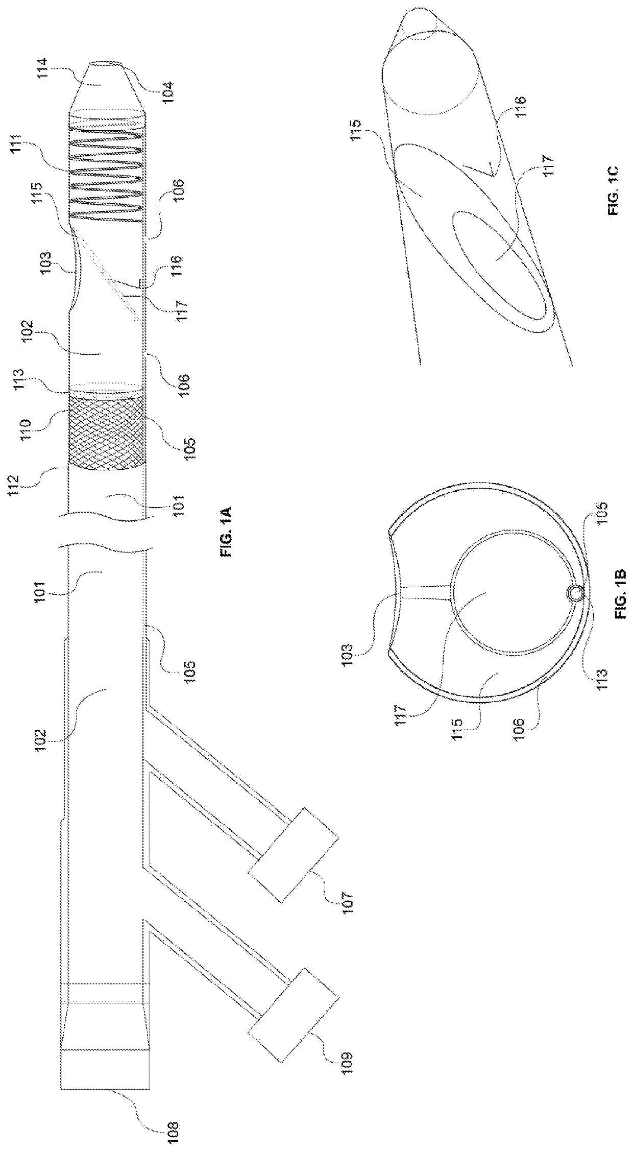
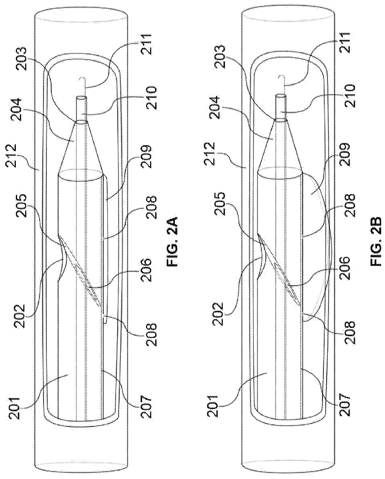
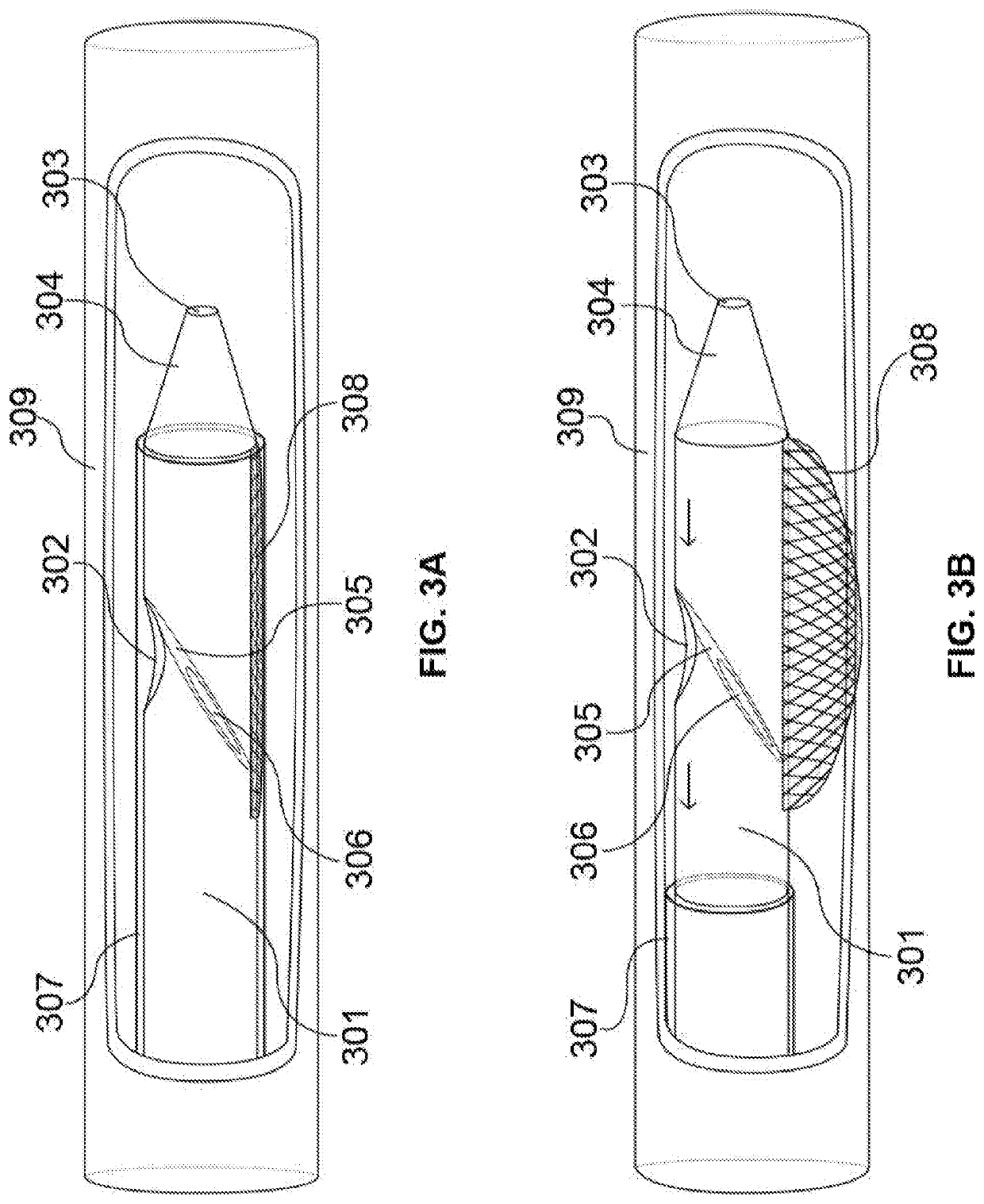
[0065]The present disclosure describes a system in which a guide / access catheter provides an endovascular conduit for co-axial catheter systems and transcatheter instrumentation to transvascularly access the subdural space, subarachnoid space, and the brain parenchyma—for targeted device delivery or implantation and / or tissue / media insertion / collection without the needs for burr holes or craniotomy. Various novel neuroendovascular transvenous access / guide catheter designs and a variety of clinical applications that the catheter(s) would enable are described herein, such as for the diagnosis and treatment of seizure disorder, pathologic brain tissue (e.g., cancer, etc.), psychiatric disease—cognitive / motor impairment, and movement disorder.

[0066]FIG. 1A-C illustrates a schematic diagram of the access catheter device comprising tubular structures featuring proximal and distal ends operably connected through lumen(s), cut-away views of various layers and inner components / features, and ...


PUM


Abstract

The present disclosure discusses a devices, systems and methods for transvascular, transvenous and / or transdural access, to the brain parenchyma, subarachnoid or subdural spaces. In some embodiments, the disclosed systems and methods may be used for local drug delivery, tissue biopsy, nanofluidic or microelectronic device / component delivery / insertion / implantation, in situ imaging, ablation of abnormal brain tissue and the like. Embodiments of the present disclosure include an access catheter system for extravascular procedures in the brain having an elongate, flexible tubular body, with at least one lumen extending axially there through between a proximal end, and a distal end. The access catheter system may include a side exit port and a distal end port. Further, the access catheter system may include a selective deflector positioned within the lumen configured to deflect a procedure catheter and permit a guide catheter.

Description

REFERENCE TO CROSS-RELATED APPLICATIONS[0001]The present disclosure claims priority to and the benefit of U.S. Provisional Application No. 62 / 871,976, filed on Jul. 9, 2019, which is hereby incorporated by reference in its entirety.TECHNICAL FIELD[0002]The present disclosure is directed towards a device, and related systems and methods for transvascular access to extravascular spaces, particularly the brain, intracranial structures, and / or the subdural or subarachnoid spaces.BACKGROUND[0003]Access to brain tissue may be required to confirm or treat neurologic diseases involving malignancy, inflammation, aberrant circuitry, or neuropathology in general, as well as to investigate fundamental properties of the brain for basic science exploration. Access to samples of intracranial tissue (i.e., for biopsy, in situ imaging, or cytological analysis) can be used to provide prognosis, tailor treatments, and monitor responses to treatment, as well as advance scientific understanding of the b...

Claims


Application Information

Patent Timeline
Patent Type & Authority Applications(United States)
IPC IPC(8): A61B18/14A61B5/293A61B5/00A61B90/00A61N1/05A61N1/36
CPCA61B18/1492A61B5/293A61B5/6853A61B5/6859A61B2018/0016A61B90/37A61N1/0534A61N1/36067A61N1/36082A61B5/6868A61B2018/00446A61B2018/00577A61B2090/363A61B2090/378A61B2090/064A61B2018/00839A61B2018/00791A61B2218/002A61B2018/00166A61B2018/00285A61B2018/0063A61B2090/3784A61M2025/1047A61M2025/0092A61M2025/0042A61B18/24
Inventor MORALES, JOSE MIGUEL
Owner VONOVA INC
Learn More