
Bioreactor wound dressing

Inactive Publication Date: 2000-03-21
REPLICATION MEDICAL
View PDF32 Cites 115 Cited by
  • Summary
  • Abstract
  • Description
  • Claims
  • Application Information

AI Technical Summary

Benefits of technology

The dressing includes a permeable transport layer which is in the direct contact with a wound, and a reservoir layer containing an aqueous liquid with substances beneficial for the healing which are transported through the transport layer into the wound. Substances undesirable for the healing may be removed from the wound into the reservoir layer through the transport layer. The transport is driven either by a concentration gradient (i.e., diffusive permeability) or by a pressure gradient (i.e., hydraulic permeability). Optionally, the dressing also includes an outside protective layer and as well as a mechanism for exchange of the liquid inside the reservoir layer. The dressing is used for protection of wounds, for control of conditions (such as water content, osmolarity, pH) in wounds, for delivery of bioactive substances into wounds, for removal of harmful substances from wounds, for cultivation of human keratinocytes and other cells in vitro and for transport of such cultured cells into wounds, and for protection and support of the human cells proliferating in wounds.

Problems solved by technology

To date, no satisfactory dressing has been developed for difficult wounds, such as deep burns or chronic wounds including decubitus ulcers, venous stasis sores, radiation ulcers, pressure sores, ischemic ulcers or diabetic ulcers.
Treatment of these wounds remains a very long and expensive process.
One of the problems of occlusive dressings is accumulation of exudate in certain wound types.
The exudate interferes with the dressing adhesion and can be a locus of an infection.
However, hydrogel occlusive dressings do have several problems.
If they are applied to the wound dehydrated, they remove water from the exudate concentrating proteins and other large biomolecules and thus causing protein denaturation and osmotic imbalance.
Hydrogels do not perform too well in the absence of an exudate where the hydrogel is supposed to maintain the moist environment of the wound bed.
A dehydrated, or partially hydrated hydrogel cannot be a source of moisture.
Furthermore, a semihydrated hydrogel typically adheres to the wound bed so that the newly formed tissue is injured during the dressing change.
From this follows that various types of occlusive dressings are suitable for some types of the wounds and some phases of the healing (i.e., the exudative phase) but they are unsuitable for others.
There is currently no dressing that could satisfactorily control the wound bed conditions, particularly due to the variability of these conditions with time.
The dressing change disturbs the healing process and exposes the wound to airborne infections.
For that and other reasons, benefits of occlusive dressings are mostly limited to clean wounds, such as surgical incisions or skin donor sites.
However, they often do not facilitate the epithelization that is essential for the wound closure.
This method is laborious and, in some cases, it is difficult to excise a sufficient amount of patient's skin without aggravating his / her condition even further.
One of the disadvantages is also the cross-hatched pattern left by the autologous graft on the newly formed skin.
These limitations of both current synthetic occlusion dressings and autografts lead to continuing search for better "biological" dressings.
However, the process of healing and integration of the matrix into the new tissue is slow and sometimes uncertain.
The keratinocyte sheet is very fragile.
The direct contact may be difficult to achieve on the irregular wound surface.
The removal of the dressings can damage the newly formed epidermal layer.
This is not very convenient since the cultivation takes approximately 14 to 21 days and is very expensive.
Consequently, even foreign keratinocytes do not elicit a strong rejection if applied as an allograft.
However, this approach has still several problems.
The first problem relates to the fact that keratinocytes grown on the polymeric support are nutrified from the solution, i.e. from the other side than in a living skin where they are fed from the dermis underneath.
This limits the efficacy of the keratinocyte cultivation and delivery.
The second problem derives from the requirement that keratinocytes should adhere well to the support to grow and multiply properly.
However, as long as they firmly adhere to the polymeric membrane, they are not released to seed the wound.
The active biomolecules from the attached keratinocytes can reach the wound bed by diffusion only and their efficacy is thus diminished.
The third problem is that as keratinocytes are applied to the wound, they are transferred from the nutrient solution optimized for their cultivation to the wound bed that is not necessarily a good environment for the keratinocyte growth.
Highly hydrated, well permeable hydrogels are typically a poor support for tissue cultures.
Tissue cells do not adhere to them and if they do, they adhere weakly, do not spread and maintain their rounded shape and are unsuitable for their proliferation.
Hydrogels with a low water content are not permeable for many of the nutrients and factors required by keratinocytes, so that they can provide only a limited support for their growth both during the cultivation and after their transfer to the wound.
Moreover, hydrogels with a low water content are typically rather rigid so that they cannot conform well to the uneven surface of the wound.
However, it has also several disadvantages, such as possibility of the infection transmission, possibility of immune reaction, limited range of mechanical and diffusion properties, high handling costs, non-transparency, etc.
The disadvantage of such dressings is very high cost and poor shelf life.

Method used


Image

  • Bioreactor wound dressing
  • Bioreactor wound dressing
  • Bioreactor wound dressing

Examples


example 1

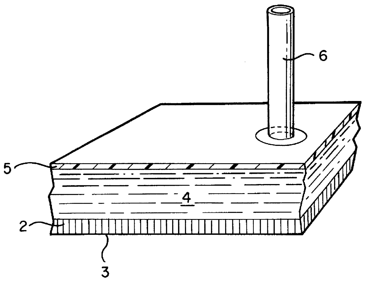
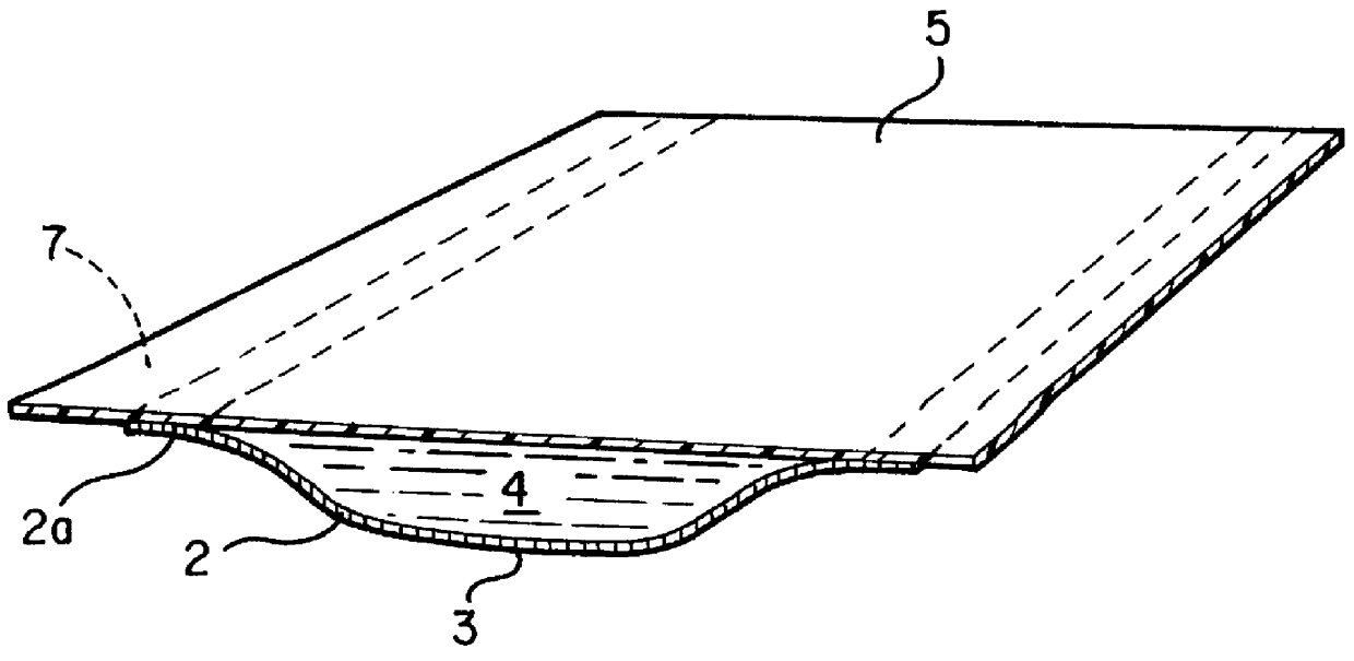
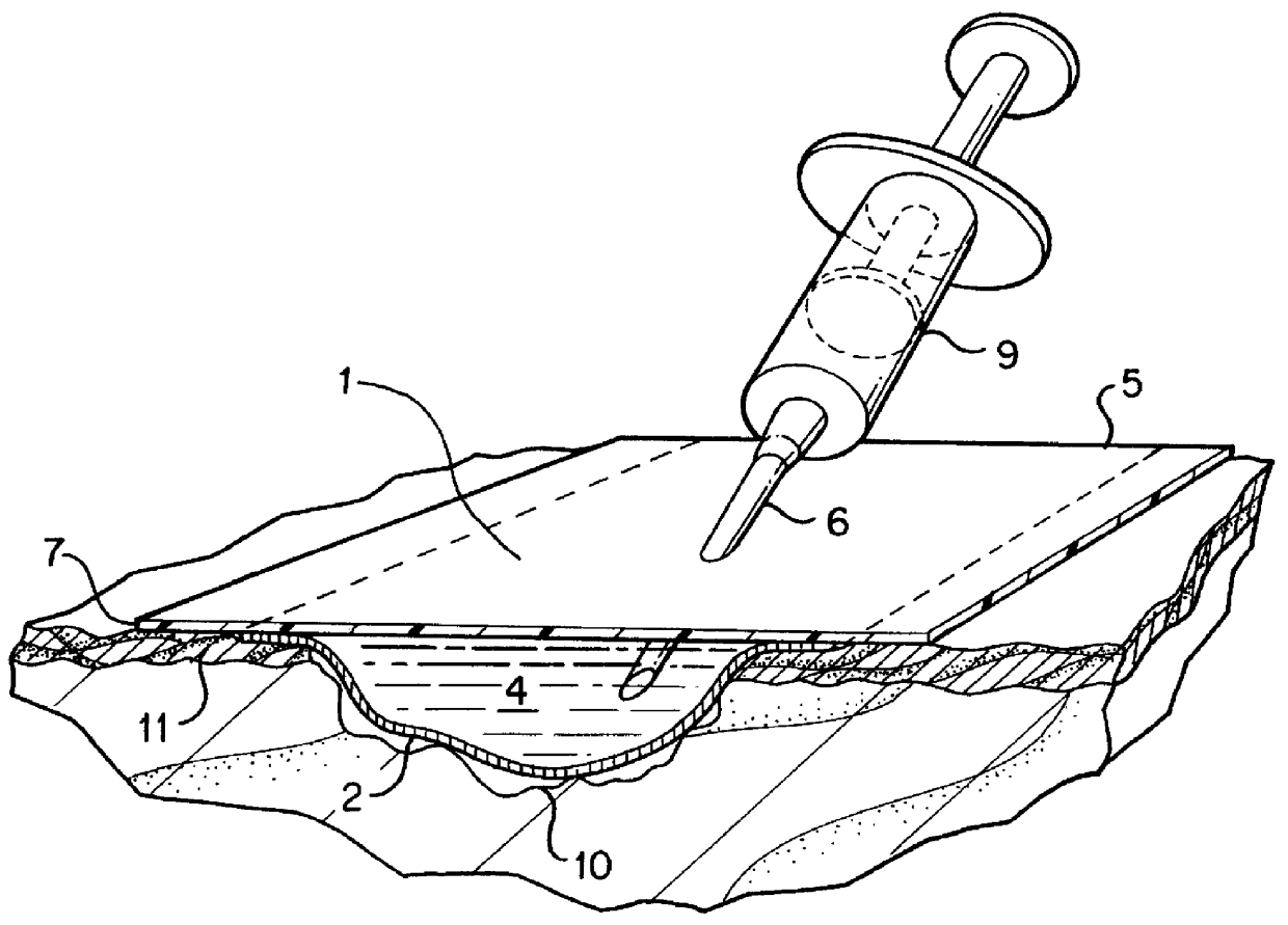
The dressing 1 is described in the FIG. 2 where 2 is the transport layer from the permeable material described below; 5 is the protective layer that is connected to the periphery zone 2a of the transport layer 2, forming a space wherein an aqueous liquid comprising a hydrogel dispersion is confined. That space is filled with the liquid forms the Reservoir Layer 4.

The protective layer 5 is made of a commercial foil of clear polyurethane polymer covered by a thin layer of acrylic pressure-sensitive adhesive.

The transport layer 2 is made from a highly permeable acrylic hydrogel based on a partially hydrolyzed polyacrylonitrile. The partial hydrolysis of polyacrylonitrile is carried out by a basic catalyst and yields a multiblock copolymer containing residual nitrile groups, sodium carboxylate groups, amidine groups and amide groups in such a proportion that the liquid content in the hydrogel is more than 90% by weight in the equilibrium with 0.9% by weight of NaCl aqueous solution. The...

example 2

The dressing from the Example 1 can be used for keratinocyte cultivation for wound healing. The transport layer is modified by exposure to bovine serum for 48 hours from the cultivation surface side. The dressing is then surface dried, packaged into a semipermeable poach and autoclaved for 30 minutes at 123.degree. C. The deposited layer of denatured proteins on the cultivation surface improves adhesion of cells in the subsequent steps. The procedure is carried out in the following steps:

1) A biopsy sample is taken from the skin of a patient suffering by a chronic or acute wound. The skin sample is treated with trypsin to release keratinocytes.

2) The dressing's 1 cavity 4 is filled with the solution A and placed into a Petri dish filled with the solution A. The dressing is turned so that the cultivation surface faces upwards. 3T3 mouse fibroblasts are irradiated by a lethal dose of Gamma radiation and seeded onto the cultivation surface.

3) After 24 hours in an incubator at 37.degree...

example 3

The transport layer is made from a hydrogel prepared by the following method:

100 weight parts (w.p.) of 2-hydroxyethylmethacrylate monomer (HEMA), 450 weight parts of water, 450 weight parts of ethyl alcohol and 3 w. p. of hydrogen peroxide was stirred for 8 hours at 65.degree. C. under reflux condenser and nitrogen protective atmosphere. The resulting viscous solution is cooled and poured into an excess of water. The white precipitate is formed and washed in deionized water for several days. The purified uncrosslinked HEMA polymer (PHEMA-S) is cut to small pieces and dried at 60.degree. C. to constant weight. The resulting glassy polymer is ground to a crude powder and stored in closed containers.

10 w.p. of the PHEMA-S powder is dissolved in 80 w.p. of ethyl alcohol, 5 parts of water, 5 parts of 1,2 propylene glycol and 0.1 part of phosphoric acid. This solution is spread into a thin layer over a polyester mesh, let dry at 60.degree. C. for 3 hours and then heated to 120.degree. C....


PUM

PropertyMeasurementUnit
Fractionaaaaaaaaaa
Fractionaaaaaaaaaa
Percent by massaaaaaaaaaa

Abstract

The present invention is a bioreactor wound dressing which includes a first layer, being a transport layer, in direct contact with a wound. It includes at least one layer of a permeable polymeric media containing, in equilibrium with body fluids, at least 40% by weight of liquid, and is impermeable for infectious agents of any kind and being permeable to water soluble substances having molecular weight up to at least 1000 Daltons. There is a second layer, being a fluid reservoir layer that is adjacent to the transport layer and is capable of containing between 40% and 100% of its volume of an aqueous liquid, wherein the transport layer and reservoir layer are permeably interconnected for aqueous solutions and are in a substantial osmotic equilibrium. The invention also includes a method of wound treatment utilizing the bioreactor wound dressing.

Description

1. Field of the Invention1. The present invention relates to wound dressings and more specifically to bioreactor wound dressings which may be used for intentional wounds--surgical incisions and graftings, as well as accidental and unintentional wounds, including cuts, penetrations, burns, sores, ulcers and other deteriorating or damaged skin and flesh wounds.2. Information Disclosure StatementTo date, no satisfactory dressing has been developed for difficult wounds, such as deep burns or chronic wounds including decubitus ulcers, venous stasis sores, radiation ulcers, pressure sores, ischemic ulcers or diabetic ulcers. Treatment of these wounds remains a very long and expensive process.It has been found that moist wounds often heal faster. This can be achieved if the desiccation of the wound is prevented by slowing down evaporation of water from the wound. Numerous types of occlusive wound dressings have been developed that are designed to maintain the favorable moist environment in...

Claims


Application Information

Patent Timeline
IPC IPC(8): A61F13/02A61L15/40A61L15/20A61L15/16A61L15/32A61L15/60A61F13/00A61F13/15
CPCA61F13/023A61L15/20A61L15/32A61L15/40A61L15/60A61L15/44A61L15/42A61F2013/00157A61F2013/00748A61F2013/00846A61F2013/15024Y10T428/249939Y10T428/249994
Inventor COOKE, RANDOLPH L.STOY, VLADIMIR A.
Owner REPLICATION MEDICAL
Learn More