Optical sensors for the detection of nitric oxide

a technology of optical sensors and nitric oxide, applied in the field of optical sensors, can solve the problems of difficult intracellular measurement, large size, difficult use for cellular nitric oxide measurement, etc., and achieve the effect of small size and good sensitivity

Inactive Publication Date: 2005-05-31
RGT UNIV OF MICHIGAN
View PDF8 Cites 25 Cited by
  • Summary
  • Abstract
  • Description
  • Claims
  • Application Information

AI Technical Summary

Benefits of technology

[0013]In another embodiment, the nitric oxide-binding compound is a porphyrin group- or heme group-containing protein. In another embodiment, the nitric oxide-binding compound is a heme-binding protein. Regardless of whether the protein is a heme-group-containing protein or a heme-binding protein, in one embodiment, the present invention contemplates that the protein (or peptide) is dye-labeled (e.g., with dyes which can be used for protein labeling that do not react to nitric oxide, such as Oregon Green dyes). This has been found to increase the signal to noise ratio of the sensors of the present invention.
[0020]The invention also contemplates optical fiberless sensors capable of detecting nitric oxide. The sensors of the present invention are: (1) small enough to enter a single mammalian cell relatively non-invasively, (2) fast and sensitive enough to catch even minor alterations in the concentration of nitric oxide and (3) mechanically stable enough to withstand the manipulation of the sensor to specific locations within the cell. Importantly, the fiberless sensors of the present invention are non-toxic and permit the simultaneous monitoring of several cellular processes.

Problems solved by technology

The drawbacks of this sensor include the following: 1) the response time (approximately 8-17 seconds) is longer than the time needed for nitric oxide in the solution to be converted to nitrite; 2) the detection of nitric oxide is complicated by interferences from dopamine, uric acid, ascorbic acid, and cysteine, 3) the sensor is relatively large in size (greater than 6 mm in diameter) and thus difficult to use for the measurement of cellular nitric oxide levels (and impossible for intracellular measurements); and 4) the sensor has relatively poor sensitivity, i.e., a relatively high limit of detection (approximately 1.3 mM of nitric oxide).
Unfortunately, the sensor reaction is reported to have taken two hours to reverse, making dynamic measurements impossible.

Method used


Image

  • Optical sensors for the detection of nitric oxide
  • Optical sensors for the detection of nitric oxide
  • Optical sensors for the detection of nitric oxide

Examples


example 1

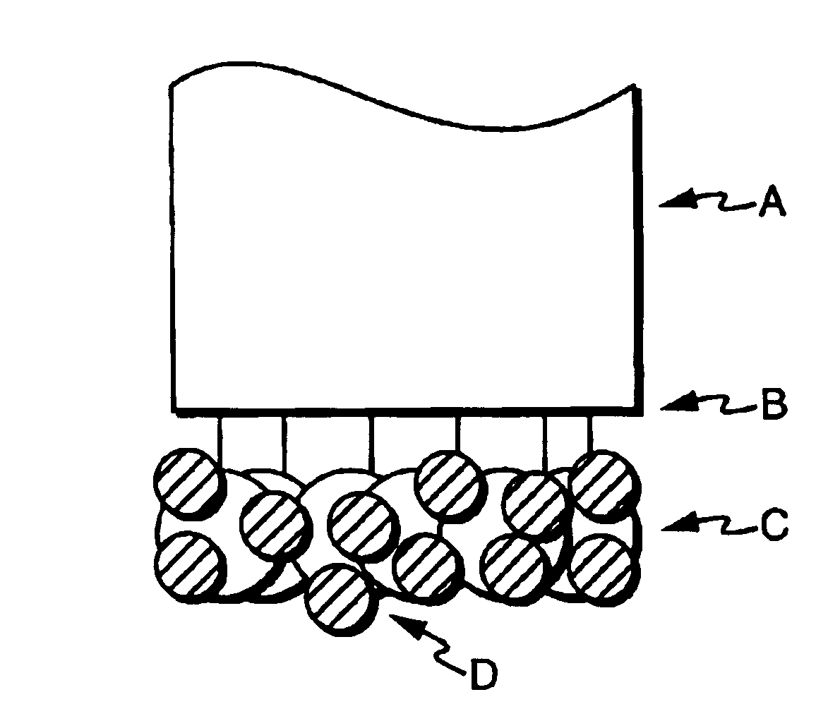
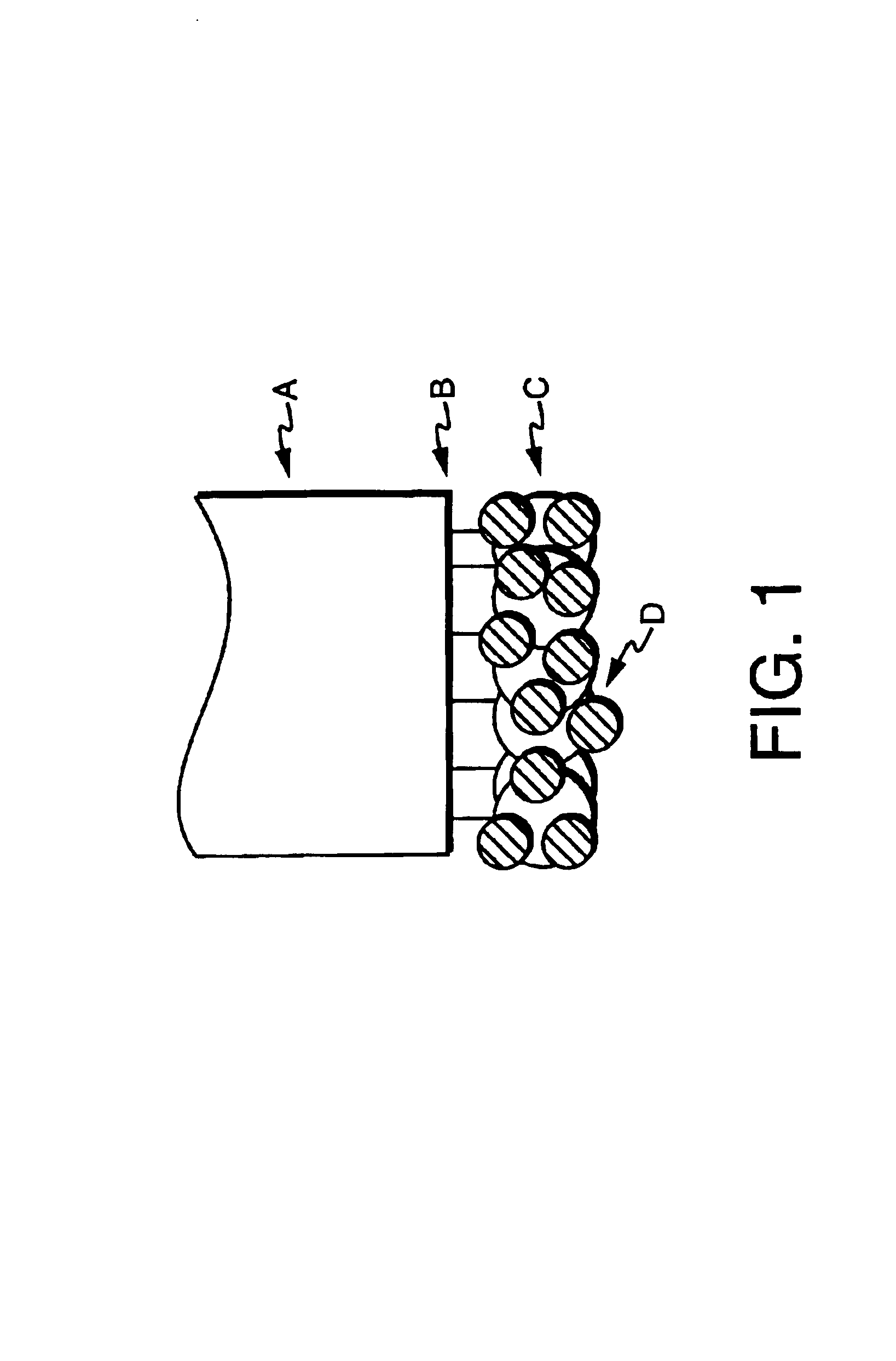
[0125]In this example, sensors were prepared by silanizing a freshly cleaved multimedia fiber by immersing the distal end for two hours in 3-(mercatopropyl) trimethoxysilane, thereby modifying the fiber with reactive groups. The end was then rinsed copiously with methanol, then tripley distilled water. The silanized fiber was placed in colloidal gold (used as received from the manufacturer) for three hours, then rinsed with water. The sensor can be stored at this point in water or air.

[0126]Two protein solutions were prepared. The first solution was prepared by dissolving 0.1% r-phycoerythrin (Molecular Probes, Eugene, Oreg.) in phosphate buffer, pH 6.0. The fiber was then immersed in the protein solution for one hour, rinsed with buffer, then used.

[0127]A second protein solution was prepared by dissolving 0.1% bovine serum albumin (BSA) in phosphate buffer, pH 6.0. The fiber was then immersed in the protein solution for one hour, rinsed with buffer, then used. After BSA attachment,...

example 2

[0130]In this example, pulled sensors are described. Sub-micron sized fiber optic sensor were prepared by pulling multimedia fiber in a home-built puller consisting of a modified pipette-puller heated with a CO2 laser. The pulled tips were then silanized, coated with aluminum in a home-built evaporator and prepared with gold colloid and protein as above.

example 3

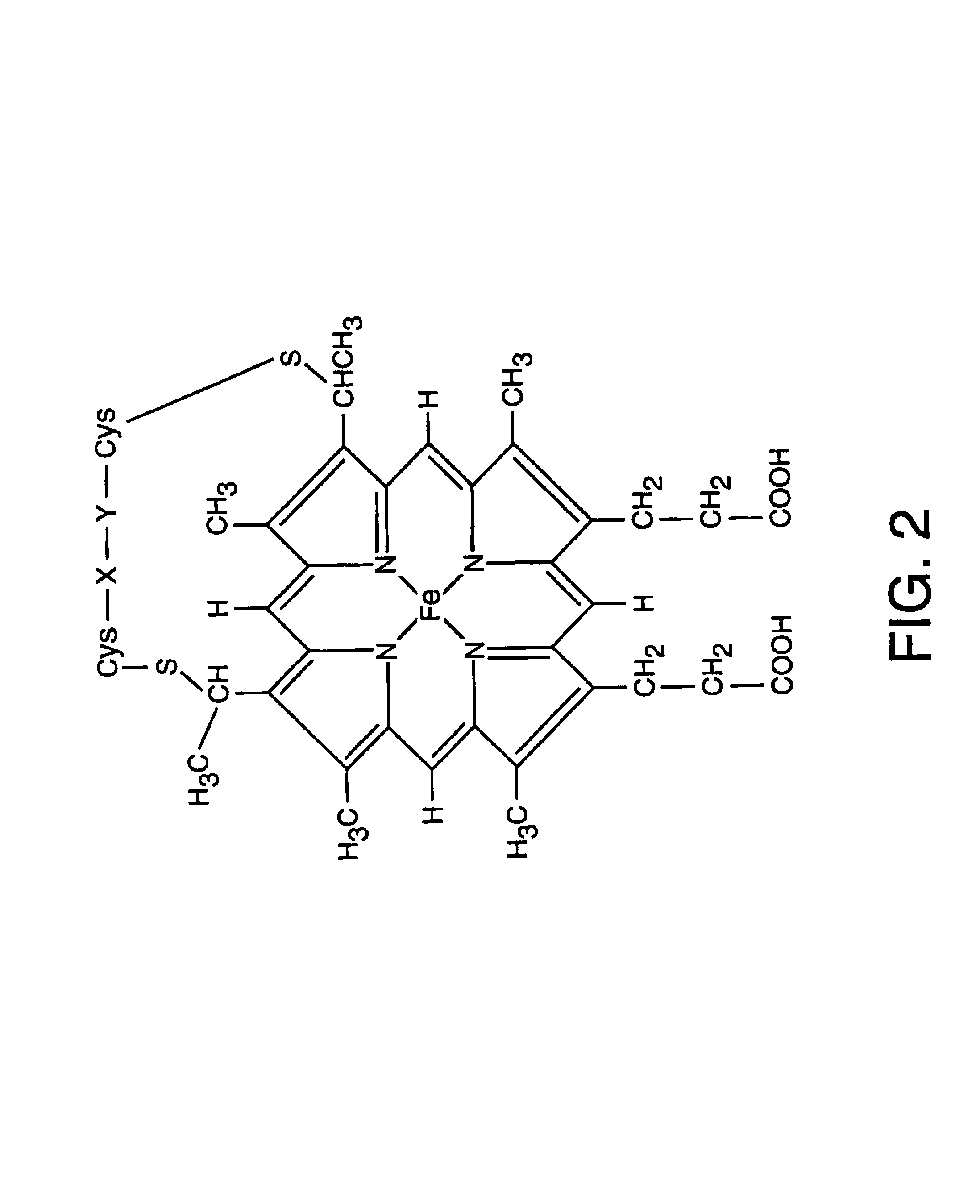
[0131]This example describes the first sensor to incorporate cytochrome c′. The heme group of this nitric oxide-binding protein is shown in FIG. 2. Cytchrome c′ was chosen as the chemical recognition element because it exhibits spectral changes upon binding nitric oxide and is highly selective. While a precise understanding of the mechanisms involved is not necessary for the practice of the invention, it is believed that, as the sixth ligand site of cytochrome c′ is buried within the protein, it is usually accessible only to carbon monoxide and nitric oxide. However, cytochrome c′ undergoes autoxidation to Fe(III), which binds nitric oxide, but not carbon monoxide.

[0132]To prepare the sensor, 100 μm core diameter multimedia fibers (General Fiber Optics, Cedar Grove, N.J.) or multimedia fibers were pulled to a submicrometer tip diameter and were silanized for 2 hrs in neat 3-mercaptopropyl-trimethoxy silane (Gelest, Inc., Tullytown, Pa.) in a well-ventilated fume hood. The fibers wer...


PUM

PropertyMeasurementUnit
response timeaaaaaaaaaa
diameteraaaaaaaaaa
sizesaaaaaaaaaa

Abstract

Optical fiber sensors and fiberless optical sensors for measuring analytes, and in particular nitric oxide, are described utilizing metals, and more particularly, metal colloids. Proteins (or fragments thereof) with selective binding are immobilized on metal particles. The proteins may be dye-labeled for increased sensitivity.

Description

[0001]“This is a Continuation of application(s) Ser. No. 09 / 365,487 filed on Aug. 2, 1999 now U.S. Pat. No. 6,636,652.”[0002]The present invention was made with government support from NIH Grant 1 RO 1 GM50300 04A1 and DARPA grant MDA972-97-0006. The government has certain rights in this invention.FIELD OF THE INVENTION[0003]The invention relates generally to optical sensors, methods of sensor fabrication and uses of such sensors, and more particularly the use of such sensors for the detection of nitric oxide.BACKGROUND[0004]Recent elucidation of the fact that nitric oxide plays many biological roles has spurred special interest in this molecule. For instance, nitric oxide is believed to play a role in vasodilation. See Marletta et al., “Unraveling the biological significance of nitric oxide”Biofactors 2:219 (1990). Nitric oxide also appears to inhibit platelet aggregation by elevating intraplatelet levels of cyclic GMP. See Diodati et al, “Complexes of Nitric Oxide with Nucleophile...

Claims


Application Information

Patent Timeline
Patent Type & Authority Patents(United States)
IPC IPC(8): G01N21/64G01N21/77
CPCB82Y15/00B82Y30/00G01N21/6408G01N21/6428G01N21/7703G01N2021/7786G01N2021/6439G01N2021/6482G01N2021/6484G01N2021/772
Inventor KOPELMAN, RAOULCLARK, HEATHERBARKER, SUSAN
Owner RGT UNIV OF MICHIGAN
Why Eureka
Try Eureka