

System and methods for electrophoretic separation of proteins on protein binding membranes
a protein and membrane technology, applied in the field of electrophoretic separation of proteins, can solve the problems of complex and time-consuming separation of protein molecules, high study and effort required to unravel the complete complement and function of human proteome, and the resolution of proteins electrophoresed through gels under non-denaturing conditions is usually poor, and achieves low conductivity of organic solvent buffer, high protein binding capacity, and easy separation
- Summary
- Abstract
- Description
- Claims
- Application Information
AI Technical Summary
Benefits of technology
Problems solved by technology
Method used
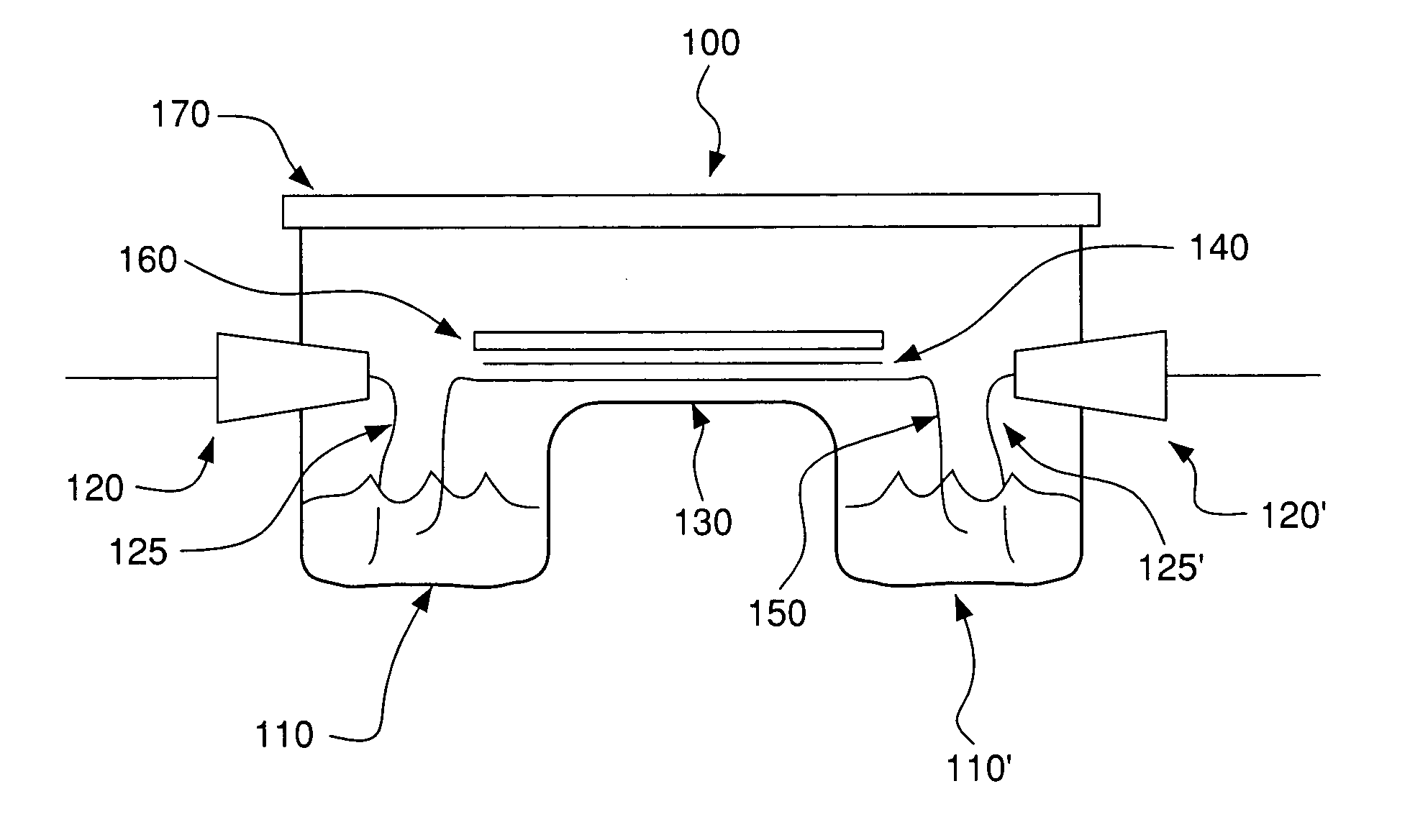
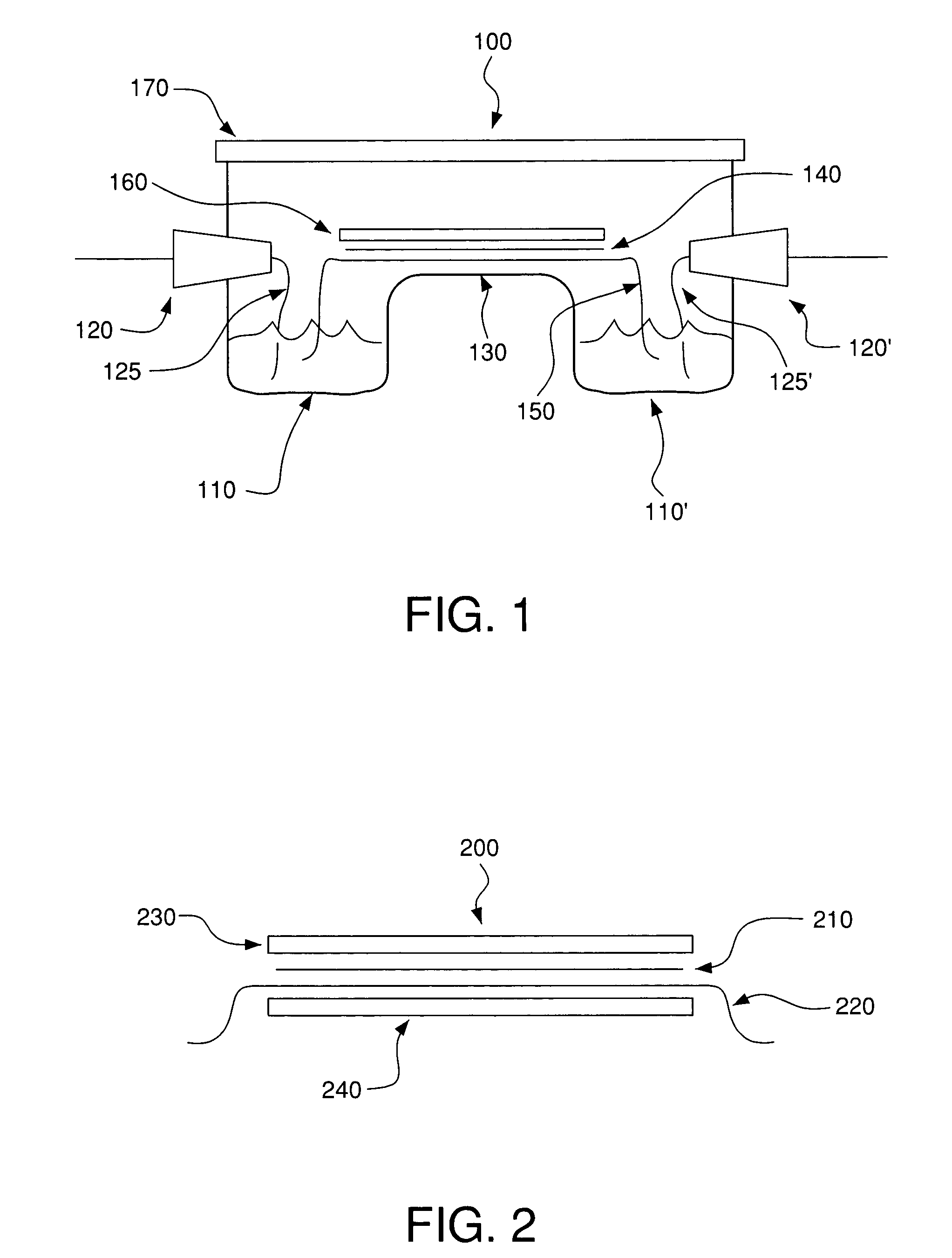
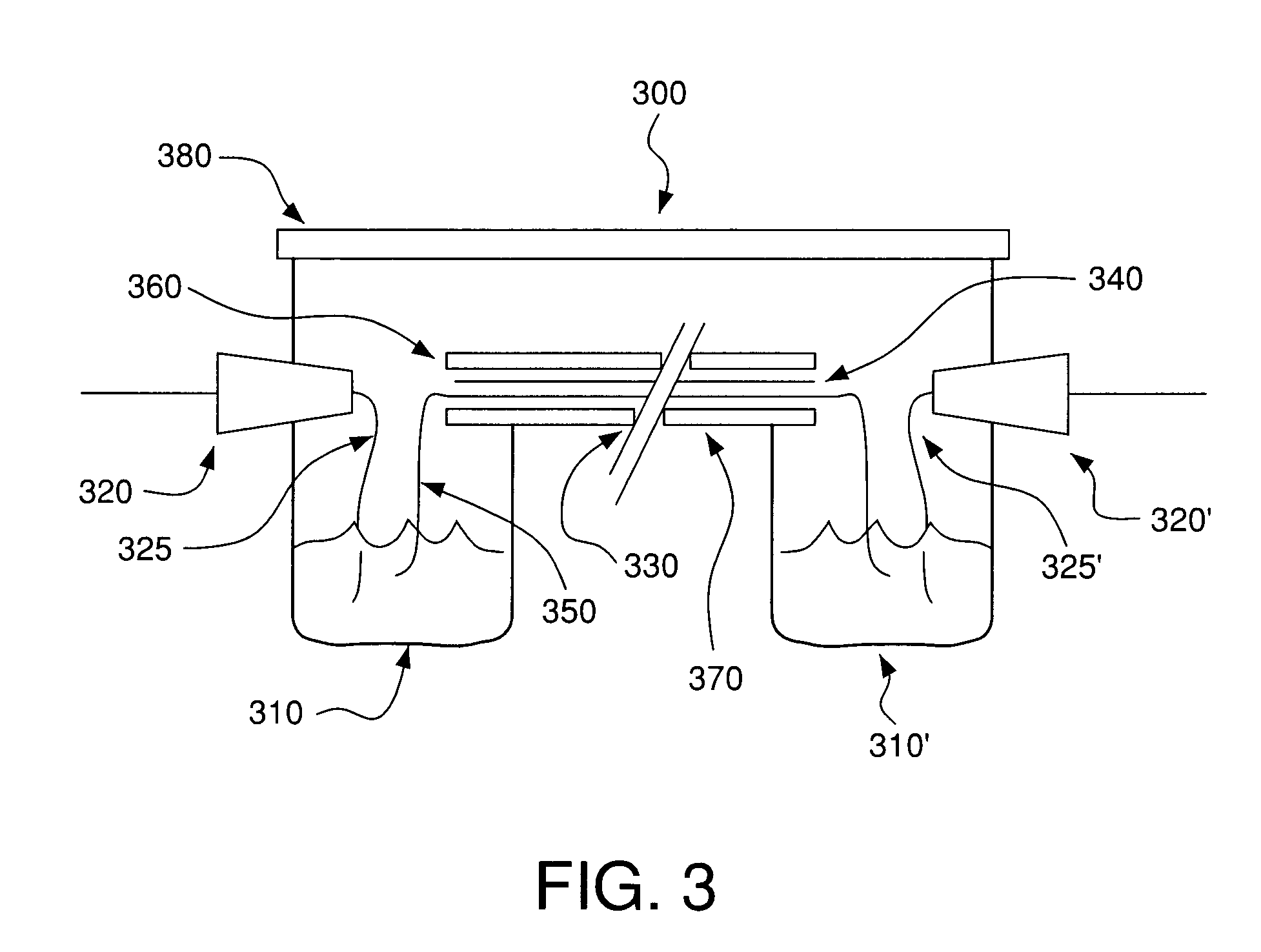
Image

Examples
example 1
Low Conductivity Organic Solvent Buffers for Membrane Electrophoresis
[0114]Low conductivity organic solvent buffers according to the invention were formulated as follows:
[0115]Buffer A—The conductivity enhancers salicylaldehyde (5 ml) and furfuryl alcohol (3 ml) were added to the base solvent ethylene cyclic carbonate (7 ml). It is necessary to melt ethylene cyclic carbonate prior to use. Electrophoresis of sample proteins at 3.5 kV on 1 cm by 8 cm strips as in Example 2 quickly produced a current over 1.5 mA and generated excessive heat. The addition of a mixture of conductivity suppressors 1,3-butanediol, dimethyl formamide and dimethyl acetamide reduced the current to 0.1 mA and eliminated the heat generation with minimal reduction in protein migration rates. The final formulation of Buffer A was:[0116]28% ethylene cyclic carbonate[0117]20% salicylaldehyde[0118]12% furfuryl alcohol[0119]8% 1,3-butanediol[0120]16% dimethylformamide[0121]16% dimethylacetamide
[0122]The pH of Buffer ...
example 2
Evaluation of Membranes for Membrane Electrophoresis
[0144]Determination of Membrane Compatibility with the Buffers—The following polymeric membranes were tested for their compatibility with Buffer A or Buffer B from Example 1: polyvinylidene difluoride (PVDF), nitrocellulose, supported nitrocellulose, cellulose acetate, DEAE-cellulose, Hybond™-N, Hybond™-NX (both are neutral nylon membranes, but as indicated earlier, they may be slightly charged), Hybond™-XL, and Hybond™-N+ (both are modified and highly positively charged nylon membranes). Each membrane was cut into 1 cm×8 cm strips and wetted with the either Buffer A or Buffer B. Of the membranes examined, the cellulose derived membranes (e.g. nitrocellulose, cellulose acetate and DEAE cellulose) were completely destroyed by the buffers soon after contact, rendering them useless for membrane electrophoresis. The remaining membranes were resistant to the organic solvents in the buffers, and were tested further for their suitability ...
example 3
One-Dimensional Membrane Electrophoresis
[0150]Six proteins having isoelectric points ranging from 3.6 to 9.6 were electrophoresed on a 7.5 cm by 8 cm PVDF membrane as in Example 2, in Buffer A at 3.5 kV for five minutes. After electrophoresis, the proteins were stained with Reactive Brown as in Example 2. The six proteins were: amyloglucosidase (pI 3.6); glucose oxidase (pI 4.2); β-lactoglobulin A (pI 5.1); myoglobin (pI 6.8 and 7.2); lentil lectin (pI 8.2, 8.6 and 8.8) and cytochrome C (pI 9.6). As shown in FIG. 5, the proteins were separated according to their isoelectric points. These results indicate that the charges on protein molecules are an important factor in their migration during membrane electrophoresis.
[0151]Since cytochrome C with a pI of 9.6 is separated using an organic solvent buffer having a pH of 4.5, this indicates that proteins with pI's that are 5 units away from the pH of the organic solvent buffer can be effectively separated by the present methods.
PUM
| Property | Measurement | Unit |
|---|---|---|
| pH | aaaaa | aaaaa |
| pH | aaaaa | aaaaa |
| thick | aaaaa | aaaaa |
Abstract
Description
Claims
Application Information