Micro CT scanners incorporating internal gain charge-coupled devices

a micro-ct scanner and charge-coupled technology, applied in the field of imaging systems, can solve the problems of cumbersome system operation, difficult use of this powerful modality for functional studies such as tumor vascular physiology or involving true whole organ physiology imaging, etc., and achieve the effect of improving imaging capabilities

Active Publication Date: 2008-04-01
RADIATION MONITORING DEVICES
View PDF24 Cites 73 Cited by
  • Summary
  • Abstract
  • Description
  • Claims
  • Application Information

AI Technical Summary

Benefits of technology

[0016]The present invention relates to a microCT system that comprises an x-ray detector that is capable of imaging at a rate of up to 300 frames per second (fps). The present invention also provides an x-ray generator that better takes advantage of the improved imaging capabilities made possible by the x-ray detector of the present invention.
[0017]The CT systems of the present invention allow for volumetric CT imaging of small animals in seconds compared to several minutes needed for the conventional CT systems. The systems of the present invention have considerable potential as a research tool for dynamic studies, particularly for the study of physiology in small animals, and in other important applications in medical imaging.
[0019]Such CCDs combine the high resolution and low noise of a CCD, and the internal gain of an avalanche photo diode (APD). The presence of an internal gain effectively reduces the read noise even when operated at high frame rates of 300 fps, thus improving the image signal-to-noise ratio (SNR).
[0020]The microcolumnar CsI(Tl) scintillator converts incident x-rays into visible light with very high efficiency and, by virtue of its structure channels the light towards the CCD. This minimizes the traditional tradeoff between spatial resolution and detection efficiency. Furthermore, with its fast scintillation decay, the CsI(Tl) allows rapid x-ray imaging without image blurring arising from residual intensity from previous exposures. Thus, the combination of a microcolumnar CsI(Tl) scintillator and an internal gain CCD can provide very high sensitivity, for example with a range of about 90% to about 98%, high spatial resolution, such as for example, from about 50 to about 70 μm, and a substantially improved SNR, such as, for example an improvement of about 10 times over currently available units.
[0021]The x-ray detectors of the present invention typically comprise a large active area and have a high speed of operation permitting high-speed data acquisition through a whole organ, such as the brain, thus enabling construction of temporal curves of the contrast passage for each individual voxel.

Problems solved by technology

However, use of this powerful modality for functional studies such as those of tumor vascular physiology or involving true whole organ physiologic imaging is difficult due to the lack of a suitable x-ray imaging detector.
The majority of the studies performed using these instruments focused on high-density tissue such as bone or teeth for which magnetic resonance imaging is less successful.
For in vivo small animal studies, particularly large population studies, these systems become cumbersome to operate because the subject must be confined in a rotating carrier designed to prevent soft-tissue organ motion.
While the above systems are capable of mass production of small animal CT systems, at present they do not incorporate high speed CT technology.
It is important to note that some of the existing microCT systems are not capable of in vivo imaging, while others having this capacity are limited by their speed of operation resulting in data acquisition times of several minutes to hours.
Thus, the existing systems cannot be used for high-speed dynamic, i.e., functional, studies, where high sensitivity and high-speed x-ray imaging is required.
Most of the current microCT systems employ CCD based detector arrays which provide adequate spatial resolution (50 to 100 μm) and imaging area (5 cm×5 cm), but offer limited imaging speeds of 1 to 5 seconds per projection.
This translates into volumetric data acquisition times in the range of 5 to 30 minutes for high resolution imaging, making it impossible to use them for functional studies.
As microCT devices are not yet capable of functional imaging, important functional and physiologic studies in animal models of various diseases such as solid tumors, stroke physiology, and acute myocardial ischemia, are performed using existing clinical CT scanners.
Additional difficulties arise from the fact that the image voxels are anisotropic, which lead to severe partial volume artifacts in small animal imaging, where each voxel represents a heterogeneous sample of various tissue components and the calculated physiologic parameters represent an average physiological behavior at best.
The limitations of current CCD based microCT x-ray imaging systems arise from two factors.
First, readout speeds are curtailed in order to minimize the system read noise, which increases with the readout rates.
Recently developed CCD based x-ray imaging systems address the speed issue to some extent, however, at the cost of reduced SNR, pixel resolution, and sensitivity.
Second, the CCDs themselves are not used for x-ray detection.
In most systems that typically use Gd2O2S (GOS) phosphors, such as the Kodak® MinR-2000, the sensitivity is limited due to the tradeoff between x-ray stopping power and spatial resolution.
Thicker GOS screens improve sensitivity, but reduce the spatial resolution because the light photons spread from the conversion point, and scatter as well.
This is due to the fact that microCT devices are not yet capable of the high processing speeds and resolution necessary for functional imaging.
However, similar advances in the field of myocardial infarction (MI) have been impeded by the challenge of performing coronary ligation during survival surgery in animals weighing 30 g or less.
Thus the surgical challenge has now given way to the technical challenge of assessing myocardial infarction, left ventricular dimensions, and cardiac function in very small animals with high heart rates (>500 bpm).
Although it is possible to non-invasively assess myocardial perfusion and infarction in patients with radionuclide techniques, the limited spatial and temporal resolution of, for example, Single Photon Emission Computed Tomography (SPECT), effectively precludes its application in mice.
Although the utility of MRI for imaging cardiac structure, function, and infarct size in mice has been well established, there are several disadvantages with MRI that limit its widespread application in biomedical research.
First, a high cost associated with high field strength (e.g., ≧4.7 T) MRI imaging limits its accessibility to researchers.
Second, the high field strength magnets preclude the use of any metallic objects in or on the animal or as part of support equipment.
Third, the overall time for setup and imaging small animals with MRI can be quite lengthy.
Finally, due to the high heart rates in small animal models, particularly mice (>500 BPM), cardiac MRI does not provide the required temporal resolution to capture the first-pass of a contrast agent in order to obtain the input function that would allow the assessment of myocardial perfusion in the mouse.

Method used


Image

  • Micro CT scanners incorporating internal gain charge-coupled devices
  • Micro CT scanners incorporating internal gain charge-coupled devices
  • Micro CT scanners incorporating internal gain charge-coupled devices

Examples


Embodiment Construction

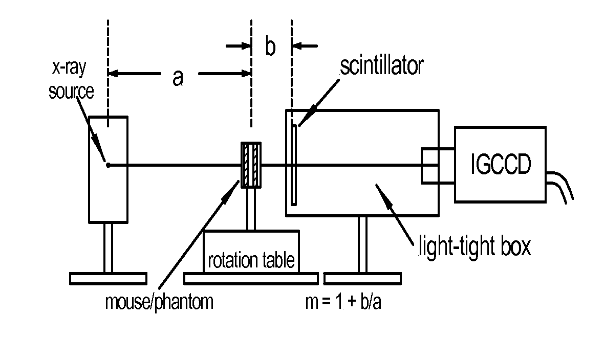
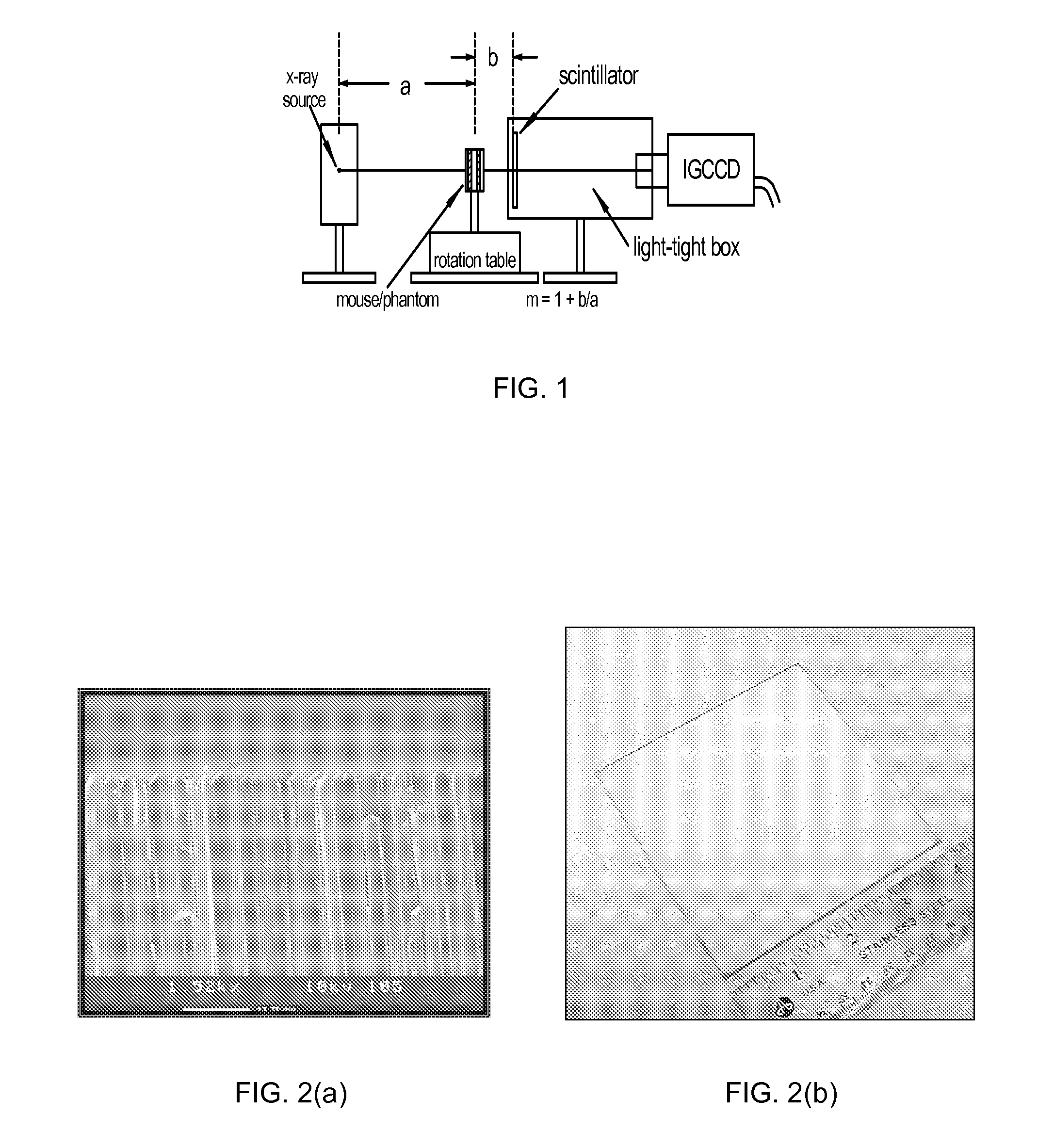
[0045]FIG. 1 schematically illustrates a high speed CT scanner system that is encompassed by the present invention. The CT scanner system generally comprises a source of penetrating radiation (e.g., x-ray source—typically an x-ray tube), an assembly for either rotating the x-ray source around the subject or for rotating the subject; a detector assembly; associated processing electronics; and a computer and software for image reconstruction, display, manipulation, post-acquisition calculations, storage and retrieval. The detectors may either be stationary or the detectors may be rotating. Alternatively, the CT system may include means to rotate a subject placed within the imaging volume and the CT assembly would then remain fixed in space.

[0046]The detector assembly can vary in the detection principles. For instance, some CT scanners have used gas detectors, where x-rays are converted to ionization electrons and positive ions in the gas, which then drift and are collected at electrod...


PUM


Abstract

The present invention provides internal gain charge coupled devices (CCD) and CT scanners that incorporate an internal gain CCD. A combined positron emission tomography and CT scanner is also provided.

Description

CROSS REFERENCE TO RELATED APPLICATIONS[0001]The present invention claims benefit to U.S. provisional patent application Ser. No. 60 / 581,478, filed Jun. 21, 2004, and is related to and claims priority to commonly owned and now abandoned provisional patent application entitled “Combined Radionuclide and X-Ray Imaging Device,” Ser. No. 60 / 581,400, filed Jun. 21, 2004, and its non-provisional U.S. patent application Ser. No. 11 / 158,938, filed Jun. 21, 2005 the complete disclosures of which are incorporated herein by reference.STATEMENT AS TO RIGHTS TO INVENTIONS MADE UNDER FEDERALLY SPONSORED RESEARCH AND DEVELOPMENT[0002]This work was supported by grant number 1 R43 CA0187-01 from the National Institutes of Health. The United States Government may have certain rights in the invention.BACKGROUND OF THE INVENTION[0003]The present invention relates generally to imaging systems. More specifically the present invention relates to novel micro-CT scanners that incorporate internal gain charg...

Claims


Application Information

Patent Timeline
Patent Type & Authority Patents(United States)
IPC IPC(8): A61B6/03G01T1/161
CPCA61B6/032A61B6/4225G01T1/1611G01T1/1615G01T1/2985H04N5/37213A61B6/507A61B6/508A61B6/037H04N5/32A61B6/583A61B2503/40H04N25/713
Inventor NAGARKAR, VIVEK V.TIPNIS, SAMEER V.
Owner RADIATION MONITORING DEVICES
Why Eureka
Try Eureka