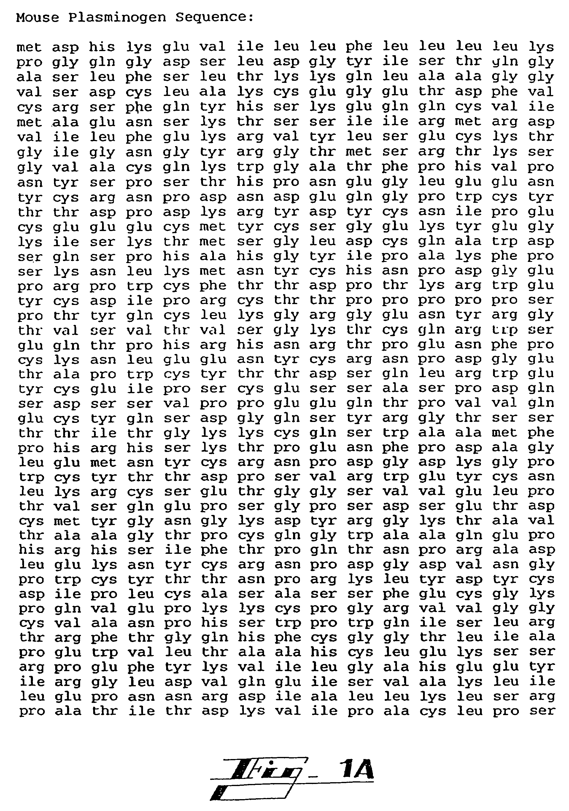
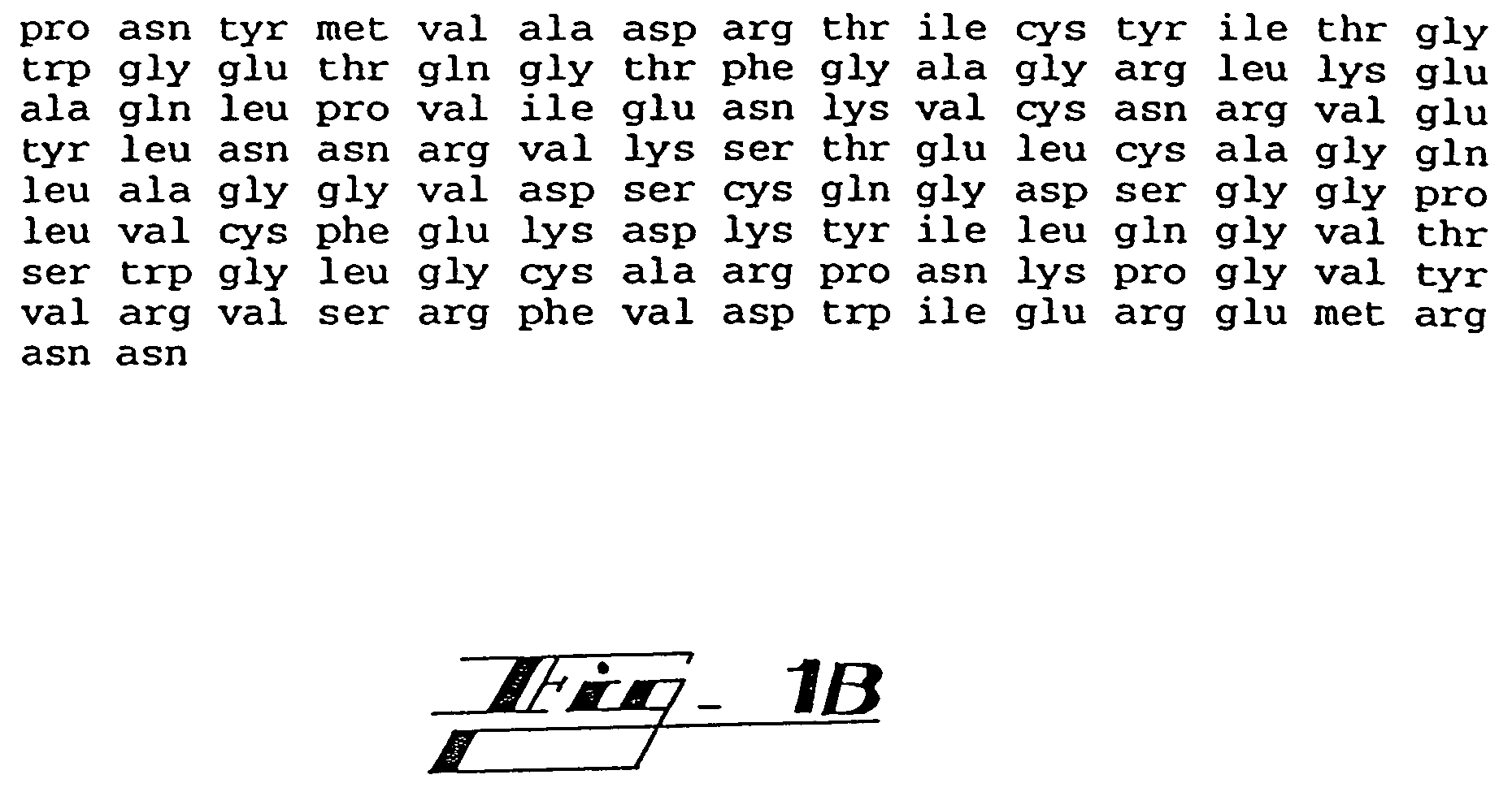
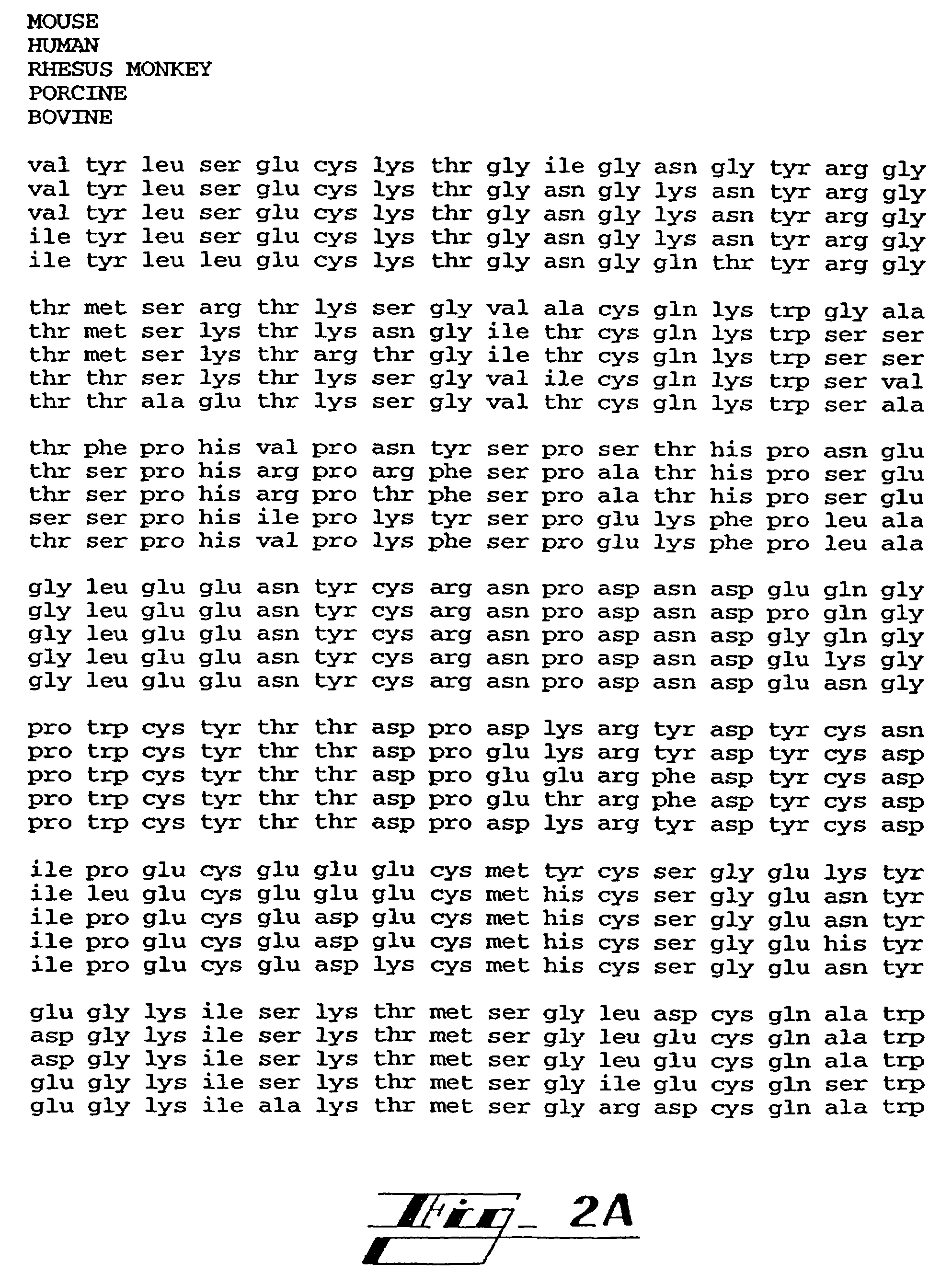
Angiostatin protein

angiostatin and protein technology, applied in the field of endothelial inhibitors, can solve the problems of increasing the thickness of the lesion, increasing the size of the lesion, and fast-expanding, and achieve the effect of minimal side effects

Inactive Publication Date: 2008-04-29
CHILDRENS MEDICAL CENT CORP
View PDF23 Cites 10 Cited by
  • Summary
  • Abstract
  • Description
  • Claims
  • Application Information

AI Technical Summary

Benefits of technology

[0049]It is yet another object of the present invention to provide a therapy for cancer that has minimal side effects.

Problems solved by technology

(1) The growth rate of tumors implanted in subcutaneous transparent chambers in mice is slow and linear before neovascularization, and rapid and nearly exponential after neovascularization. Vascular reactions of normal and malignant tumors in vivo. I. Vascular reactions of mice to wounds and to normal and neoplastic transplants. J. Natl. Cancer Inst.
(2) Tumors grown in isolated perfused organs where blood vessels do not proliferate are limited to 1-2 mm3 but expand rapidly to >1000 times this volume when they are transplanted to mice and become neovascularized. (Folkman J, et al., Tumor behavior in isolated perfused organs: In vitro growth and metastasis of biopsy material in rabbit thyroid and canine intestinal segments. Annals of Surgery 164:491-502, 1966)
(3) Tumor growth in the avascular cornea proceeds slowly and at a linear rate, but switches to exponential growth after neovascularization. (Gimbrone, M. A., Jr. et al., Tumor growth and neovascularization: An experimental model using the rabbit cornea. J. Natl. Cancer Institute 52:41-427, 1974)
(4) Tumors suspended in the aqueous fluid of the anterior chamber of the rabbit eye, remain viable, avascular and limited in size to 3. Once they are implanted on the iris vascular bed, they become neovascularized and grow rapidly, reaching 16,000 times their original volume within 2 weeks. (Gimbrone M A Jr., et al., Tumor dormancy in vivo by prevention of neovascularization.
(5) When tumors are implanted on the chick embryo chorioallantoic membrane, they grow slowly during an avascular phase of >72 hours, but do not exceed a mean diameter of 0.93+0.29 mm. Rapid tumor expansion occurs within 24 hours after the onset of neovascularization, and by day 7 these vascularized tumors reach a mean diameter of 8.0+2.5 mm. (Knighton D., Avascular and vascular phases of tumor growth in the chick embryo. British J. Cancer, 35:347-356, 1977)
(6) Vascular casts of metastases in the rabbit liver reveal heterogeneity in size of the metastases, but show a relatively uniform cut-off point for the size at which vascularization is present. Tumors are generally avascular up to 1 mm in diameter, but are neovascularized beyond that diameter. (Lien W., et al., The blood supply of experimental liver metastases. A microcirculatory study of normal and tumor vessels of the liver with the use of perfused silicone rubber. Surgery 68:334-340, 1970)
(7) In transgenic mice which develop carcinomas in the beta cells of the pancreatic islets, pre-vascular hyperplastic islets are limited in size to Nature 339:58-61, 1989)
(8) A specific antibody against VEGF (vascular endothelial growth factor) reduces microvessel density and causes “significant or dramatic” inhibition of growth of three human tumors which rely on VEGF as their sole mediator of angiogenesis (in nude mice). The antibody does not inhibit growth of the tumor cells in vitro. (Kim K J, et al., Inhibition of vascular endothelial growth factor-induced angiogenesis suppresses tumor growth in vivo. Nature 362:841-844, 1993)
(9) Anti-bFGF monoclonal antibody causes 70% inhibition of growth of a mouse tumor which is dependent upon secretion of bFGF as its only mediator of angiogenesis. The antibody does not inhibit growth of the tumor cells in vitro. (Hori A, et al., Suppression of solid tumor growth by immunoneutralizing monoclonal antibody against human basic fibroblast growth factor. Cancer Research, 51:6180-6184, 1991)
(10) Intraperitoneal injection of bFGF enhances growth of a primary tumor and its metastases by stimulating growth of capillary endothelial cells in the tumor. The tumor cells themselves lack receptors for bFGF, and bFGF is not a mitogen for the tumors cells in vitro. Modulation of solid tumor growth in vivo by bFGF.
(11) A specific angiogenesis inhibitor (AGM-1470) inhibits tumor growth and metastases in vivo, but is much less active in inhibiting tumor cell proliferation in vitro. It inhibits vascular endothelial cell proliferation half-maximally at 4 logs lower concentration than it inhibits tumor cell proliferation. (Ingber D, et al., Angioinhibins: Synthetic analogues of fumagillin which inhibit angiogenesis and suppress tumor growth. Nature, 48:555-557, 1990). There is also indirect clinical evidence that tumor growth is angiogenesis dependent.
(12) Human retinoblastomas that are metastatic to the vitreous develop into avascular spheroids which are restricted to less than 1 mm3 despite the fact that they are viable and incorporate 3H-thymidine (when removed from an enucleated eye and analyzed in vitro).
(13) Carcinoma of the ovary metastasizes to the peritoneal membrane as tiny avascular white seeds (1-3 mm3). These implants rarely grow larger until one or more of them becomes neovascularized.
(14) Intens-ity of neovascularization in breast cancer (Weidner N, et al., Tumor angiogenesis correlates with metastasis in invasive breast carcinoma. 324:1-8, 1991, and Weidner N, et al., Tumor angiogenesis: A new significant and independent prognostic indicator in early-stage breast carcinoma, J Natl. 84:1875-1887, 1992) and in prostate cancer (Weidner N, Carroll P R, Flax J, Blumenfeld W, Folkman J. Tumor angiogenesis correlates with metastasis in invasive prostate carcinoma. American Journal of Pathology, 143(2):401-409, 1993) correlates highly with risk of future metastasis.
(15) Metastasis from human cutaneous melanoma is rare prior to neovascularization. The onset of neovascularization leads to increased thickness of the lesion and an increasing risk of metastasis. (Srivastava A, et al., The prognostic significance of tumor vascularity in intermediate thickness (0.76-4.0 mm thick) skin melanoma.
(16) In bladder cancer, the urinary level of an angiogenic protein, bFGF, is a more sensitive indicator of status and extent of disease than is cytology. (Nguyen M, et al., Elevated levels of an angiogenic protein, basic fibroblast growth factor, in urine of bladder cancer patients. J. Natl. Cancer Inst.

Method used


Image

  • Angiostatin protein
  • Angiostatin protein
  • Angiostatin protein

Examples


example 1

Choice of an Animal-Tumor System in Which Growth of Metastasis is Inhibited by the Primary Tumor and is Accelerated After Removal of the Primary Tumor.

[0179]By screening a variety of murine tumors capable of inhibiting their own metastases, a Lewis lung carcinoma was selected in which the primary tumor most efficiently inhibited lung metastasis. Syngeneic C57BI6 / J six-week-old male mice were injected (subcutaneous dorsum) with 1×10⁶ tumor cells. Visible tumors first appeared after 3-4 days. When tumors were approximately 1500 mm3 in size, mice were randomized into two groups. The primary tumor was completely excised in the first group and left intact in the second group after a sham operation. Although tumors from 500 mm3 to 3000 mm3 inhibited growth of metastases, 1500 mm3 was the largest primary tumor that could be safely resected with high survival and no local recurrence.

[0180]After 21 days, all mice were sacrificed and autopsied. In mice with an intact primary tumor, there were...

example 2

Isolation of a Variant of Lewis Lung Carcinoma Tumor That is Highly Metastatic, Whether or Not the Primary Tumor is Removed.

[0182]A highly metastatic variant of Lewis lung carcinoma arose spontaneously from the LLC-Low cell line of Example 1 in one group of mice and has been isolated according to the methods described in Example 1 and repeatedly transplanted. This tumor (LLC-High) forms more than 30 visible lung metastases whether or not the primary tumor is present.

example 3

Size of Metastases and Proliferation Rate of Tumor Cells Within Them. Effect of the Primary Tumor That Inhibits Metastases (LLC-Low).

[0183]C57BI6 / J mice were used in all experiments. Mice were inoculated subcutaneously with LLC-Low cells, and 14 days later the primary tumor was removed in half of the mice. At 5, 10 and 15 days after the tumor had been removed, mice were sacrificed. Histological sections of lung metastases were obtained. Mice with an intact primary tumor had micrometastases in the lung which were not neovascularized. These metastases were restricted to a diameter of 12-15 cell layers and did not show a significant size increase even 15 days after tumor removal. In contrast, animals from which the primary tumor was removed, revealed large vascularized metastases as early as 5 days after operation. These metastases underwent a further 4-fold increase in volume by the 15th day after the tumor was removed (as reflected by lung weight and histology). Approximately 50% of ...


PUM

PropertyMeasurementUnit
volumeaaaaaaaaaa
diameteraaaaaaaaaa
sizeaaaaaaaaaa

Abstract

The present invention provides plasminogen fragments containing kringle region domains having anti-angiogenic activity and termed angiostatin protein. The plasminogen fragments of the present invention may be used for the treatment of angiogenesis-dependent diseases such as cancer.

Description

[0001]This application is a continuation application of U.S. patent application Ser. No. 09 / 788,142, filed Feb. 16, 2001, now abandoned, which is a continuation application of U.S. patent application Ser. No. 09 / 338,387, filed Jun. 22, 1999, now abandoned, which is a continuation of U.S. patent application Ser. No. 09 / 309,821, filed May 11, 1999, now abandoned, which is a division of U.S. patent application Ser. No. 08 / 866,735, filed May 30, 1997, issued as U.S. Pat. No. 5,945,403. U.S. patent application Ser. No. 09 / 338,387, filed Jun. 22, 1999, now abandoned, is also a continuation-in-part application of U.S. patent application Ser. No. 08 / 989,477, filed Dec. 12, 1997, now abandoned, which is a continuation-in-part application of U.S. patent application Ser. No. 08 / 429,743, filed Apr. 26, 1995, issued as U.S. Pat. No. 5,885,795, which is a continuation-in-part application of U.S. patent application Ser. No. 08 / 326,785, filed Oct. 20, 1994, issued as U.S. Pat. No. 5,792,845, which ...

Claims


Application Information

Patent Timeline
Patent Type & Authority Patents(United States)
IPC IPC(8): C07K14/47C12N15/12C12N15/70C12N15/09A61K38/00A61K48/00A61P35/00A61P43/00C12N5/07C12N5/071C12N5/10C12N9/68G01N33/574
CPCA61K38/00A61K48/00C12N9/6435G01N33/574Y10S530/828C12Y304/21007A61P35/00A61P43/00C07K14/435
Inventor O'REILLY, MICHAEL S.FOLKMAN, M. JUDAHCAO
Owner CHILDRENS MEDICAL CENT CORP
Why Eureka
Try Eureka