Aligned scaffolds for improved myocardial regeneration

a technology of aligned scaffolds and myocardial cells, which is applied in the direction of prosthesis, skeletal/connective tissue cells, bandages, etc., can solve the problems of difficult processing methods, limited success of materials, and failure of the body to integrate and/or remodel these materials into surrounding tissue, etc., to promote cell migration and proliferation, promote cell adhesion and promote cell adhesion

Active Publication Date: 2008-06-10
BOSTON SCI SCIMED INC
View PDF3 Cites 64 Cited by
  • Summary
  • Abstract
  • Description
  • Claims
  • Application Information

AI Technical Summary

Benefits of technology

[0011]The design of the scaffolds can be varied in relation to different tissue architectures by varying the dimensions of the elements of the scaffolds, and / or the materials that the elements of the scaffold are made of. The elements of the scaffold are the A-strips, S-strips and the separating layers, and their dimensions as to width, length, thickness and porosity are fixed according to a scaffold's intended use in the repair, regeneration or growth of a particular tissue or cell type, in vivo or in vitro. The width of the A-strips and S-strips are fixed to promote the ordered alignment of cells within a layer so that cells can receive spatial- and orientation-dependent signals for cell growth and / or differentiation from each other and from the strip materials. The materials of the scaffold are also chosen such that the A-strips promote the adhesion of cells more than the S-strips and the separating layers, and such that the porosity of the S-strips and separating layers allow a homogeneous distribution of cells throughout the scaffold. Further, the scaffold can be supplemented or treated with bioactive agents such as those, for example, that promote cell migration and proliferation.

Problems solved by technology

However, these materials have enjoyed limited success because of their poor biocompatibility.
Synthetic materials frequently cause persistent inflammatory reactions and are further limited by their lack of biodegradation, which results in the failure of the body to integrate and / or remodel these materials into the surrounding tissue.
However, in order for these structures to promote functional tissue growth / reconstruction, a complex micro-architecture must be produced, and this requires difficult processing methods.
Efforts to use animal materials have been unsatisfactory when these materials are cross-linked by formaldehyde or glutaraldehyde, for example.
This form of generalized aldehydic crosslinking tends to render biomaterials sufficiently unrecognizable to tissue cells such that normal remodeling and integration are not promoted.
Similarly, other types of chemical processing of animal or human biomaterials, such as extraction with detergents, hypertonic buffers or hypotonic buffers can alter them to the degree that leave these biomaterials toxic to tissue cells, ineffective in promoting angiogenesis, and ineffective in stimulating repair and remodeling processes needed for the conversion of an implant into a functional substitute for the tissue or organ being replaced.
While tissue-like constructs have been fabricated and been shown to have some resemblance to their natural counterparts, these constructs do not readily develop the matrix complexity characteristic of the actual tissues that they are meant to imitate (U.S. Pat. Nos. 4,485,096 and 4,485,097).
However, a limitation of these natural forms is that it is difficult to fashion three-dimensional tissue structures that mimic the endogenous tissue architecture where the graft or scaffold is to be used, for example, to form the complex shape of a valve.
Cardiac tissue repair presents challenges to tissue-engineering methods.
Cardiac tissues are difficult to repair as the tissue has a limited ability to regenerate and because the tissue has a complex architecture that is difficult to mimic.
However, cultured cardiomyocytes typically spread on a flat substrate to form an unorganized monolayer with disorganized myofibrils and junctions.
However, such methods provide two-dimensional tissue layers that are difficult to transform into a three-dimensional tissue repair scaffold.

Method used


Image

  • Aligned scaffolds for improved myocardial regeneration
  • Aligned scaffolds for improved myocardial regeneration

Examples


example 1

Method of Making the Scaffold

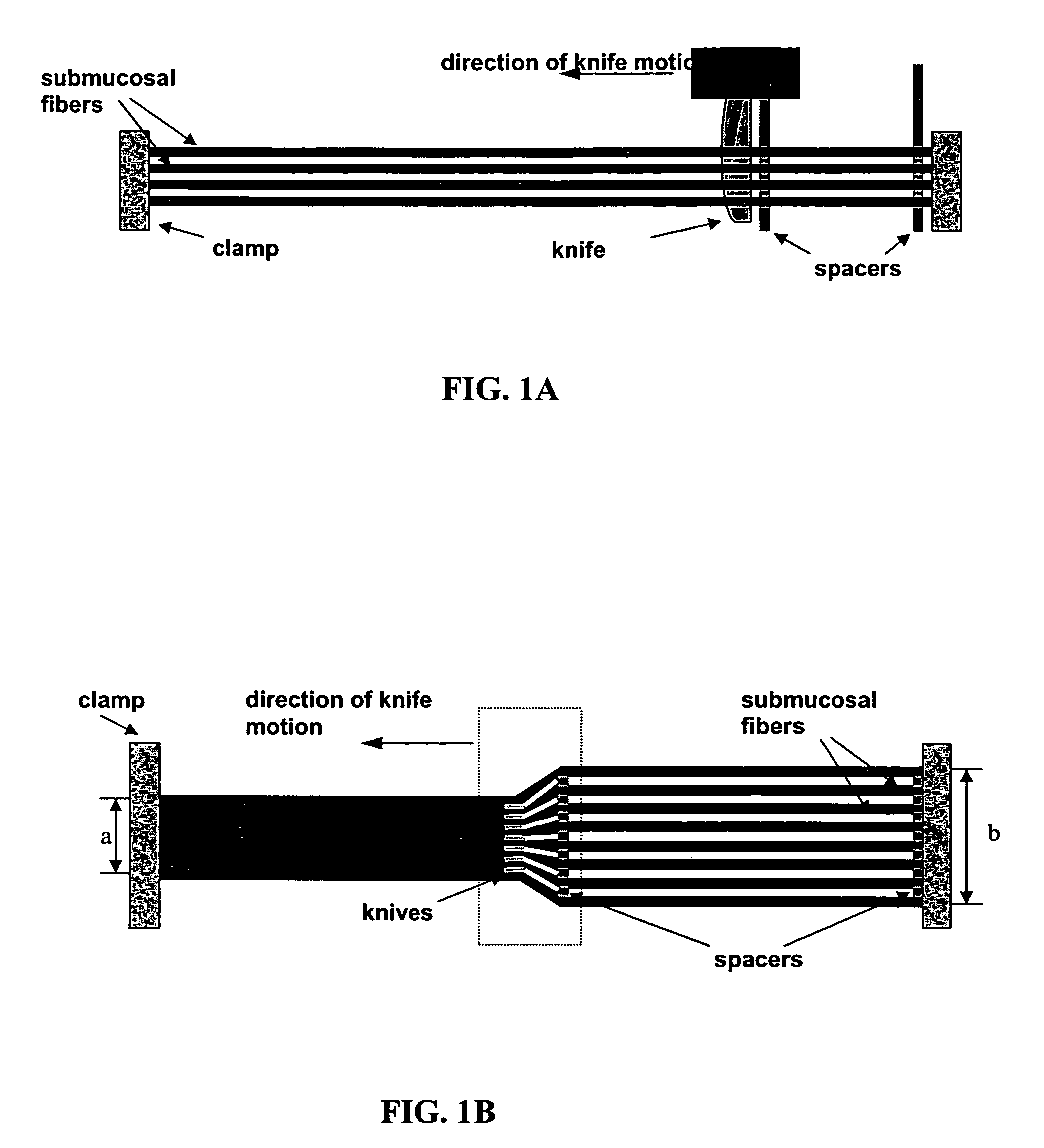
[0070]The instruments used in this example are pictured in FIG. 1.

[0071]A porcine small-intestine submucosa (SIS) sheet is obtained (vivoSIS™ cell culture disk, Cook Biotech). The disk has a diameter of 12 millimeters and a nominal thickness of 115 micrometers. The disks are sterile, acellular, and endotoxin free.

[0072]The disks are rehydrated with at least two changes of phosphate buffered saline (PBS) pH 7.4. Upon rehydration, the disks become pliable and easily fold upon themselves. The disks are cut into squares and clamped in layers as shown in FIG. 1A at a fixed distance between layers of about 100 micrometers.

[0073]A comb of knives is inserted at one end while another comb of spacers follows the knives. The distances between each knife in the comb of knives are set at a fixed distance of about 30 micrometers. The distance between each spacer in the comb of spacers are set at a fixed distance of about 100 micrometers (width). As the comb of knives ...

example 2

In Vitro Seeded Scaffolds

[0075]The scaffold according to Example 1 is seeded with any cell-type (or combinations of) in vitro. A scaffold is placed in a tissue culture dish or bioreactor with a serum-containing culture medium appropriate for the cells intended for seeding. Generally, cells at a concentration of 1-5×10⁶ cells / ml are added to the dish or bioreactor and incubated for 24-72 hours at 37° C. at 5% CO2.

[0076]For example, embryonic stem cell-derived cardiomyocytes are incubated with a scaffold that is 5 millimeters thick (25 layers) and 2 centimeters square with 106 cells in 25 ml of culture medium. This incubation occurs under shaking at 50 rpm at 37° C. at 5% CO2 for 48 hours. This seeded-scaffold is further cultured in media for ten additional days to allow cell spreading.

[0077]When bioreactors are used for seeding cells onto scaffolds, some examples are: flasks (static or mixed at 50 or 90 rpm) (Bellco, Vineland, N.J.), 6-well dishes mounted on an xyz gyrator operated a...

example 3

Testing of Scaffolds for Cardiac Tissue Repair

[0105]To test the scaffolds of the present invention for the ability to promote cell differentiation and growth, a variety of methods are known in the art and can be used. The methods vary relative to the cell seeded in the scaffold, either in vitro or in vivo. This example provides some methods that can be used to test the scaffold's ability to promote cell growth and differentiation for cardiac tissue using cardiomyocyte cells isolated from heart tissue seeded onto scaffolds according to Example 1.

[0106]For in vitro scaffolds, medium samples (1 mL) are taken at the end of the seeding period (see Example 2) and every two days during cultivation. pH and partial pressures of oxygen (ρ-O2) and carbon dioxide (ρ-CO2) are measured immediately after sampling using a gas blood analyzer (Model 1610, Instrumentation Laboratory, Lexington, Mass.) with an accuracy of 0.1% for pH and 2% for ρ-O2 and ρ-CO2. Cell death and damage during seeding and c...


PUM

PropertyMeasurementUnit
thicknessaaaaaaaaaa
widthaaaaaaaaaa
thicknessaaaaaaaaaa

Abstract

The present invention relates to a biocompatible, three-dimensional scaffold useful to grow cells and to regenerate or repair tissue in predetermined orientations. The scaffold is particularly useful for regeneration and repair of cardiac tissue. The scaffold contains layers of alternating A-strips and S-strips, wherein the A-strips within each layer are aligned parallel to each other and preferentially promote cellular attachment over attachment to the S-strips. Methods of producing and implanting the scaffold are also provided.

Description

[0001]This application claims benefit of priority to U.S. Provisional Application Ser. No. 60 / 487,909, filed Jul. 16, 2003, the entire contents of which are hereby incorporated by reference in their entirety.BACKGROUND OF THE INVENTION[0002]Biocompatible and biodegradable scaffolds are used in tissue reconstruction and repair. The scaffold, for example, can serve as both a physical support and adhesive substrate for cells during in vitro culturing and subsequent implantation in vivo. Scaffolds are utilized to deliver cells to desired sites in the body, to define a potential space for engineered tissue, and to guide the process of tissue development. Cell transplantation on scaffolds has been explored for the regeneration of skin, heart, nerve, liver, pancreas, cartilage, and bone tissue using various biological and synthetic materials. Scaffolds have also been implanted directly into patients without prior culturing of cells in vitro. In this case, the initially cell-free scaffold n...

Claims


Application Information

Patent Timeline
Patent Type & Authority Patents(United States)
IPC IPC(8): C12N5/00A61K9/00A61K35/12A61L27/36A61L27/54A61L27/60C12N5/02C12N5/071C12N5/077
CPCA61L27/3604A61L27/367A61L27/54A61L27/60C12N5/0068C12N5/0657C12N5/0698A61K35/12A61L2300/252A61L2300/254A61L2300/256A61L2300/414A61L2300/426A61L2300/43A61L2300/604C12N2502/094C12N2502/1323C12N2533/74C12N2533/92C12N2535/10A61L27/3804A61L27/3834C12N5/00
Inventor FREYMAN, TOBYPALASIS, MARIAUNGS, MARK
Owner BOSTON SCI SCIMED INC
Why Eureka
Try Eureka