Compositions and methods for detection and isolation of phosphorylated molecules

a technology of phosphorylation status and composition, which is applied in the direction of instruments, peptides/protein ingredients, peptides, etc., can solve the problems of insufficient research capacity to simply identify, inability to perform systematic parallel analysis of the phosphorylation status of large sets of proteins involved in the regulatory circuitry of cells and tissues, and inability to achieve simple identification. , to achieve the effect of simplifying the subsequent analysis of the sampl

Active Publication Date: 2008-11-04
MOLECULAR PROBES
View PDF118 Cites 24 Cited by
  • Summary
  • Abstract
  • Description
  • Claims
  • Application Information

AI Technical Summary

Benefits of technology

[0026]Alternatively, when the method is utilized to selectively isolate phosphorylated target molecules from solution, visualizing the complex may not be necessary. To isolate the phosphorylated target molecules, the ternary complex can be precipitated, immobilized, separated by a chromatographic or electrophoretic technique or by a magnetic field or remain in solution. In some cases, organic extraction can be used to separate the metal-chelating moiety from the phosphorylated target molecule. When the ternary complex is precipitated or otherwise immobilized, the phosphorylated target molecules can be separated from the nonphosphorylated target molecules and other components of the sample by affinity chromatography, such as by simple washing with an aqueous, organic or mixed aqueous / organic wash solution. In some cases, it is advantageous to further analyze the extracted phosphorylated target molecules while they are still immobilized on a matrix. Isolation of phosphorylated target molecules is useful for further analysis of the target molecules, such as by liquid chromatography / mass spectrography, an electrophoretic separation technique, by detection of the bound target molecules by an antibody to any part of the target molecule or by a variety of other techniques. In particular, isolation of the phosphorylated target molecules simplifies the subsequent analysis of the sample by removing interfering components of the original sample.

Problems solved by technology

Phosphorylation reactions are key features of protein function, and thus phosphorylated proteins must be able to be identified if the proteome is to be fully understood; however, to date no practical methods exist for the systematic parallel analysis of the phosphorylation status of large sets of proteins involved in the regulatory circuitry of cells and tissues.
It is not enough for researchers to simply identify whether a protein is a phosphorylated protein or not.
Such approaches are restricted to a limited range of biological materials, such as tissue culture samples and analysis of clinical samples would require in vivo labeling of patients, which is not feasible.
Immunoblotting is best performed after blocking unoccupied sites on the solid-phase support with protein solutions, which interferes with microchemical analysis.
This also poses problems with subsequent use of the protein for sequencing and mass spectrometry.
Though high-quality antibodies to phosphotyrosine are commercially available, antibodies that specifically recognize phosphoserine and phosphothreonine residues have been more problematic, often being sensitive to amino acid sequence context.
The reliability of these antibodies has been questioned because of potential steric hindrances between the interaction of these antibodies and the phosphoproteins.
Moreover, when phosphoproteins are not enriched prior to detection with the antibody, the presence of unrelated proteins co-migrating with the protein of interest may lead to false positive signals.
Therefore, identification of phosphorylated proteins using immunoblotting and immunoprecipitation techniques is effectively limited to proteins containing phosphorylated tyrosine residues.
Alternatively, phosphorylated proteins can be identified using chromogenic dyes, but with limited success.
Stains-All is not routinely used to detect phosphoproteins due to poor specificity and low sensitivity.
), is designed to detect phosphoproteins in gels; however, this method has many limitations.
The detection sensitivity of the staining method is considerably poorer than Coomassie Blue staining, with 80-160 ng of phosvitin, a protein containing roughly 100 phosphoserine residues, being detectable by the commercialized kit.
The staining procedure is quite complex (involving seven different reagents) and alkaline hydrolysis requires heating gels to 65° C., which causes the gel matrix to hydrolyze and swell considerably.
These methods are time consuming, require purified phosphopeptides, and are limiting in what can be isolated.
Both procedures identified the monophosphorylated trypsin peptide fragment from the test protein β-casein, but both failed to detect the tetraphosphorylated peptide fragment.
However, currently, there is no satisfactory method for the specific and reversible fluorescent detection of gel-separated phosphoproteins from complex samples.
However, neither method has been shown to be suitable for detection of phosphoproteins directly in gels.
One problem with the approach is that a delicate balance must be struck between the base and the ethanedithiol in order to achieve elimination of the phosphate group from the serine residue and addition of the ethanedithiol to the resulting dehydroalanine residue without hydrolysis of the peptide backbone.
This is limiting in that researchers must have purified amounts of both proteins.
While these procedures accurately characterize the phosphorylation status of proteins and peptides, they are unsuitable for high-throughput screening of phosphorylated substrates.
However, mass spectroscopic analysis of proteolytic digests of proteins rarely provides full coverage of the protein sequence and regions of interests often go undetected.
However, there are several complications using current techniques, including loss of phosphopeptides that do not bind to the column (low affinity), difficulty in the subsequent elution of phosphorylated peptides, and background from non-phosphorylated peptides that have affinity for immobilized metal ions (low specificity).
The technology has unsurpassed quantitative characteristics, particularly when used in combination with reagents for the detection of total proteins.

Method used


Image

  • Compositions and methods for detection and isolation of phosphorylated molecules
  • Compositions and methods for detection and isolation of phosphorylated molecules
  • Compositions and methods for detection and isolation of phosphorylated molecules

Examples


example 1

Determination of BAPTA Selectivity for Gallium and Gallium Ions for Phosphorylated Target Molecules and a Screening Method for Phosphate-Binding Compounds

[0332](A) BAPTA with trivalent gallium ions selectively detects phosphoproteins.

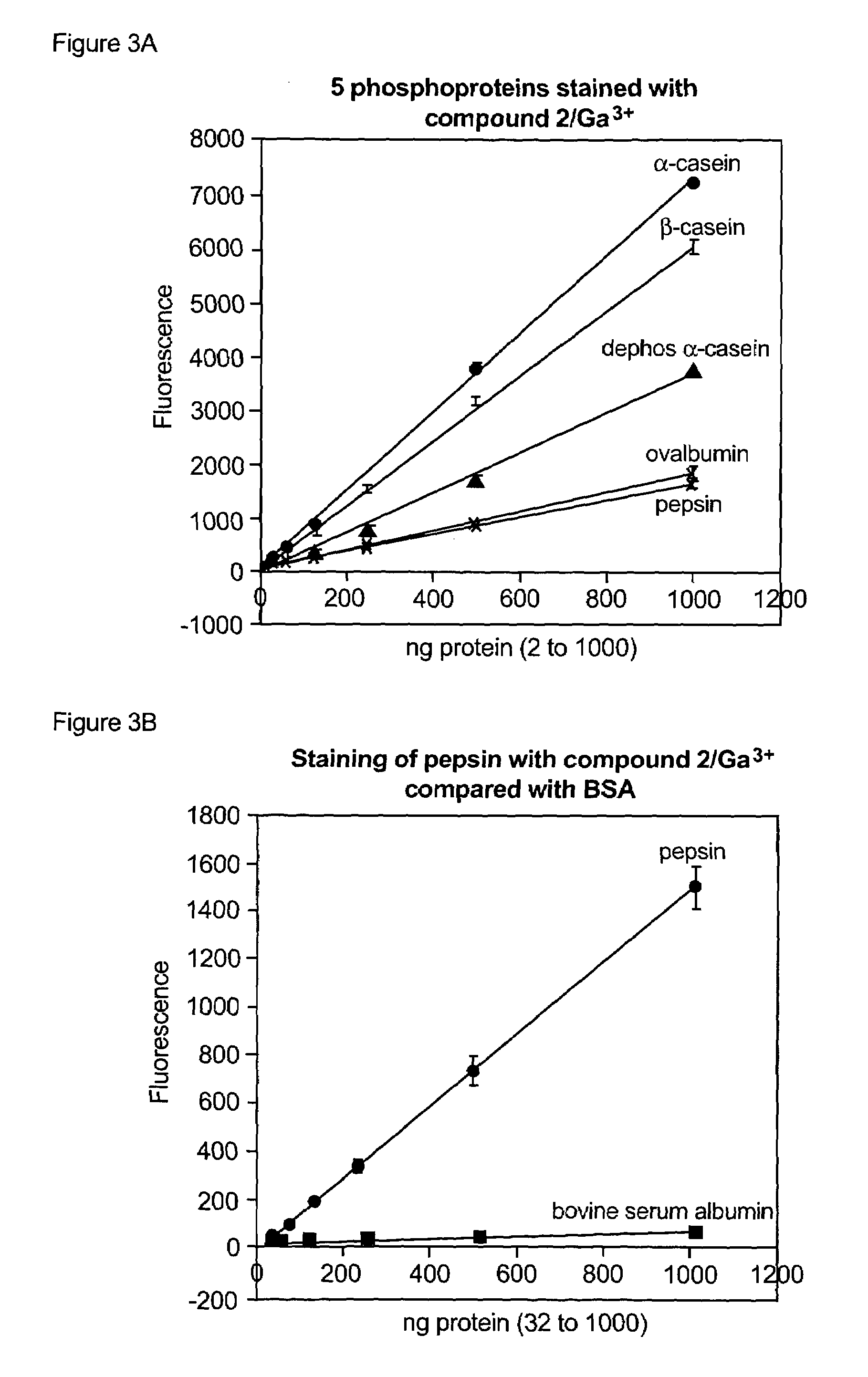
[0333]A comprehensive search of metal-chelating compounds was performed to identify fluorescent reagents that when combined with a gallium salt (gallium chloride) would selectively detect phosphorylated target molecules (particularly phosphopeptides and phosphoproteins) in a mixture of phosphorylated and nonphosphorylated target molecules. The compounds were tested in a fluorescence spectrophotometer for their ability to bind gallium (III) ion and selectively detect the phosphoprotein ovalbumin. Binding to gallium (III) ion was determined by a fluorescence increase of the same compound in the presence of up to 5 μM gallium chloride in 75 mM NaOAc (pH 4.0) and 140 mM NaCl. Ovalbumin detection was also judged by a fluorescence increase; however, the compo...

example 2

Detection of Phosphoproteins in SDS-Polyacrylamide Gels

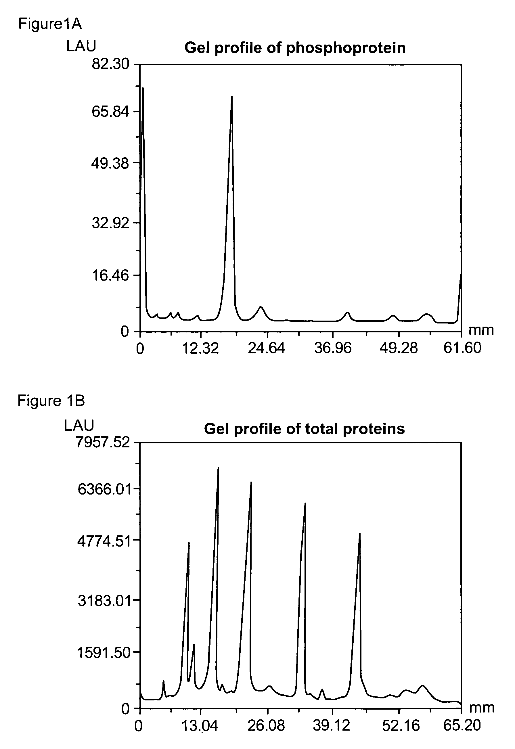
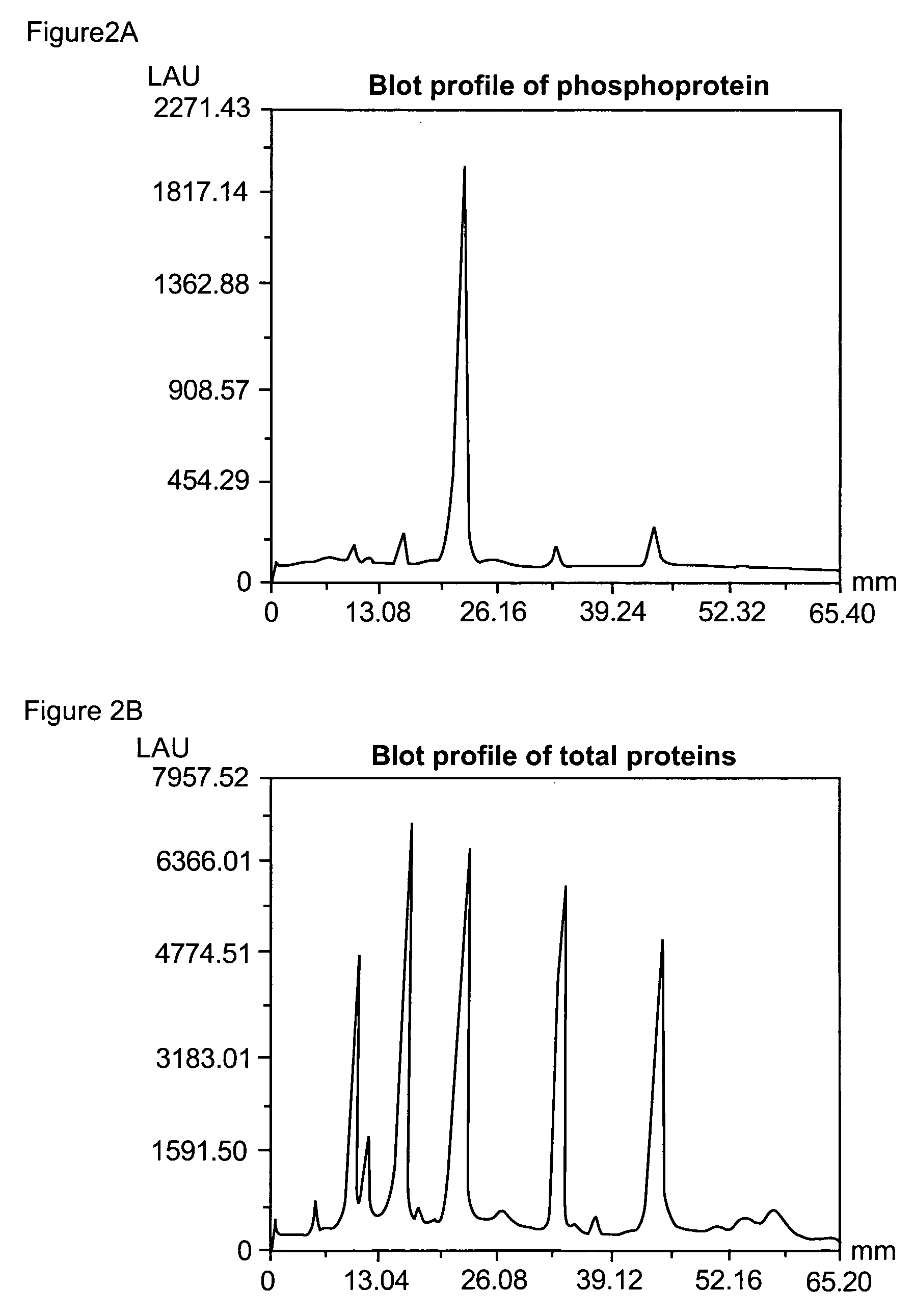
[0341]Phosphoproteins were separated by SDS-polyacrylamide gel electrophoresis utilizing a 4% T, 2.6% C stacking gel, pH 6.8 and 13% T, 2.6% C separating gel, and pH 8.8, according to standard procedures. % T is the total monomer concentration (acrylamide+crosslinker) expressed in grams per 100 mL and % C is the percentage crosslinker (e.g., N,N′-methylene-bis-acrylamide, N,N′-diacryloylpiperazine or other suitable agent). The separating gels were 8 cm wide by 5 cm high and 0.75 cm in thickness. After electrophoresis, the gels were fixed by immersing them in 100 mL 45% methanol and 5% acetic acid for 90 minutes. The gels were washed twice in water for a total of 30 minutes. The gels were then added to a binding solution of the invention (Example 1E) and incubated for 120 minutes at room temperature with gentle orbital shaking, typically 50 rpm. The binding buffer contained 50 mM NaOAc (pH 4.0), 250 mM sodium chloride, 20% v / v 1,...

example 3

Serial Dichromatic Detection of Phosphoproteins and Total Protein in SDS Polyacrylamide Gels

[0344]After detection of the phosphoproteins as in Example 2, the gel was incubated overnight with 60 ml SYPRO® Ruby protein gel stain (Molecular Probes, Eugene, Oreg.) with gentle orbital shaking, typically 50 rpm. The gel was then incubated in 7% acetic acid, 10% methanol for 30 minutes, also at 50 rpm. The orange signal from the phosphorylated and non-phosphorylated proteins was collected with a standard CCD camera-based imaging system with 300 nm UV light excitation and a 600 nm bandpass filter.


PUM

PropertyMeasurementUnit
pHaaaaaaaaaa
pHaaaaaaaaaa
pHaaaaaaaaaa

Abstract

The present invention relates to phosphate-binding compounds that find use in binding, detecting and isolating phosphorylated target molecules including the subsequent identification of target molecules that interact with phosphorylated target molecules or molecules capable of being phosphorylated. A binding solution is provide that comprises a phosphate-binding compound, an acid and a metal ion wherein the metal ion simultaneously interacts with an exposed phosphate group on a target molecule and the metal chelating moiety of the phosphate-binding compound forming a bridge between the phosphate-binding compound and a phosphorylated target molecule resulting in a ternary complex. The binding solution of the present invention finds use in binding and detecting immobilized and solubilized phosphorylated target molecules, isolation of phosphorylated target molecules from a complex mixture and aiding in proteomic analysis wherein kinase and phosphatase substrates and enzymes can be identified.

Description

CROSS-REFERENCE TO RELATED APPLICATIONS[0001]This application is a continuation-in-part of U.S. Ser. No. 10 / 703,816, filed Nov. 7, 2003, now pending, which is a continuation-in-part of U.S. Ser. No. 10 / 428,192, filed May 2, 2003, now U.S. Pat. No. 7,102,005, which claims priority to U.S. Ser. No. 60 / 377,733, filed May 3, 2002, abandoned; U.S. Ser. No. 60 / 393,059, filed Jun. 28, 2002, abandoned; U.S. Ser. No. 60 / 407,255, filed Aug. 30, 2002, abandoned; and U.S. Ser. No. 60 / 440,252, filed Jan. 14, 2003, abandoned, which disclosures are herein incorporated by reference.STATEMENT REGARDING FEDERALLY SPONSORED RESEARCH[0002]This invention was made in part with government support under grant number 1 R33 CA093292-01, awarded by the National Cancer Institute. The United States Government may have certain rights in this invention.FIELD OF THE INVENTION[0003]The present invention relates to metal-chelating compositions and methods for use in the detection and isolation of phosphorylated targ...

Claims


Application Information

Patent Timeline
Patent Type & Authority Patents(United States)
IPC IPC(8): G01N33/50C12Q1/48G01N33/53
CPCG01N33/5008G01N33/502G01N33/5091G01N2333/4709
Inventor AGNEW, BRIANGEE, KYLE R.MARTIN, VLADIMIR V.
Owner MOLECULAR PROBES
Why Eureka
Try Eureka