Magnetic resonance apparatus for use with active electrode and drug deliver catheter

a magnetic resonance and active electrode technology, applied in the field of magnetic resonance apparatus for use with active electrodes and drug delivery catheters, can solve the problems of inability to achieve the target receptors of brain parenchyma, inability to accurately navigate the process, and inability to accurately diagnose the accuracy of the diagnostic procedure, so as to improve the alterations in the magnetic field. effect of

Inactive Publication Date: 2009-03-17
RGT UNIV OF MINNESOTA +1
View PDF18 Cites 44 Cited by
  • Summary
  • Abstract
  • Description
  • Claims
  • Application Information

AI Technical Summary

Benefits of technology

[0026]While the ADC is preferred means within the present invention of mapping the delivery of drug in tissue, other embodiments of the invention allow for additional tissue contrast parameters to track the delivery of a drug into tissue. In other words, the delivery of a drug into tissue will cause other MRI-observable changes which can be mapped (as is done for ADC) and which can be used to spatially track the delivery and extent of a drug into a tissue. While some of these observations may be larger in magnitude than others, any of the effects can be used as a tracking mechanism. The tissue contrast changes apparent on an MR image can arise from ADC, from alteration in the BO magnetic field (often referred to as magnetic susceptibility or ABO produced by the presence of a substance in said tissue), from alterations in local tissue T1 relaxation times, from local T2 relaxation times, from T2* relaxation times (which can be created by susceptiblity differences), from the magnetization transfer coefficients (MTC is an effect produced by local communication between free water protons and those of nearby macromolecular structures), from the ADC anistropty observed in oriented matter, and also from local differences in temperature which will affect in varying degrees all of the included tissue contrast parameters. In addition, the delivery of drug can also be tracked from magnetic field frequency shifts caused by the drug or arising from agents added with unique frequency shifts from those of the local protons (such as that created from F-19 or fluorine-19 agents formed in or added to the drug).
[0027]MR imaging of the alterations in the BO magnetic field (also known as imaging of the local magnetic susceptiblity) can reveal the spatial distribution of a drug from the interaction of the drug with the otherwise homogeneous magnetic field found in MRI. To enhance the alterations in the magnetic field BO caused by the drug, small amounts of a BO-altering added agents can be added to the drug during delivery. This can include iron oxide particles, or materials comprising lanthanide-, manganese-, and iron-chelates. In addition, vehicles containing differing gases (N2, O2, CO2) will also alter the local magnetic field and thus produce a magnetic susceptibility effect which can be imaged.

Problems solved by technology

Although endoscopic, arthroscopic, and endovascular therapies have produced significant advances in healthcare, the diagnostic accuracy and clinical utility of these procedures is ultimately “surface limited” by what the surgeon can see through the device itself or otherwise visualize during the course of the procedure.
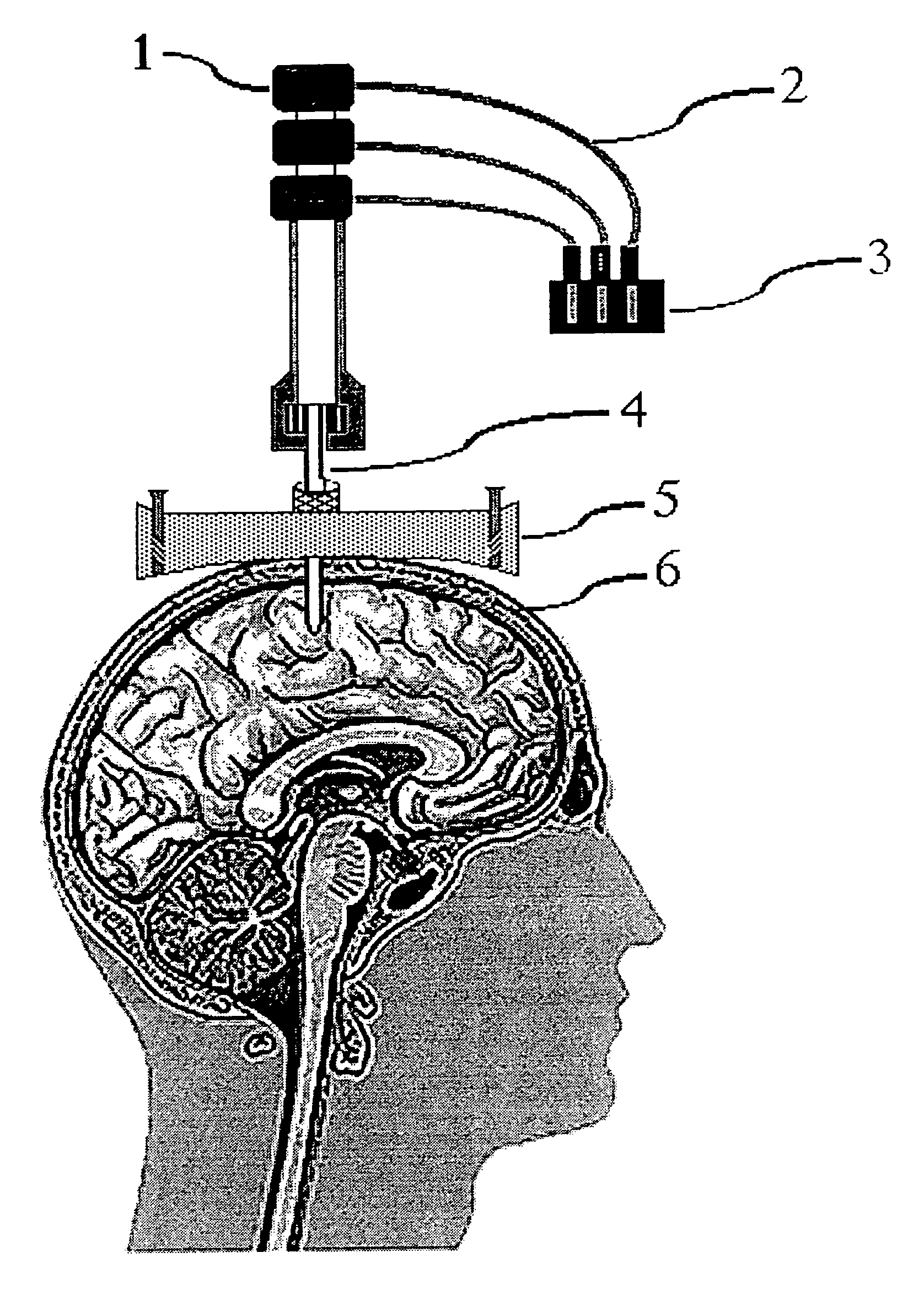
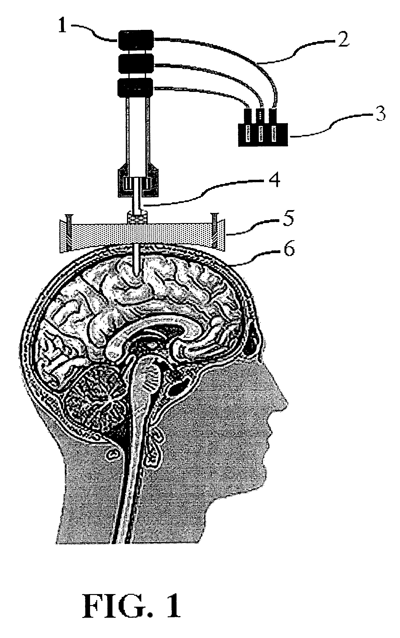
However, following intracerebroventricular injection, many therapeutic drug agents, particularly large-molecular weight hydrophobic drugs, fail to reach their target receptors in brain parenchyma because of metabolic inactivation and inability to diffuse the brain tissues, which may be up to 18 mm from a cerebrospinal fluid surface.
An important issue in targeted drug delivery is the accuracy of the navigational process used to direct the movement of the drug delivery device.
To date, direct intraparenchymal delivery of purified or synthetic dopamine, or its precursors, analogs or inhibitors has not demonstrated clear therapeutic benefit because of various problems associated with drug delivery, stability, dosage and cytotoxicity.
However, similar difficulties with reliable continuous delivery of these agents in actual clinical settings have yet to be resolved.
Reservoir limitations as well as drug solubility and stability have, however, restricted the usefulness of this technology.
However, this method, appears to rely on surface erosion of the bioabsorbable polymer, which is in turn influenced by various hydrolytic events, thereby increasing the likelihood of drug degradation, and rendering predictable release rate difficult.
A farther problem appears to be attributable to limited diffusional surface area per unit volume of larger size microspheres, such that only a limited volume of cells can be loaded into a single microcapsule.
The patented inventions referenced above provide useful methods for introducing, delivering, or applying a drug agent to a specific target tissue, but each invention also has inherent problems.
For example, some catheter systems which provide endovascular drug delivery require temporary blocking of a segment of the vessel, thereby transiently disrupting brain perfusion.
Microencapsulated coatings on catheters permit longer exposure of the tissue to the drug agent, but the physical limitations imposed by microcapsules restrict the volume and concentration of drug that can be effectively applied to any tissue area.
The sheath and any catheter components required to physically manipulate the sheath greatly increase the profile of the catheter, and thereby limit the variety of applications for which the drug delivery system can be employed.
Furthermore, the binders or adhesives used in catheter coatings may themselves significantly dilute the concentration of the therapeutic agent.
Finally, thermal and light energy required to melt and bond coatings such as macroaggregated albumin, to reduce tissue mass by ablation, and to inhibit restenosis by cytotoxic irradiation may also cause damage to blood vessels.
Although U.S. Pat. No. 5,470,307 to Lindall describes significant improvements over previous catheter-based drug delivery systems, there are nonetheless some problems.
First, in common with other currently used endovascular access devices, such as catheters, microcatheters, and guidewires, the catheter tip is difficult to see on MRI because of inadequate contrast with respect to surrounding tissues and structures.
This makes accurate localization difficult and degrades the quality of the diagnostic information obtained from the image.
Also, the mere observation of the location of the catheter in the drug delivery system does not reliably or consistently identify the position, movement and / or efficient delivery of drugs provided through the system.
In either case, the safety and efficacy of the procedure might be jeopardized, with resulting increased risk to the patient.
Although the mechanical effects of the magnetic field on ferromagnetic devices present the greatest danger to patients through possible unintended movement of the devices, tissue and device heating may also result form radio-frequency power deposition in electrically conductive material located within the imaging volume.
However, the presence of conductive elements in the catheter also introduces increased electronic noise and the possibility of Ohmic heating, and these factors have the overall effect of degrading the quality of the MR image and raising concerns about patient safety.
Thus, in all of these examples of implantable medical probes, the presence of MR-incompatible wire materials causes large imaging artifacts.
The lack of clinically adequate MR visibility and / or imaging artifact contamination caused by the device is also a problem for most commercially available catheters, microcatheters and shunts.
However, these patents do not provide the artifact-free MR visibility in the presence of rapidly alternating magnetic fields, such as would be produced during echo-planar MR imaging pulse sequences used in real-time MR guidance of intracranial drug delivery procedures.
Nor do these patents teach a method for monitoring with MR-visible catheters the outcomes of therapeutic interventions, such as, for example, drug delivery into brain tissues, cerebral ventricles, or subarachnoid space.
Image distortion may include general signal loss, regional signal loss, general signal enhancement, regional signal enhancement, and increased background noise.
The magnetic susceptibility artifact produced by the device should be small enough not to obscure surrounding anatomy, or mask low-threshold physiological events that have an MR signature, and thereby compromise the physician's ability to perform the intervention.
At the same time, the eddy currents are limited due to the ultra-thin conductive coating on the device.
At the same time, the limited distribution of drug injected from a single catheter tip presents other problems.
Delivery of a drug from a single point source also limits the distribution of the drug by decreasing the effective radius of penetration of the drug agent into the surrounding tissue receptor population.

Method used


Image

  • Magnetic resonance apparatus for use with active electrode and drug deliver catheter
  • Magnetic resonance apparatus for use with active electrode and drug deliver catheter
  • Magnetic resonance apparatus for use with active electrode and drug deliver catheter

Examples


Embodiment Construction

[0047]One of the significant difficulties with delivery of materials such as drugs, hormones, or neurotransmitters to living tissue is assuring that the materials are delivered to the target receptor location in the intended amount. Many materials which are delivered to a patient, even though beneficial in the treatment of a specific condition, may be moderately or even strongly noxious or damaging to healthy tissue. It is therefore an object of conventional materials application treatment to maximize dosage to a desired location and to minimize dosage to locations which do not require the delivery of the material. Additionally, newer medical treatments may include procedures which remove unwanted deposits of materials with an expectation that the removal will be assisted by physical removal (by a withdrawal system) or natural bodily function removal (e.g., the circulatory system), or which may attempt to stimulate the body to produce natural chemicals (of which a patient may be def...


PUM


Abstract

The invention is an apparatus and method for treatments and targeted drug delivery into a living patient, particularly but not exclusively using magnetic resonance (MR) imaging. The apparatus and method are useful in delivery to all types of living tissue and uses MR Imaging to track the location of drug delivery and estimating the rate of drug delivery. An MR-visible drug delivery device positioned at a target site (e.g., intracranial delivery) delivers a diagnostic or therapeutic drug solution into the tissue (e.g., the brain). The spinal distribution kinetics of the injected or infused drug agent are monitored quantitatively and non-invasively using water proton directional diffusion MR imaging to establish the efficacy of drug delivery at a targeted location.

Description

CROSS REFERENCE TO RELATED APPLICATION[0001]This application is a continuation-in-part of U.S. patent application Ser. No. 08 / 856,894, filed on May 15, 1997, now U.S. Pat. No. 6,061,587 which is incorporated herein by reference.BACKGROUND OF THE INVENTION[0002]1. Field of the Invention[0003]This invention relates to the design, construction, and use of a catheter with multiple functionality including at least an active electrode and a drug deliver system, with the electrode of the catheter providing at least one functionality different from stimulating or effecting drug delivery or release. The catheter is particularly useful in conjunction with magnetic resonance (MR) imaging to identify areas within a patient where changes in a molecular environment are occurring, as from chemical concentration changes effected by medical procedures. The invention also describes a drug delivery device for targeted drug delivery into a patient using magnetic resonance (MR) imaging combined with con...

Claims


Application Information

Patent Timeline
Patent Type & Authority Patents(United States)
IPC IPC(8): A61B5/055
CPCA61B5/14525A61B19/201A61M31/005G01R33/285G01R33/286G01R33/287G01R33/5601G01R33/56341A61B17/3421A61B2019/5251A61B2019/5454A61B2019/5458A61M2025/0057A61M2025/0073A61M2210/06A61M2210/0693G01R33/281G01R33/563A61B90/11A61B2034/2051A61B2090/3954A61B2090/3958A61B2562/17
Inventor KUCHARCZYK, JOHNMOSELEY, MICHAEL E.
Owner RGT UNIV OF MINNESOTA
Why Eureka
Try Eureka