
Mass spectrometric quantitation method for biomolecules based on metabolically labeled internal standards

a mass spectrometric and internal standard technology, applied in chemical methods analysis, instruments, material analysis, etc., to achieve the effect of easy comparison between three or more samples, good accuracy and good accuracy

Inactive Publication Date: 2010-07-20
EISIA R&D MANAGEMENT CO LTD
View PDF11 Cites 13 Cited by
  • Summary
  • Abstract
  • Description
  • Claims
  • Application Information

AI Technical Summary

Benefits of technology

The present invention relates to a method for quantitating biological molecules in a sample using an isotopically labeled internal standard substance. This method can be used in proteomics to measure the expression levels of proteins in a sample. The invention also provides a program for carrying out this analysis. The technical effect of this invention is to improve the accuracy and reproducibility of quantitative analysis of biological molecules by comparing the mass spectrometry data of the isotopically labeled internal standard substance with the data of the unlabeled substance. This method can be used in drug development, lipid analysis, and protein analysis, among others.

Problems solved by technology

However, nucleotide sequence information is not sufficient to elucidate biological phenomena, and although the total genomes of Escherichia coli, yeast, Caenorhabditis elegans, drosophila and the like have actually been elucidated, it does not mean that the entirety of the gene function of these model living organisms has been elucidated.
However, merely identifying a protein, or merely elucidating a component, is insufficient as functional analysis.
Two-dimensional electrophoresis is routinely used as means for proteome analysis; however, with such a method that compares spots of protein stained on a gel, comparing accurately the quantity of individual protein present in two samples to be compared is extremely difficult.
The reasons being, issues exist in:1. reproducibility of extraction from cell and reproducibility due to pretreatment manipulation such as cell fractionation / crude purification2. reproducibility of protein separation in two-dimensional electrophoresis and the like3. reproducibility of gel staining and the like; furthermore,4. when a plurality of proteins are mixed in one spot / band, the quantitative aspect with respect to an individual protein is lost.
Thus, currently, capturing a small variation is difficult with a pattern comparison by gel staining, except when the expression amount varies dramatically, i.e., 10 to 100 times or more.
Obtaining quantitative data in an analysis by the mass spectroscopic device necessarily requires correction with an internal standard substance, as reproducibility of ionization rate is poor.
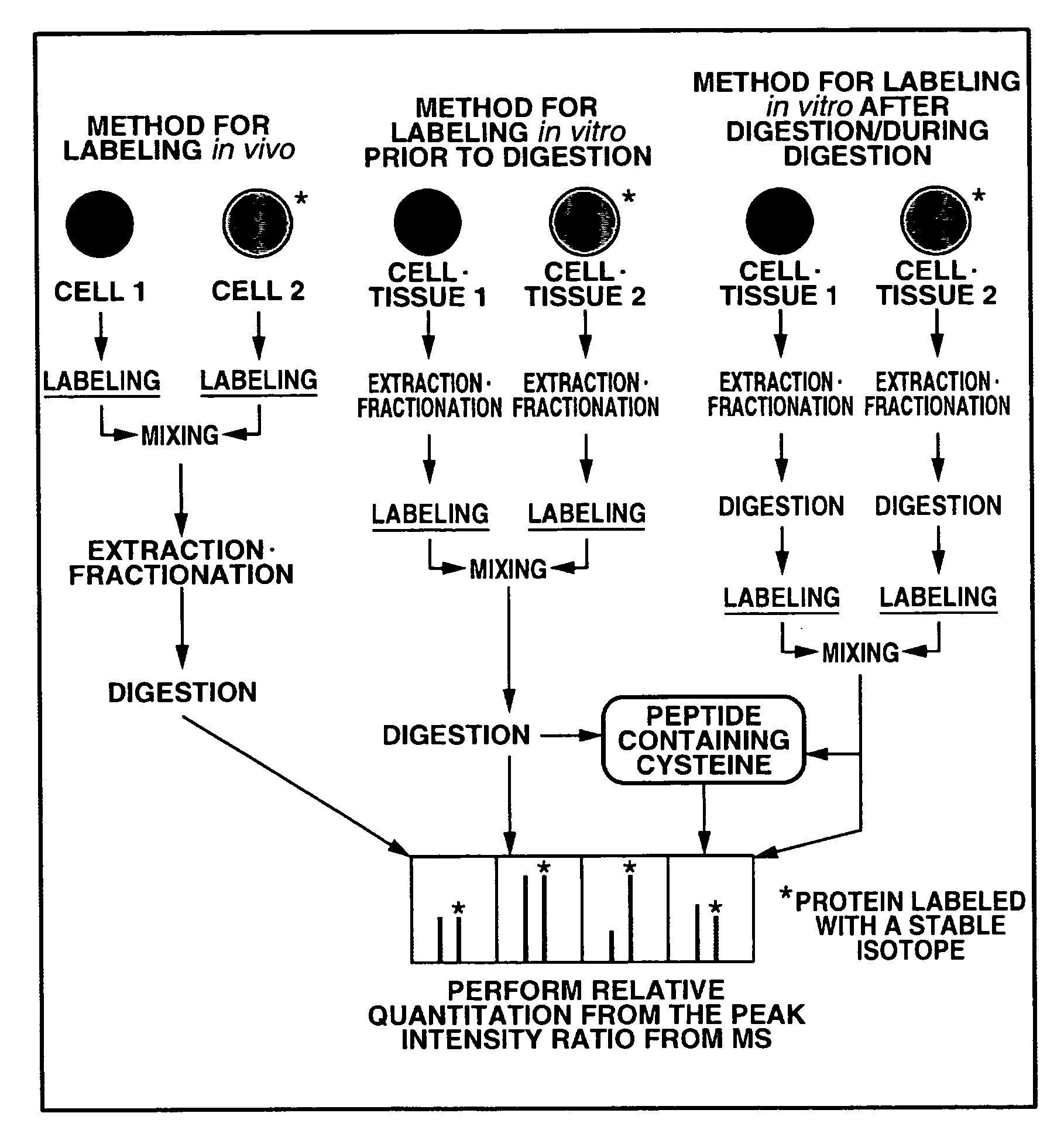
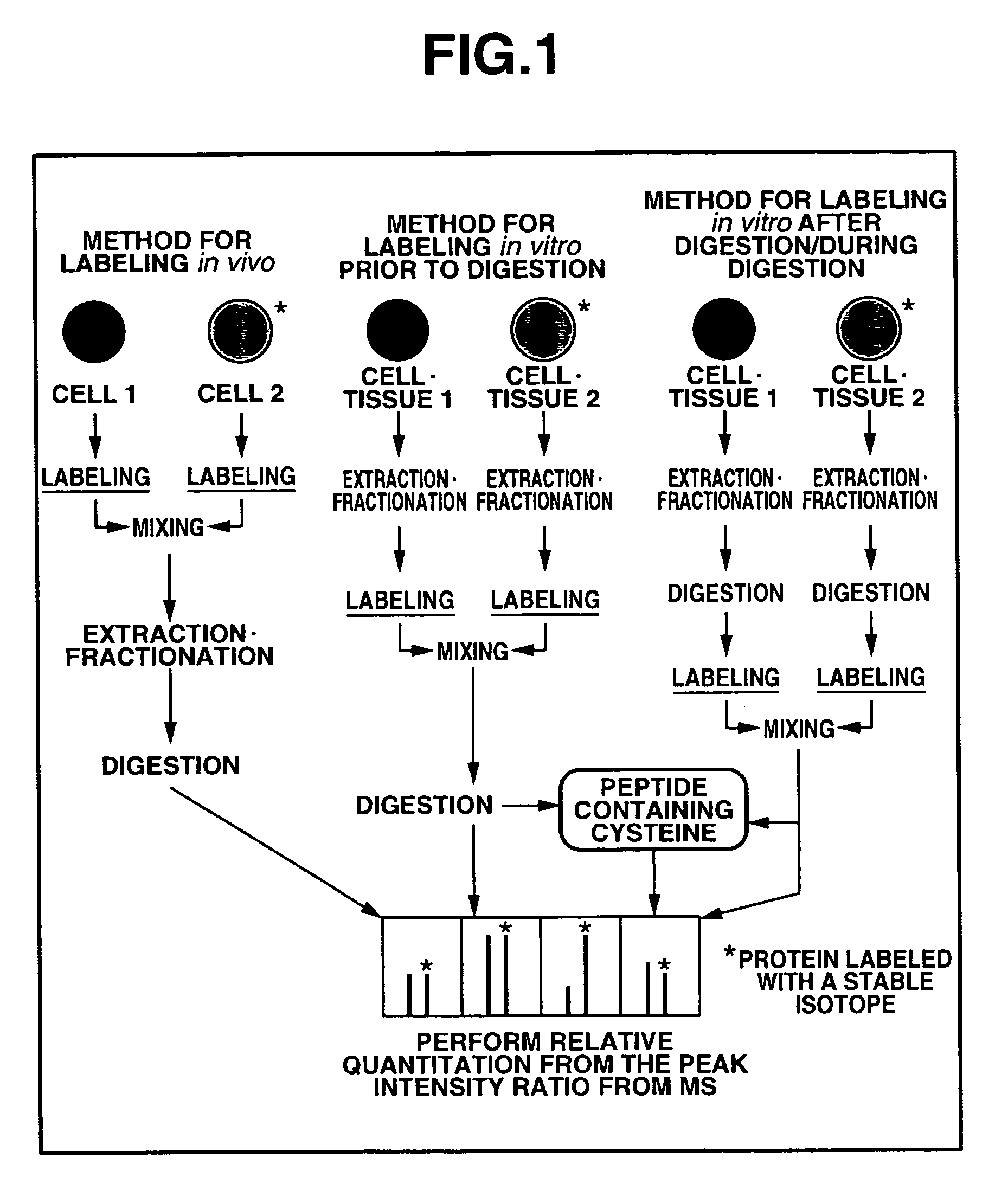
However, while this in vivo labeling method is in principle applicable to every cell as long as they are cultured cells, in reality, such as large change in cell growth rate due to the change in culture conditions, culture in a culture medium containing a stable isotope is often difficult.
In addition, tissue from animal and human or the like, cannot be cultured easily, and cannot be measured with this method.
However, proteins that do not contain a cysteine residue cannot be quantified with this method, in addition, the variation of quantitative value at each experiment in the pretreatment stage such as purification cannot be corrected.
However, with these methods, the variation of quantitative value at each experiment in the pretreatment stage, such as digestion, fractionation and purification of the protein contained in the sample, cannot be corrected.
In the former, labeling is simple, and highly reproducible data can be expected; however, when the sample is a tissue, a biological fluid, a cell organ, a protein complex, a cell incapable of growing in a culture medium containing a stable isotope, or the like, it cannot be applied to proteins that do not incorporate stable isotope contained therein.
The latter can be applied to every sample; however, the variation of quantitative value at each experiment in the pretreatment stage until labeling, such as sample disruption, extraction, digestion, fractionation and purification, cannot be corrected.
In addition, the labeling reaction per se is often not simple such that differences in the experience of the experimenters show.
Further, carrying out comparison of three or more samples was not simple, which also decreased accuracy.
Furthermore, when the measurement subject changed the internal standard substance to be labeled also changed, each time requiring the labeling conditions to be examined; in addition, comparison between experiments was not possible.
On the other hand, quantitating the internal standard substance biological molecule was not simple; therefore, absolute quantitation was very difficult.
However, with this method, an isotope labeled synthetic peptide must always be prepared for each experiment, quantification must also be carried out at each experiment, such that comprehensive quantitation is difficult.
In addition, it is influenced by the recovery rate in the purification process of the objective protein, and also influenced by the efficiency of in-gel digestion generally told to be 30-50%, such that accurate quantitation is difficult.
However, it has been pointed out that two-dimensional electrophoresis was not suitable for a quantitative analysis of protein owing to the quantity of protein developed being very small and the recovery rate at analytical time being prone to errors.
However, merely identifying a protein, or elucidating components by mass spectroscopy is insufficient as functional analysis of protein.

Method used


Image

  • Mass spectrometric quantitation method for biomolecules based on metabolically labeled internal standards
  • Mass spectrometric quantitation method for biomolecules based on metabolically labeled internal standards
  • Mass spectrometric quantitation method for biomolecules based on metabolically labeled internal standards

Examples


first embodiment

OF THE PRESENT INVENTION

[0252]The present invention provides an internal standard substance that is a metabolically isotope labeled biological molecule, a cell containing an internal standard substance that is a metabolically isotope labeled biological molecule, a reagent containing the internal standard substance, a reagent containing the cell that contains the internal standard substance, and the like.

[0253]In the following, a method for preparing an internal standard substance that is a metabolically isotope labeled biological molecule and a cell containing an internal standard substance that is a metabolically isotope labeled biological molecule will be described in particular.

[0254]The cell containing an internal standard substance that is a metabolically isotope labeled biological molecule is a cell of the same species as the sample to be measured, preferably derived from the same tissue, in particular preferably easy to grow in a culture medium containing an isotope, but is n...

second embodiment

OF THE PRESENT INVENTION

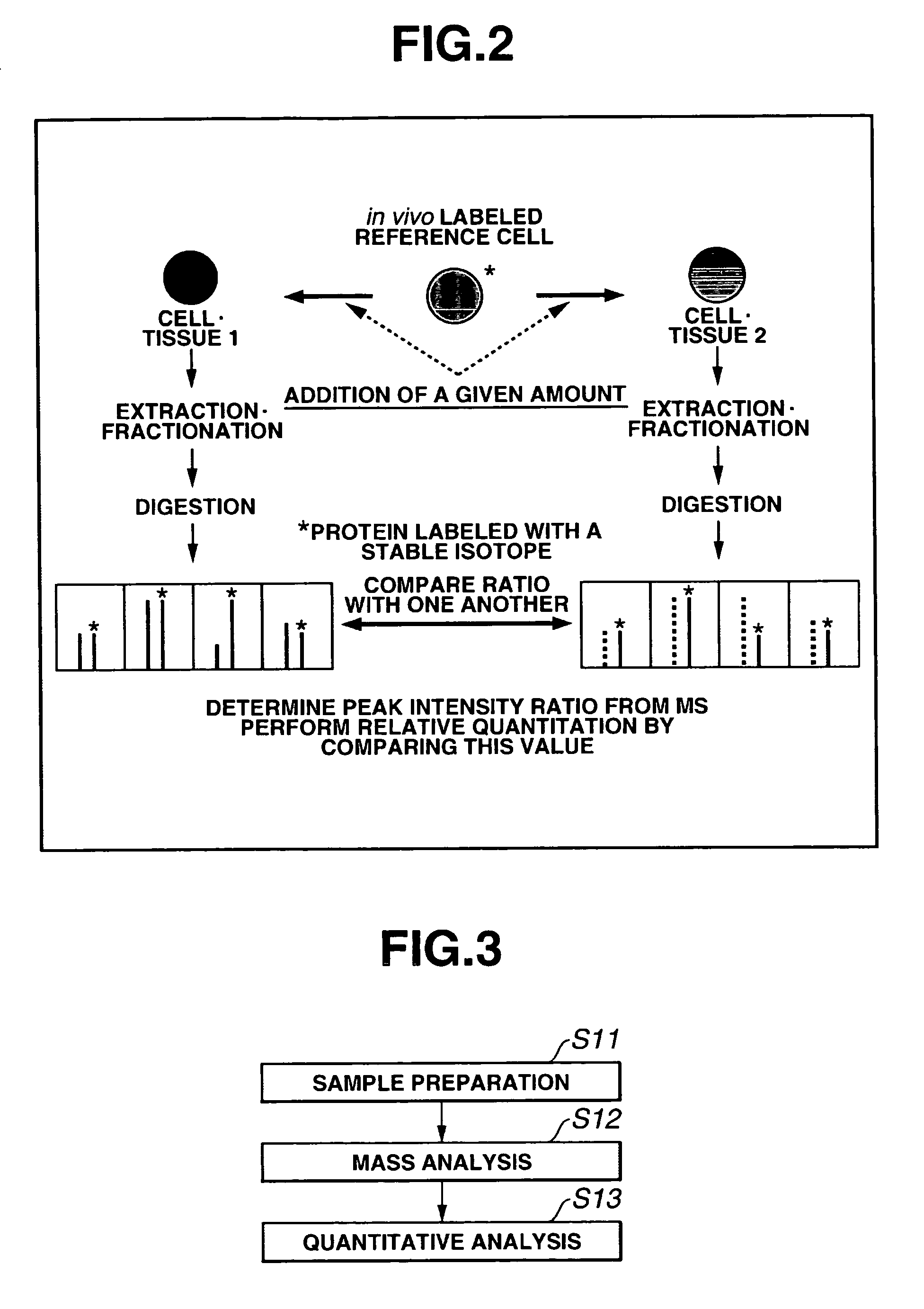
[0289]FIG. 3 shows an overall scheme summarizing a method according to the second embodiment of the present invention for a quantitatively analyzing a protein as one example of biological molecule. As shown in FIG. 3, first, a measurement sample preparation is carried out, mass analysis is then carried out using a mass spectroscopic device. Next, the quantitative analysis method according to the present invention is executed.

[0290]The measurement sample preparation shown in step S11 of FIG. 3 comprises the steps of: isotope labeling a sample; extracting and fractionating a biological molecule from each sample and the like. Examples of the methods for isotope labeling the sample include the method of culturing in a culture medium containing an isotope labeled amino acid, the method of isotope labeling chemically or enzymically in vitro, and the like.

[0291]The method of culturing in the culture medium containing the isotope labeled amino acid comprises the step...

example 1

[0356]The following operations were carried out to prepare a cell containing an internal standard substance that was a metabolically isotope labeled biological molecule.

[0357]Mouse neuroblastoma Neuro2A was cultured. RPMI-1640 (Sigma, R-7130) containing 10% fetal bovine serum (MOREGATE, BATCH 32300102), 100 U / ml of penicillin G and 100 μg / ml of streptomycin (GIBCO, 15140-122) was used as a culture medium. As RPMI-1640, a powder culture medium not containing L-glutamine, L-lysine, L-methionine, L-leucine and sodium hydrogencarbonate was selected, and components missing from the culture medium were added (L-glutamine (G-8540 manufactured by Sigma), L-lysine (L-9037 manufactured by Sigma), L-methionine (M-5308 manufactured by Sigma) and sodium bicarbonate (Wako Pure Chemical Industries, 191-01305) were added in the amounts of 0.3 g / L, 0.04 g / L, 0.015 g / L and 2 g / L respectively. In addition, 0.05 g / L of L-leucine labeled with a stable isotope (Cambridge Isotope Laboratories, CLM-2262) w...


PUM

PropertyMeasurementUnit
concentrationaaaaaaaaaa
pHaaaaaaaaaa
pHaaaaaaaaaa

Abstract

An object of the present invention is to quantitate with good accuracy, furthermore, quantitate absolutely, one or a plurality of biological molecules in a sample such as a tissue, a biological fluid, a cell, a cell organ or protein complex.By adding a metabolically isotope labeled biological molecule as an internal standard substance and measuring with a mass spectrometer, quantitating with good accuracy one or a plurality of target molecules in a sample has become possible. In addition, by performing waveform separation processing during mass analysis, a highly accurate quantitative analysis method of mass analysis is provided.

Description

TECHNICAL FIELD[0001]The present invention relates to a method for quantitating one or a plurality of biological molecules in a sample by adding to the sample an internal standard substance that is a metabolically isotope labeled biological molecule, or a cell containing an internal standard substance that is a metabolically isotope labeled biological molecule, and measuring with a mass spectrometer.[0002]In addition, the present invention relates to a quantitative analysis method for a biological molecule; more specifically, the present invention relates to an analysis method using an isotope labeled biological molecule and to a program for this analysis.BACKGROUND ART[0003]A genome is the total DNA contained in one cell, containing the entirety of the genes and the intergenic regions of a living organism, and organisms known to us are defined by the genome. Currently, reading of the human nucleotide sequences has already been completed, and the number of genes is being predicted. ...

Claims


Application Information

Patent Timeline
Patent Type & Authority Patents(United States)
IPC IPC(8): G01N24/00G01N27/62G01N33/68
CPCG01N33/6848Y10T436/105831Y10T436/104165Y10T436/104998Y10T436/10Y10T436/24
Inventor ODA, YOSHIYAISHIHAMA, YASUSHITABATA, TSUYOSHI
Owner EISIA R&D MANAGEMENT CO LTD
Learn More