Variably crosslinked tissue
a tissue and cross-linked technology, applied in the field of human or animal tissue fixing, can solve the problems of tissue shrinkage during fixation, stenosis and regurgitation, slow release of glutaraldehyde from the implanted device, etc., and achieve the effect of improving resistance to calcification and minimising shrinkag
- Summary
- Abstract
- Description
- Claims
- Application Information
AI Technical Summary
Benefits of technology
Problems solved by technology
Method used
Image
Examples
example 1
[0076]This first experiment was carried out to show the effect of 1-6 hexane diamine concentration on surface reduction during cross linking.
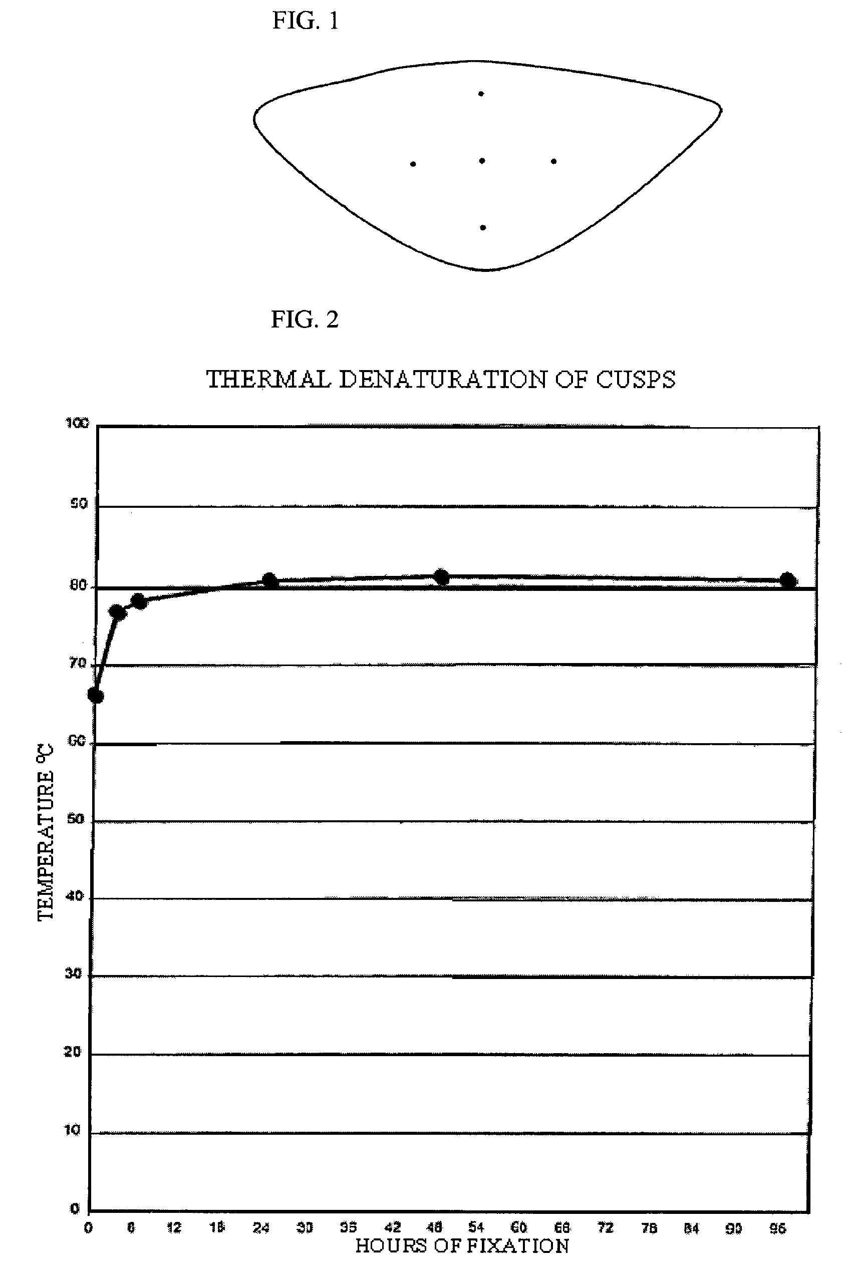
[0077]Eighty leaflets were excised from fresh porcine aortic roots. Each leaflet was blotted to remove excess buffer and placed flat, outflow side down, on a glass surface. Under a dissection microscope equipped with a reticule, the cusps were then marked in the radial and circumferential directions as shown in FIG. 1, with tissue marking dye following the manufacturer's recommendations. The distance between the center dot and each of the four exterior dots was 5 mm before cross linking; therefore, the maximum distance between markers was 10 mm. The leaflets were then randomly distributed in 4 groups of 20 cusps each and cross-linked by incubation under the following conditions:
[0078]Group 1. Aqueous solution of 11.25 mM 1,6-hexanediamine (DIA) in 20 mM HEPES buffer, pH 6.5, containing 20 mM 1-Ethyl-3-(3-Dimethylaminopropy-1)-Carbodiimide (EDC)...
example 2
[0085]The following experiment was carried out to determine the effect of DIA concentration greater than 112.5 mM in cross linking.
[0086]Twenty-one leaflets for each condition were excised from fresh porcine aortic roots. For each condition, seven roots plus their 3 respective excised leaflets were incubated for 96 hours at room temperature in 250 ml of an aqueous solution of 20 mM HEPES and either 112.5 mM or 160 mM DIA, in the presence of 20 mM EDC and 1 mM Sulfo-NHS. After incubation, the samples were washed with sterile saline and then sterilized 3 times for 48 hours at 4° C. with 25 mM EDC in the absence (0%) of, or in the presence of either 5% or 20% isopropyl alcohol. For one condition, 100 mM ethanolamine was added as a blocker during sterilization with a solution containing 20% isopropanol.
[0087]The results are presented in Table 2 and demonstrate that 3 consecutive sterilization treatments of the leaflets fixed with 112.5 mM DIA, in the absence of isopropyl alcohol, do not...
example 3
[0089]The next experiment was carried out to determine the effect of the duration of incubation on cross linking of porcine aortic valves from three different standpoints.
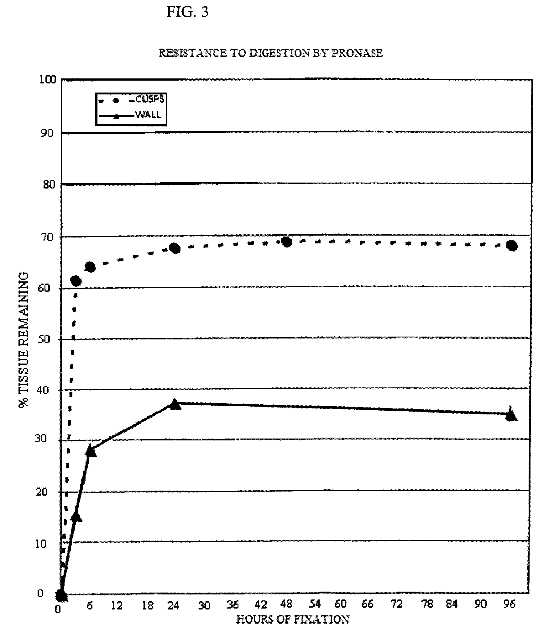
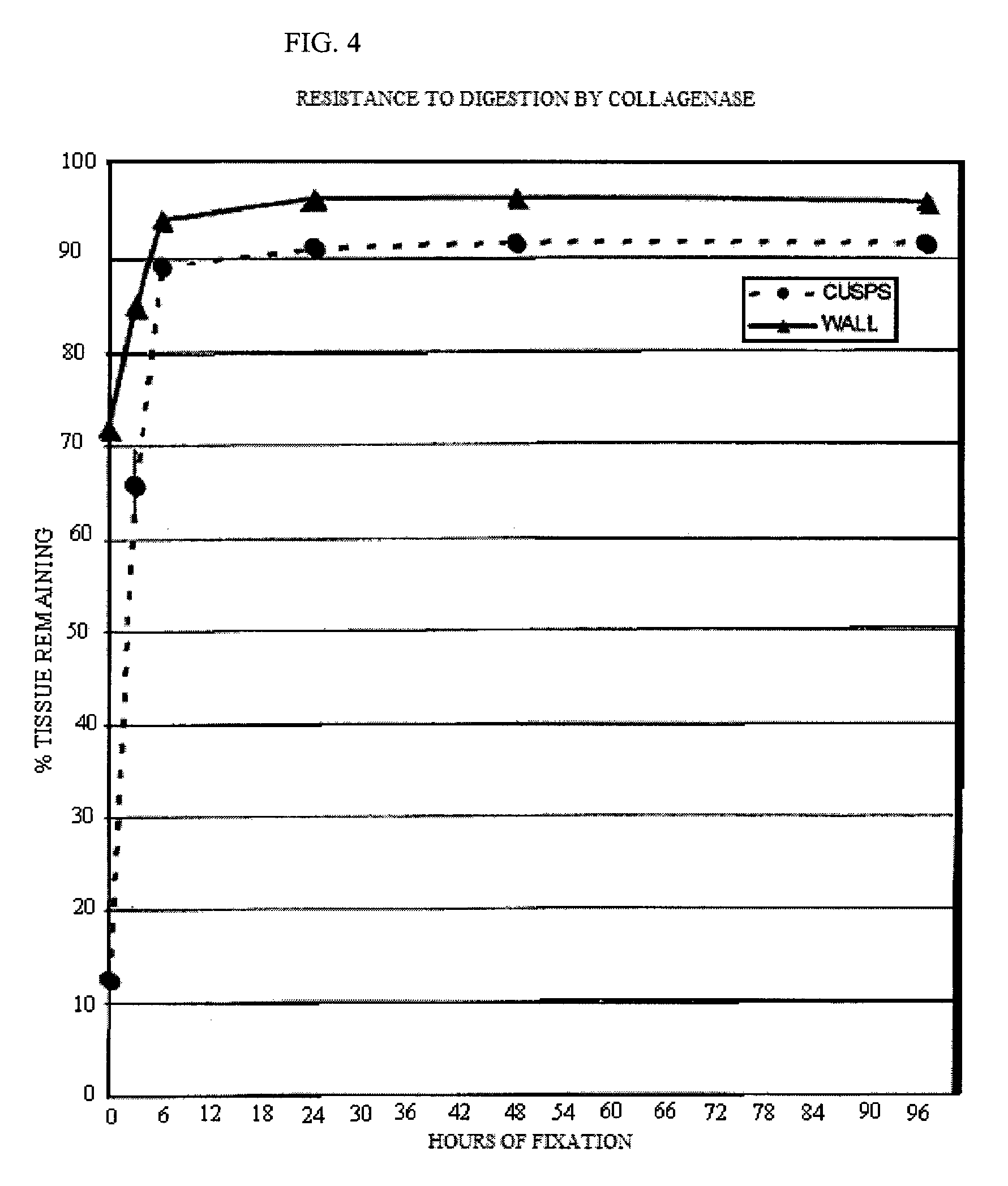
[0090]Five groups of 4 valves each were incubated in an aqueous solution containing 20 mM HEPES, 112.5 mM DIA, pH 6.5, 20 mM EDC and 1 mM Sulfo-NHS for 3, 6, 24, 48 and 96 hours. The valves were then washed with sterile saline to eliminate any reaction byproducts. They were stored in 10 mM HEPES, 0.85% sodium chloride, pH 7.4, and 20% isopropyl alcohol until use. Thermal denaturation tests and tests for resistance to proteolytic degradation by collagenase and by protease were performed for the determination of cross linking efficacy. These test procedures are described in the J. Heart Valve Dis. 1996; 5(5):518-25. Fresh porcine aortic roots were used as a control.
[0091]a. Thermal Denaturation
[0092]The leaflets were excised from the aortic roots and submitted (n=3 per condition) to thermal stability testing as descr...
PUM
| Property | Measurement | Unit |
|---|---|---|
| shrinkage temperature | aaaaa | aaaaa |
| shrinkage temperature | aaaaa | aaaaa |
| reaction temperature | aaaaa | aaaaa |
Abstract
Description
Claims
Application Information
 - R&D
- Intellectual Property
- Life Sciences
- Materials
- Tech Scout
- Unparalleled Data Quality
- Higher Quality Content
- 60% Fewer Hallucinations
Browse by: Latest US Patents, China's latest patents, Technical Efficacy Thesaurus, Application Domain, Technology Topic, Popular Technical Reports.
© 2025 PatSnap. All rights reserved.Legal|Privacy policy|Modern Slavery Act Transparency Statement|Sitemap|About US| Contact US: help@patsnap.com


