

Method for diagnosing immunoglobulin A nephropathy and TGBM nephropathy
a nephropathy and immunoglobulin technology, applied in the field of protein development, can solve the problems of inability to accurately diagnose iga nephropathy, damage caused by mediators, and difficulty in directly determining the degree of progression of diabetic nephropathy, so as to diagnose diabetic nephropathy early, easy to diagnose iga nephropathy, and high specificity and sensitivity
- Summary
- Abstract
- Description
- Claims
- Application Information
AI Technical Summary
Benefits of technology
Problems solved by technology
Method used
Image

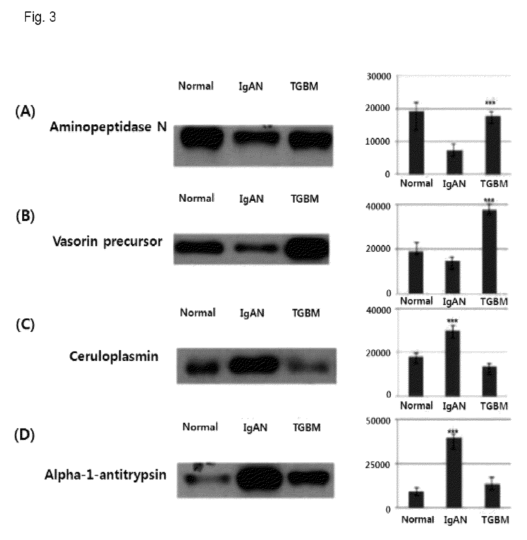
Examples
example 2
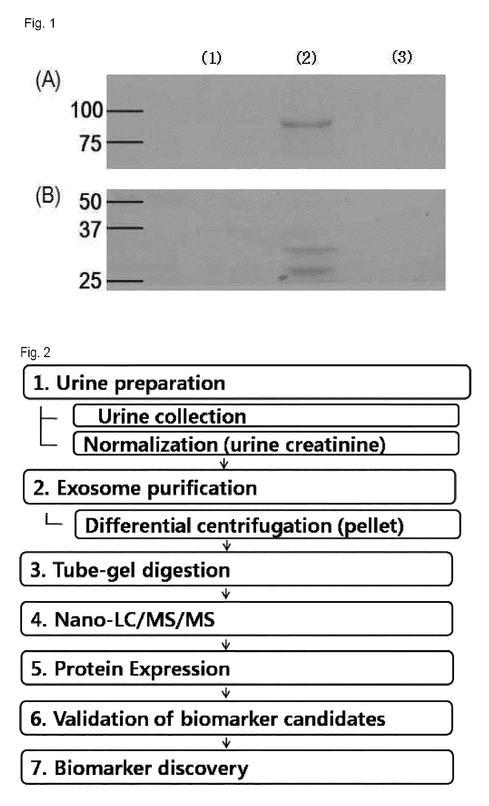
Purification of Exosome
[0030]2-1. Purification of Exosome
[0031]The urine creatinine level of each urine sample was recognized by a body fluid analyzer. Then, urine samples of three groups were put together after their volumes were set in such a manner that the same creatinine level was included. The put-together urine sample was subjected to first centrifugation (17,000 g for 15 min at 4° C.) to remove fragments such as whole cells or large membrane proteins. The supernatant of the first centrifugation was subjected to second centrifugation (200,000×g for 1 h at 4° C.) to precipitate low-density membrane vesicle protein. The precipitated resultant product was resuspended by using 50 ul of isolation buffer (250 mM sucrose / 10 mM triethanolamine / 0.5 mM PMSF / 1 μM leupeptin), transferred to 1.5 ml polypropylene tube, added with 60 mg / ml of DTT, and reacted at 60° C. for 10 minutes to separate THP. Then, the isolation buffer was added to carry out centrifugation (200,000 g for 1 h at 4° C...
example 3
[0034]The obtained exosome sediment was suspended in 14 ul of 0.3% SDS solution, mixed with 5 ul of acrylamide solution (30%), 0.7 ul of 1% ammonium persulfate, and 0.3 ul of TEMED, and polymerized at room temperature for about 30 minutes. A small gel was cut into pieces with a size of 1 mm3 by a surgeon's knife, and washed with 25 mM of ABC buffer (pH 8.0) for 20 min and 25 mM of ABC (pH 8.0) / 50% ACN buffer for 20 min. Then, the gel was dried by vacuum-drying, and subjected to a general in-gel digestion method. Each protein was added with 10 mM of DTT, reduced at 56° C. for 30 minutes, and then added with 55 mM of iodoacetamide (IAA), and alkylated by dark reaction at room temperature. Then, the gel pieces were washed with 25 mM of ABC (pH 8.0) buffer for 20 min and 25 mM of ABC (pH 8.0) / 50% ACN buffer for 20 min, and dried by vacuum-drying. Trypsin was dissolved in 25 mM of ABC buffer to a concentration of 100 ng / ul, and reacted with gel pieces at 37° C. for 16 hou...
example 4
Protein Identification
[0035]In nano-size liquid chromatography analysis, a nanoAcquity system (Waters Corporation, Milford, Mass.) was used, and herein, C18 trap column of 5 μm, 2 mm 180 μm (Symmetry), and C18 analyzing reversed-phase column of 1.7 μm, 25 cm×5 μm (BEH) (Waters Corporation) were used. The obtained peptide was analyzed by being dissolved in 0.1% formic acid. A mobile phase A (0.1% formic / water), and a mobile phase B (0.1% formic acid / CAN) were used. The phase was flowed into the trap column at a flow rate of 10 μL / min for 3 min. The mobile phase B was flowed at a concentration of 3 to 40% and a flow rate of 300 nl / min, for 360 min to separate peptide. The column was washed with 90% mobile phase B for 15 min, and then restabilized with 3% mobile phase B for 20 min. The temperature of the column was maintained at 35° C., and an auxiliary pump was used to move 100 fmol [Glu1]-Fibrinopeptide B / μL at a flow rate of 300 mL / min to a nano-lock reference spray of a mass spectr...
PUM
| Property | Measurement | Unit |
|---|---|---|
| pH | aaaaa | aaaaa |
| temperature | aaaaa | aaaaa |
| concentration | aaaaa | aaaaa |
Abstract
Description
Claims
Application Information