
Porous SERS analytical devices and methods of detecting a target analyte

a technology of raman spectroscopy and analytical devices, which is applied in the direction of spectrometry/spectrophotometry/monochromators, optical radiation measurement, instruments, etc., can solve the problems of high cost, time-consuming, and laborious methods, and achieve the effects of avoiding the high cost of nanofabricated sers substrates and the complexity associated, improving detection limits, and low variation

Active Publication Date: 2016-02-09
UNIV OF MARYLAND
View PDF25 Cites 3 Cited by
  • Summary
  • Abstract
  • Description
  • Claims
  • Application Information

AI Technical Summary

Benefits of technology

The present invention is a new method for fabricating a versatile and cost-effective surface-enhanced Raman scattering (SERS) substrate. The method utilizes a consumer inkjet printer to modify paper substrates with a sizing agent to create hydrophobic regions that concentrate the analyte, allowing for low-level chemical and biomolecular detection. The invention also introduces a novel technique called filter SERS that leverages the filtration process to create a SERS-active substrate and concentrate the analyte, resulting in significantly improved detection limits and a simplified procedure. This new approach makes SERS more accessible and cost-effective, paving the way for practical applications in on-site and field-based detection.

Problems solved by technology

While sensitive, such methods are labor intensive, time consuming, and costly.
They also require expensive and bulky equipment, and hence are not portable.
Unfortunately, the high cost and complications associated with the fabrication of conventional SERS-active substrates have prevented the wide use of SERS.
Furthermore, these substrates exhibit limited shelf life, with progressive reduction in SERS activity due to oxidation of the nanostructures (see Erol M. et al.
However, such microfluidic SERS devices introduce additional complexity to their fabrication, and may decrease sensitivity due to difficulties in the optical coupling, decreased sample volume, and the inability to concentrate the analyte by drying.
As a result, chemical and biomolecular detection using SERS has been costly on a per-sample basis.
Furthermore, SERS-active substrates produced through such conventional techniques have a short shelf life and must be used quickly.
Thus, such conventional SERS systems have not been found practical for routine laboratory analysis of chemicals and biomolecules, and are not an option for field-based applications.
Conventional SERS methodologies are therefore limited to laboratory settings due to their high cost and short shelf life.

Method used


Image

  • Porous SERS analytical devices and methods of detecting a target analyte
  • Porous SERS analytical devices and methods of detecting a target analyte
  • Porous SERS analytical devices and methods of detecting a target analyte

Examples


example 1

[0058]Materials

[0059]Fisherbrand chromatography paper, 0.19 mm in thickness, was used for the substrate. Hexadecenyl succinic anhydride (ASA) from Thermo-Fisher Scientific (Pittsburgh, Pa.) was used to create a hydrophobic surface on the paper. Silver nitrate, sodium citrate, glycerol, and hexanol were obtained from Sigma-Aldrich (St. Louis, Mo.). Rhodamine 590 chloride, also known as Rhodamine 6G (R6G), was purchased from Exciton (Dayton, Ohio).

[0060]Nanoparticle Synthesis

[0061]Silver nanoparticles were synthesized using the method of Lee and Meisel (Lee P. C. and Meisel D. (1982) J. Phys. Chem. 86:3391-3395). Briefly, 90 mg of silver nitrate was added to 500 mL of ultrapure water (18.2 MΩ), which was then brought to a boil in a flask under vigorous stirring. Sodium citrate (100 mg) was added, and the solution was left to boil for an additional 10 min. After the solution turned greenish brown, which indicated the formation of silver colloid, it was then removed from the heat.

[0062]...

example 2

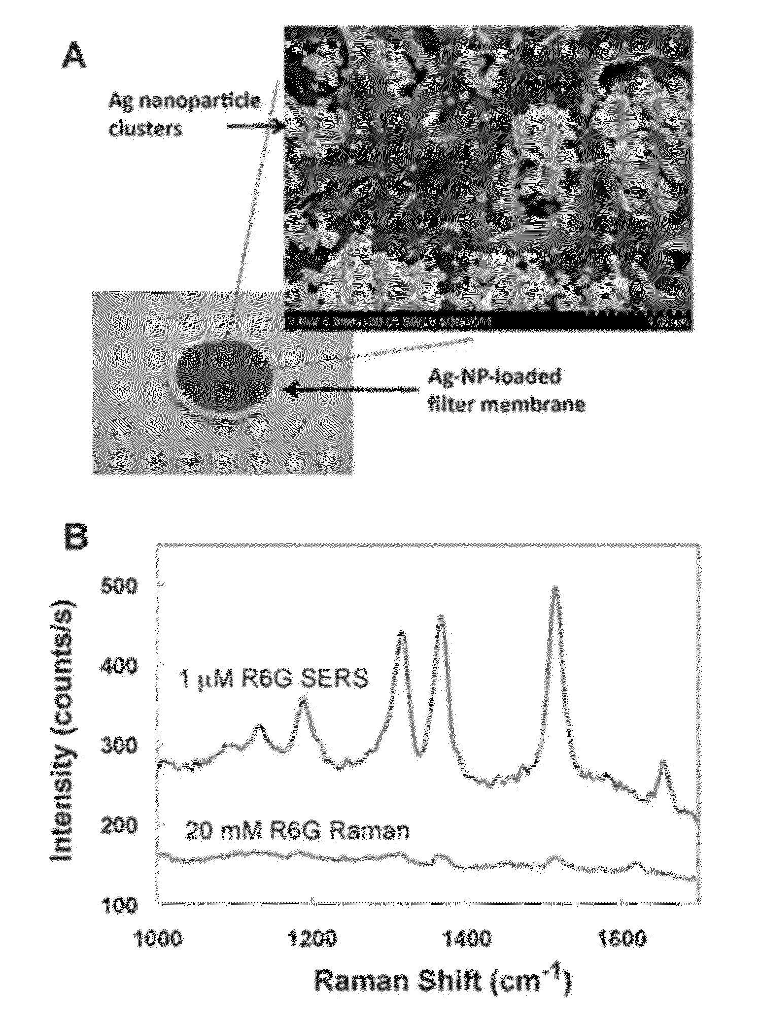
[0081]We demonstrate the detection performance of the filter SERS technique of at least 1-2 orders of magnitude better than the typical approach of drying a sample in silver colloid onto a surface. We achieved a detection limit of 10 nM for the common model SERS analyte Rhodamine 6G (R6G) using a portable spectrometer and diode laser. Furthermore, we demonstrate the utility of the filter SERS technique for field based applications by detecting parts per billion (ppb) concentrations of melamine, a food contaminant, as well as ppb concentrations of malathion, a widely used pesticide, in aqueous solution. The disclosed technique shows relatively low variability, and all acquired data sets could be fit well with a Langmuir isotherm. This indicates that the disclosed field based technique is not only simple and sensitive, but it can also be quantitative.

[0082]Materials

[0083]Nylon and Millipore PVDF filter membranes, 13 mm diameter, 0.22 μm pore sizes were purchased from Fisher Scientific...

example 3

[0109]Fabrication and Operation of Optofluidic SERS Dipstick

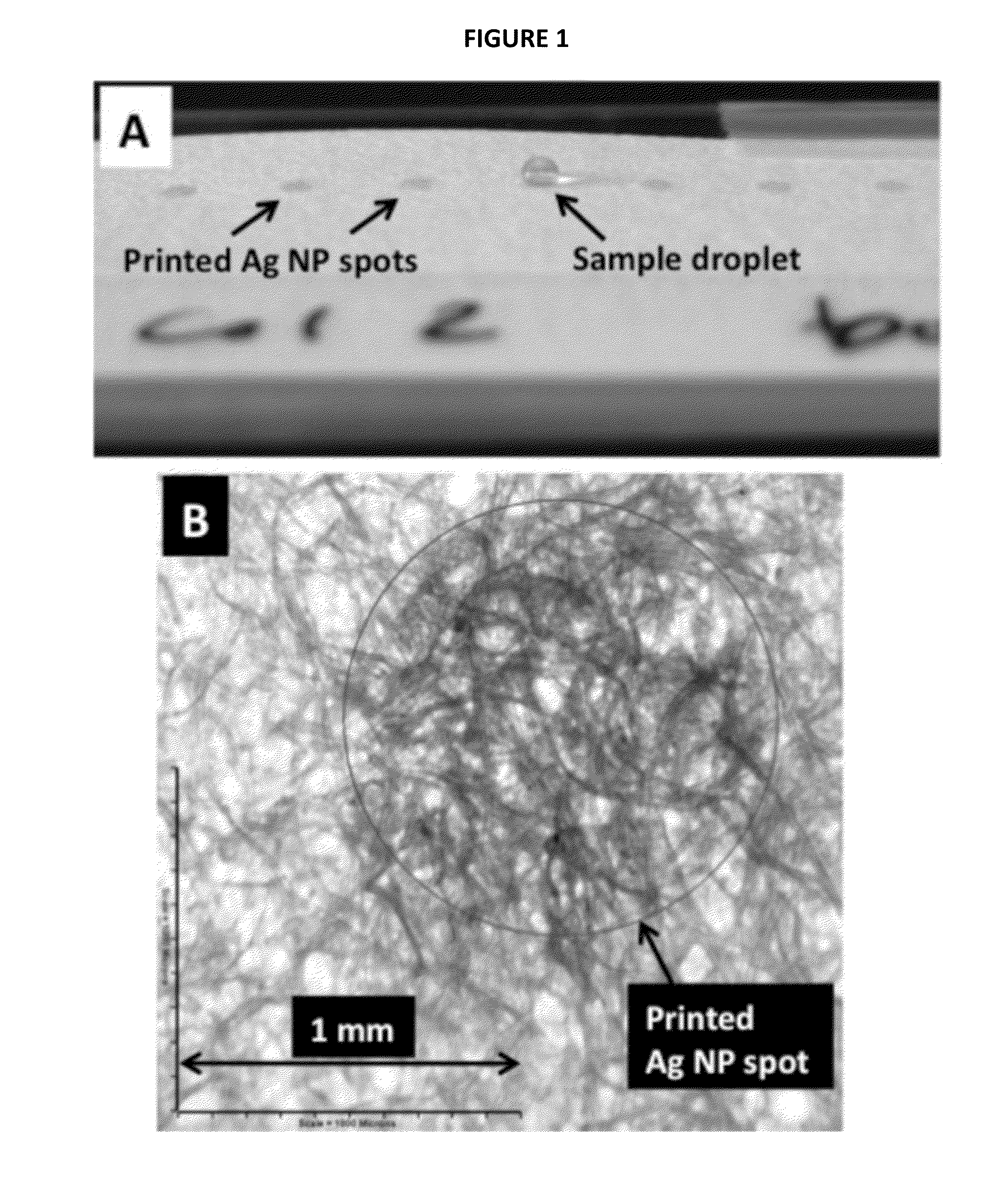
[0110]A low-cost commercial piezo-based EPSON inkjet printer was used to print the SERS active substrates on chromatography paper. To form the ink, a silver colloid, prepared by the method of Lee and Meisel (1982), supra, J. Phys. Chem. 86:3391-3395, was concentrated 100× by centrifugation; glycerol and ethanol were added (40% and 10% by volume respectively) to optimize the surface tension and viscosity for optimal printing. The ink was added into re-usable cartridges and printed 10 times onto the desired regions of the paper (FIG. 12a). After printing silver nanoparticles onto the paper, individual dipsticks were cut out of the sheet. The end of the dipstick containing the silver nanoparticles serves as the detection region, while the rest of the paper strip serves as the sample collection zone.

[0111]For the dipstick experiments reported here, 5 μL of Rhodamine 6G (R6G) in methanol was added to the sample collection zone. ...


PUM

PropertyMeasurementUnit
volumeaaaaaaaaaa
diameteraaaaaaaaaa
optical densityaaaaaaaaaa

Abstract

A surface enhanced Raman spectroscopy (SERS) analytical device includes a substrate having a porous structure, and a plurality of plasmonic nanoparticles embedding in the porous structure and forming a sensing region. A method of detecting a target analyte in a sample includes contacting the sample with the substrate, whereby the target analyte, if present in the sample, is concentrated in the sensing matrix. The substrate may then be analyzed using SERS detection equipment.

Description

STATEMENT REGARDING FEDERALLY SPONSORED RESEARCH OR DEVELOPMENT[0001]This work was supported by the National Institutes of Health as provided for by the terms of K25EB006011. The US government has certain rights in this invention.CROSS-REFERENCE TO RELATED APPLICATIONS[0002]This application is based on U.S. Provisional Patent Application Ser. No. 61 / 539,334, filed Sep. 26, 2011, application Ser. No. 61 / 585,990, filed Jan. 12, 2012, and application Ser. No. 61 / 603,800, filed Feb. 27, 2012, which applications are all incorporated herein by reference in their entireties and to which priority is claimed.FIELD OF THE INVENTION[0003]The present invention relates to a low-cost surface enhanced Raman spectroscopy (SERS) analytical device, methods of forming a SERS analytical device, and methods of detecting a target analyte.BACKGROUND OF THE INVENTION[0004]Detection of trace chemicals in solid and liquid samples may be achieved by methods that combine chromatography techniques with mass spe...

Claims


Application Information

Patent Timeline
Patent Type & Authority Patents(United States)
IPC IPC(8): G01J3/44G01N21/552G01N21/65B82Y30/00
CPCG01J3/44G01N21/554G01N21/658B82Y30/00
Inventor YU, WEI WENWHITE, IAN M.
Owner UNIV OF MARYLAND
Learn More