Biology polypeptide medicament blood vessel bracket and preparation method thereof

A technology of biological polypeptides and vascular stents, applied in the field of medical devices, can solve the problems of delaying vascular endothelial repair, ignoring the specific inhibition of smooth muscle cells, etc., and achieve the effect of promoting differentiation and proliferation, inhibiting proliferation and migration of vascular smooth muscle cells, and promoting repair

Active Publication Date: 2008-12-03
乐普(深圳)国际发展中心有限公司
View PDF0 Cites 15 Cited by
  • Summary
  • Abstract
  • Description
  • Claims
  • Application Information

AI Technical Summary

Problems solved by technology

[0003] The wide application of drug-eluting stents has greatly reduced the occurrence of in-stent restenosis. Although drug-eluting stents can effectively inhibit the proliferation of smooth muscle cells and reduce and prevent intimal hyperplasia, they delay the repair of vascular endothelium; CD34 antibody-engineered stents Although endothelial progenitor cells can be captured in blood vessels based on the antigen-antibody binding theory and rapidly endothelialized on the surface of vascular stents, the specific inhibition of smooth muscle cells is ignored

Method used


Image

  • Biology polypeptide medicament blood vessel bracket and preparation method thereof
  • Biology polypeptide medicament blood vessel bracket and preparation method thereof

Examples


preparation example Construction

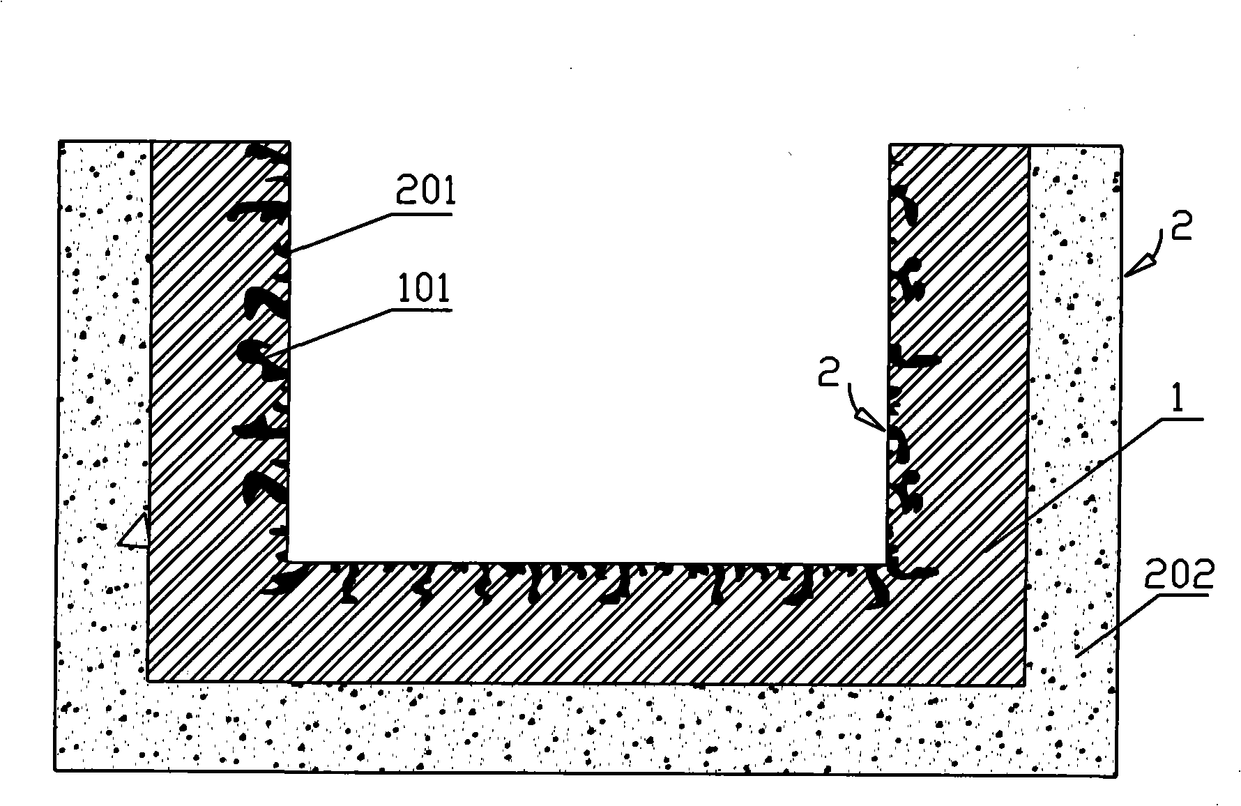
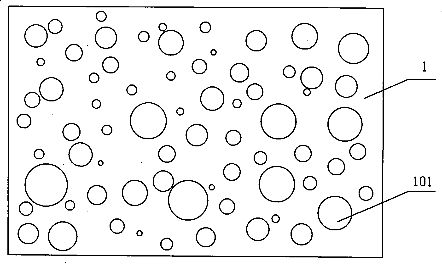
[0031] A method for preparing a biological polypeptide drug vascular stent, firstly prepare a large number of holes 101 on the stent body 1 through chemical or physical methods such as corrosion, anodic oxidation, micro-arc oxidation, micro-arc nitriding, etc., or a combination of these methods, or A layer of coating with holes 101 is formed on the surface of the stent, and the stent body is made of medical materials with good biocompatibility. Metal materials such as stainless steel, cobalt-based alloys, titanium alloys, nickel-titanium alloys, and polylactic acid can also be used. Biomacromolecule material; then use a special process to protect the inner surface of the stent, reserve holes on the inner surface of the stent to fix the biological polypeptide, and at the same time embed, fix and coat one or more kinds of substances that can inhibit blood vessels on the outer surface of the stent body 1 Active drug 202 for smooth muscle cell proliferation and migration, such as a...

Embodiment 1

[0047] In this embodiment, an arginine-glycine-aspartic acid (RGD) polypeptide drug 201 is fixed on the inner surface of the stent body 1 with holes 101, and rapamycin anti-smooth muscle cell proliferation drug is coated on the outer surface of the stent body 1 202;

[0048] ①Pretreatment of the surface of the stent body, ②Preparation of holes, ③Post-treatment process steps of the surface of the stent body are the same as described above;

[0049] ④ Drug preparation: add 0.2g rapamycin to 10ml tetrahydrofuran solution;

[0050] ⑤External surface coating: use a balloon to protect the inner surface of the stent body with holes, and disperse the above-mentioned prepared drug evenly at room temperature, then spray it on the outer surface of the stent, and cure it in the air for 60 minutes; repeat the ⑤ outer surface coating step , until the drug loading reaches 2.2μg / mm 2 ; Place the bracket in a vacuum oven to dry;

[0051] 6. Internal surface immobilization: the biological po...

Embodiment 2

[0056] ①Pretreatment of the surface of the stent body, ②Preparation of holes, ③Post-treatment process steps of the surface of the stent body are the same as described above;

[0057] ④ Drug preparation: Add 0.1g polylactic acid (PLA) into 10ml tetrahydrofuran solution to prepare biological polypeptide drug 201, then add 0.5g rapamycin, rapamycin and arginine-glycine-aspartic acid ( RGD) The mixing ratio of polypeptide drugs is 0.1~10;

[0058] ⑤ Coating on the outer surface: protect the inner surface of the stent body 1 with holes with a balloon, mix and disperse the above-mentioned drug prepared evenly at room temperature, and then evenly coat the polymer dispersion wrapped with the above-mentioned drug on the stent Cured in the air for 30min; repeat the above operation until the weight of the drug-loaded layer reaches 2.5μg / mm 2 ; Then place the coated bracket 1 in a vacuum oven to dry;

[0059]⑥Inner surface immobilization: Weigh 0.5g of penicillamine cyclic peptide (cycl...


PUM


Abstract

The invention relates to a biologic polypeptide medicine blood vessel support and a preparation method thereof. The biologic polypeptide medicine blood vessel support comprises a support body and active medicines. The support body which is medical material with pores and good biocompatibility is made from stainless steel, cobalt-base alloy, titanium alloy, nickel-titanium alloy or polylactic acid biopolymer; the active medicines comprise a biologic polypeptide medicine and a smooth muscle cell proliferation inhibition medicine. The support is characterized in that: the support body with the pores has the biologic polypeptide medicine fixed on the internal surface of the body and the smooth muscle cell proliferation inhibition medicine coated on the external surface of the body. The preparation me(3) carrying out the after-treatment of the surface of the support body; (4) preparing the medicines; (5) coating the external surface; (6) fixing the medicine onto the internal surface; (7) carrying out the process step of low temperature drying. The support can selectively absorb endothelium progenitor cells which quickly differentiate into endothelium cells to promote the restoration of the endothelium after the support is built in; the support can also effectively inhibit the proliferation and migration of vascular smooth muscle cells, persistently and effectively reduce the formation of a new inner membrane, effectively prevent restenosis inside the support, and avoid the risk of potentially fatal late thrombosis.

Description

technical field [0001] The invention belongs to the field of medical devices, and relates to a biological polypeptide drug vascular stent and a preparation method thereof. Background technique [0002] Since 1987, when Sigwart et al first applied intravascular metal stents to coronary arteries, it has provided a good way to treat vascular occlusive diseases. The main reason for the efficacy of treatment (PCI). The main mechanism of in-stent restenosis is the hyperplasia of vascular intima and the delay of vascular endothelialization. Therefore, the prevention and treatment of in-stent restenosis is mainly to promote the rapid repair of vascular endothelium and inhibit the excessive proliferation of smooth muscle cells. [0003] The wide application of drug-eluting stents has greatly reduced the occurrence of in-stent restenosis. Although drug-eluting stents can effectively inhibit the proliferation of smooth muscle cells and reduce and prevent intimal hyperplasia, they dela...

Claims


Application Information

Patent Timeline
Patent Type & Authority Applications(China)
IPC IPC(8): A61F2/82A61L31/00A61L31/16A61K31/436A61K45/00A61P7/02A61P9/10
Inventor 余占江蒲忠杰
Owner 乐普(深圳)国际发展中心有限公司
Why Eureka
Try Eureka