
Rapid detection method employing ultrasensitive quantum dot microsphere immunity-chromatograph test paper strips

A technology of immunochromatography and detection method, which is applied in the field of detection, can solve the problems of strict detection limit, quenching of fluorescence intensity, unobtainable and other problems, and achieve the effect of simple detection method, improved sensitivity and accurate results

Inactive Publication Date: 2010-11-24
SHANGHAI NORMAL UNIVERSITY
View PDF6 Cites 47 Cited by
  • Summary
  • Abstract
  • Description
  • Claims
  • Application Information

AI Technical Summary

Problems solved by technology

The disadvantages of the existing immunochromatographic detection technology are: due to the extremely strict requirements for food safety testing, pesticide and veterinary drug residue detection limits; at the same time, food substances such as meat, poultry, fruits and vegetables, grains and other complex components make pretreatment difficult; colloidal gold Immunochromatographic detection sensitivity often cannot meet people's requirements
The silicon-coated quantum dot microspheres obtained by this method can only wrap a small amount of quantum dots due to electrostatic repulsion, and at the same time, due to the effect of reagents in the reaction, the fluorescence intensity is greatly quenched, and high sensitivity cannot be obtained.
[0006] Chinese patents 200610024086.7, 200810045548.2, 200810041133.8, 200810227473.X, and 200810186010.3 all involve quantum dot immunochromatographic test strip detection methods to varying degrees. Since all of them are single quantum dots or core-shell quantum dots, the sensitivity is low even if the packaging technology is applied. are also severely restricted

Method used

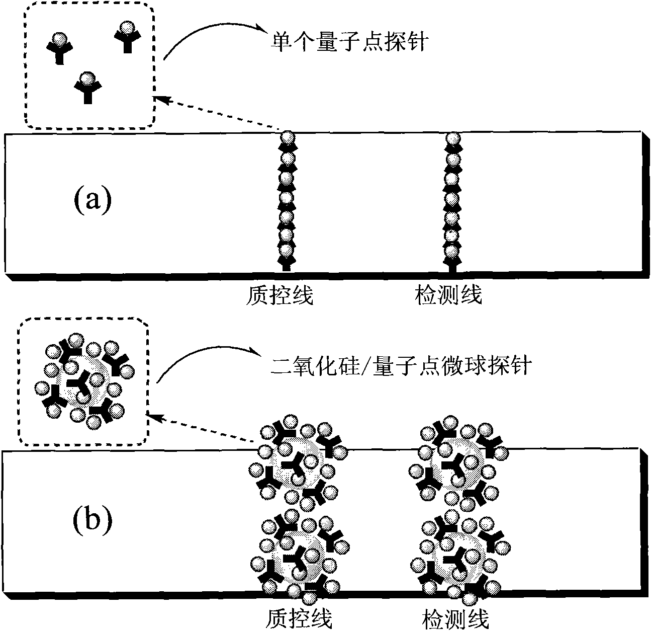

Image

  • Rapid detection method employing ultrasensitive quantum dot microsphere immunity-chromatograph test paper strips
  • Rapid detection method employing ultrasensitive quantum dot microsphere immunity-chromatograph test paper strips
  • Rapid detection method employing ultrasensitive quantum dot microsphere immunity-chromatograph test paper strips

Examples


Embodiment 1

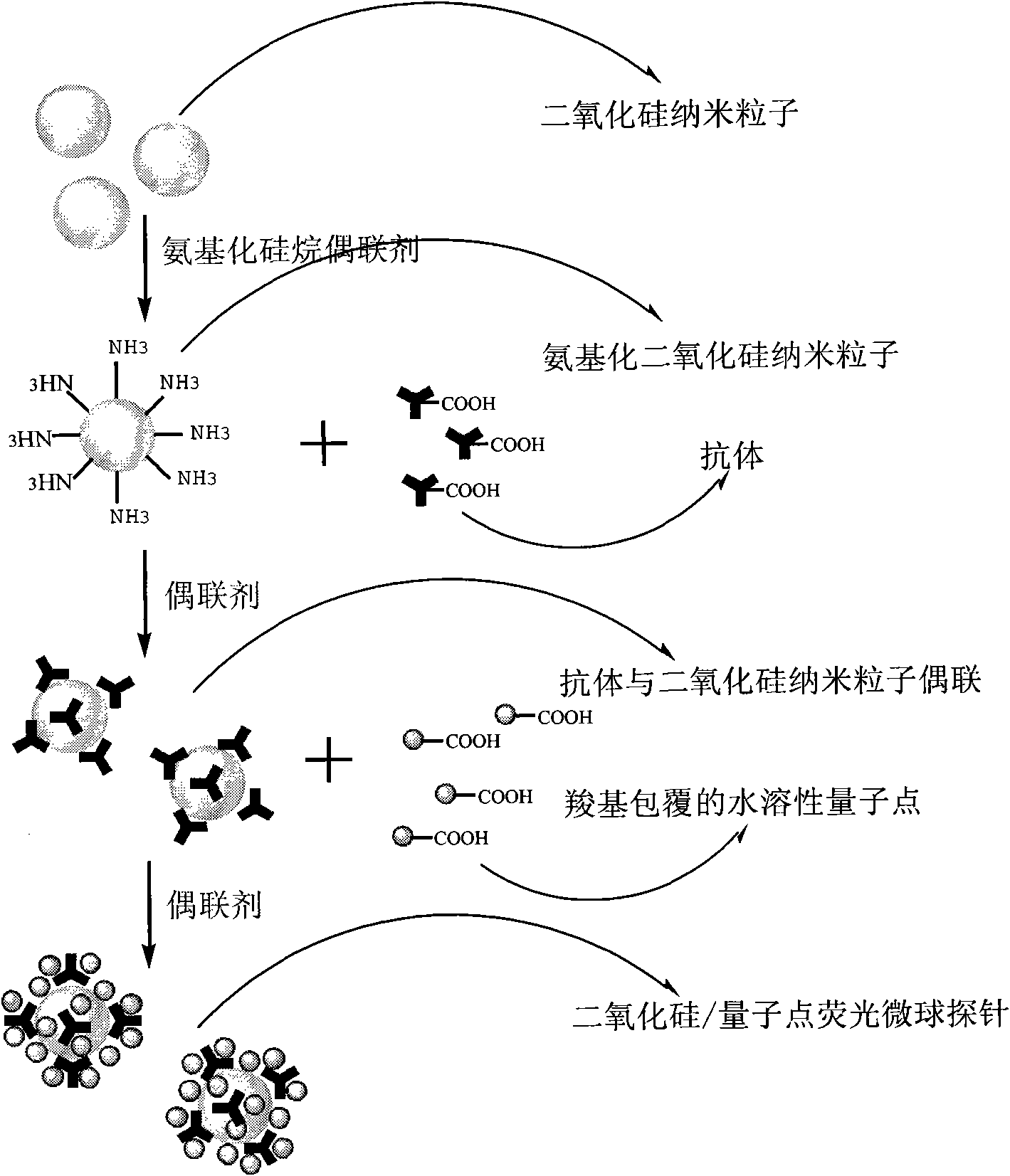
[0055] Preparation of aminated silica: Weigh 8.85ml Triton X-100, 37.5ml cyclohexane, 9ml n-hexanol and an appropriate amount of water to form an inverse microemulsion system, respectively add 0.5ml TEOS and 0.3ml ammonia water under magnetic stirring, After reacting for 24 hours, 0.1ml APTES and 0.05ml THPMP were added to continue the reaction for 12 hours, acetone was demulsified, purified by high-speed centrifugation and dried. Characterized by field emission scanning electron microscopy, the particle size is 115nm.

[0056] Aqueous phase synthesis of CdTe quantum dots: 2×10 in nitrogen saturated 244mL -2 mol / LCdCl 2 2.5H 2 O solution (114.2 mg) was added with 104.4 uL of mercaptopropionic acid, and the pH of the solution was adjusted to about 9.2 with 1 mol / L NaOH. Inject freshly prepared NaHTe. Under vigorous stirring, deoxidize with N2 for 20 minutes, then quickly add the above-mentioned newly prepared sodium telluride hydride solution into the reaction system, and f...

Embodiment 2

[0061] Preparation of aminated silica: In the ethanol / water mixed system, add 2ml tetraethyl orthosilicate and 2ml ammonia water, stir magnetically for 12 hours to obtain silica nanoparticles, remove impurities by high-speed centrifugation, and add 1ml of APTES was refluxed at 90°C for 12 hours to obtain aminated silica nanoparticles with a particle size of 231nm.

[0062] Synthesis of CdTe / ZnSe quantum dots: Synthesize oil-soluble CdTe / CdSe core-shell quantum dots in a high-temperature organic phase, and then use MPA for ligand exchange to transfer the oil-soluble quantum dots to the water phase. Water-soluble CdTe / CdSe quantum dots coated with carboxyl groups are obtained.

[0063] The green anti-penicillin monoclonal antibody silica / quantum dot microsphere probe and the red anti-chloramphenicol monoclonal antibody silica / quantum dot microsphere probe were prepared according to the method in Example 1. Spray on the binding pads respectively, and arrange the two binding pads...

Embodiment 3

[0066] The preparation of aminated silica nanoparticles and water-soluble quantum dots is the same as in Example 1.

[0067] According to the method of Example 1, the silica / red quantum dot microsphere probe coupled with anti-alpha-fetoprotein monoclonal antibody was prepared.

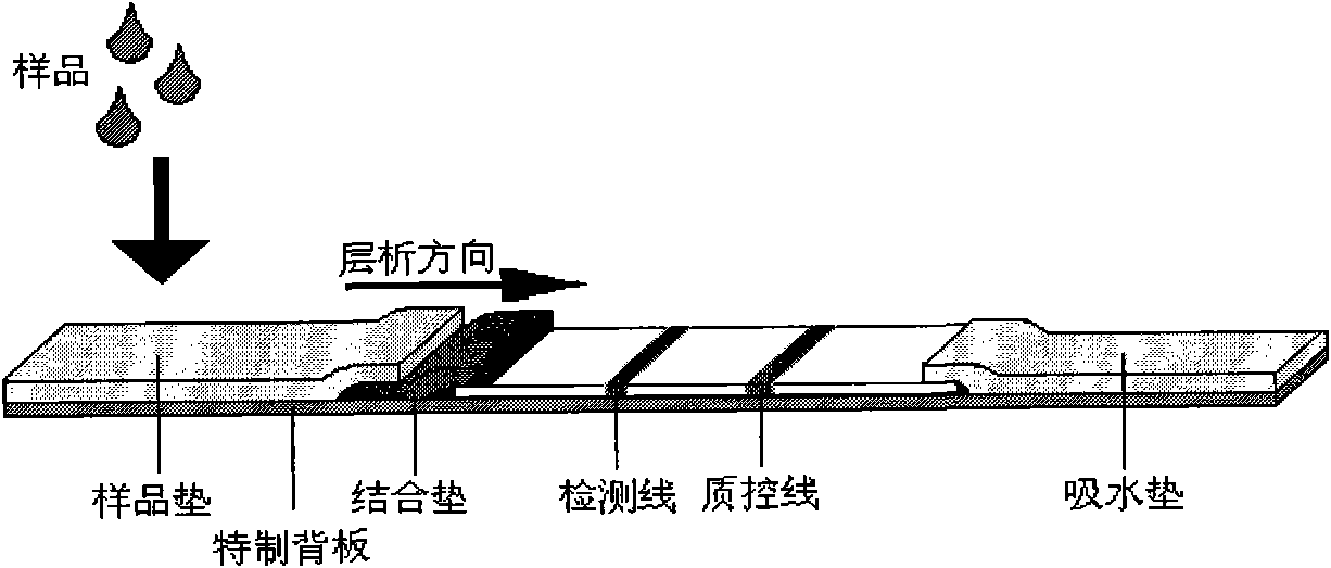
[0068] On the special backboard, paste NC film (with the goat anti-mouse quality control line and the anti-alpha-fetoprotein polyclonal antibody detection line drawn with a film-drawing instrument), water-absorbing pads, and secondary antibodies sprayed with anti-alpha-fetoprotein monoclonal antibody. The bonding pad of the silicon oxide / red quantum dot microsphere probe, the sample pad, and then cut into 4mm wide strips with a strip cutter.

[0069] In semi-quantitative testing, if a red line appears on the test line, it is positive, otherwise it is negative. If the quality control line does not show red, the test is invalid.

[0070]For quantitative detection, prepare a series of alpha-fetoprotein s...


PUM


Abstract

The invention relates to a detection method, in particular to a rapid detection method employing ultrasensitive quantum dot microsphere immunity-chromatograph test paper strips. The prior art has the disadvantage that the detection sensitivity is unable to satisfy demands of users because of high detection limit of samples, complex composition of the samples and great pretreatment difficulty. The rapid detection method of the invention comprises the steps of: preparing silicon dioxide nanoparticles, incubating the amino silicon dioxide nanoparticles with antibody for 1-10 hours in the presence of a condensation agent at 37 DEG C, and removing the antibody and the condensation agent which are failed to bond 1-4 times through high-speed centrifugation, redissolving precipitates obtained after centrifugation, adding the condensation agent and carboxylic water-soluble quantum dots, and removing free quantum dots and the condensation agent through high-speed centrifugation; preparing a silica / quantum dot composite microsphere probe; and constructing an immunity-chromatograph test paper strip system. The invention has the advantages of simple and convenient detection method, accurate and visual results, high sensitivity, low price, and wide application and can be used for semi-quantitative detection and quantitative detection.

Description

technical field [0001] The invention relates to a detection method, in particular to a rapid detection method of an ultrasensitive quantum dot microsphere immunochromatographic test strip. Background technique [0002] Immunochromatographic test strip detection is a fast, simple, sensitive, intuitive, low-cost method that can be used for on-site detection at any time as needed. It has advantages that other detection methods such as gas chromatography, high performance liquid chromatography, gas chromatography-mass chromatography, liquid chromatography-mass chromatography, capillary electrophoresis and other instrument detection methods do not have. It plays an important role in detection technology, and it is also a good supplement to traditional detection and instrument detection. Especially with the development of science and technology and the improvement of living standards, major human diseases, environmental pollution, food safety and other issues are increasingly con...

Claims


Application Information

Patent Timeline
IPC IPC(8): G01N33/543G01N33/531G01N33/558
Inventor 王元凤白亚龙魏新林
Owner SHANGHAI NORMAL UNIVERSITY
Learn More