Acute myocardial infarction diagnosis-oriented biosensor and preparation method thereof
An acute myocardial infarction and biosensor technology, applied in the field of biomedicine, can solve the problems of low-concentration protein detection and long time-consuming detection sensitivity, and achieve important clinical practical value, fast response speed, and high diagnosis rate.
- Summary
- Abstract
- Description
- Claims
- Application Information
AI Technical Summary
Problems solved by technology
Method used
Image

Examples
Embodiment 1
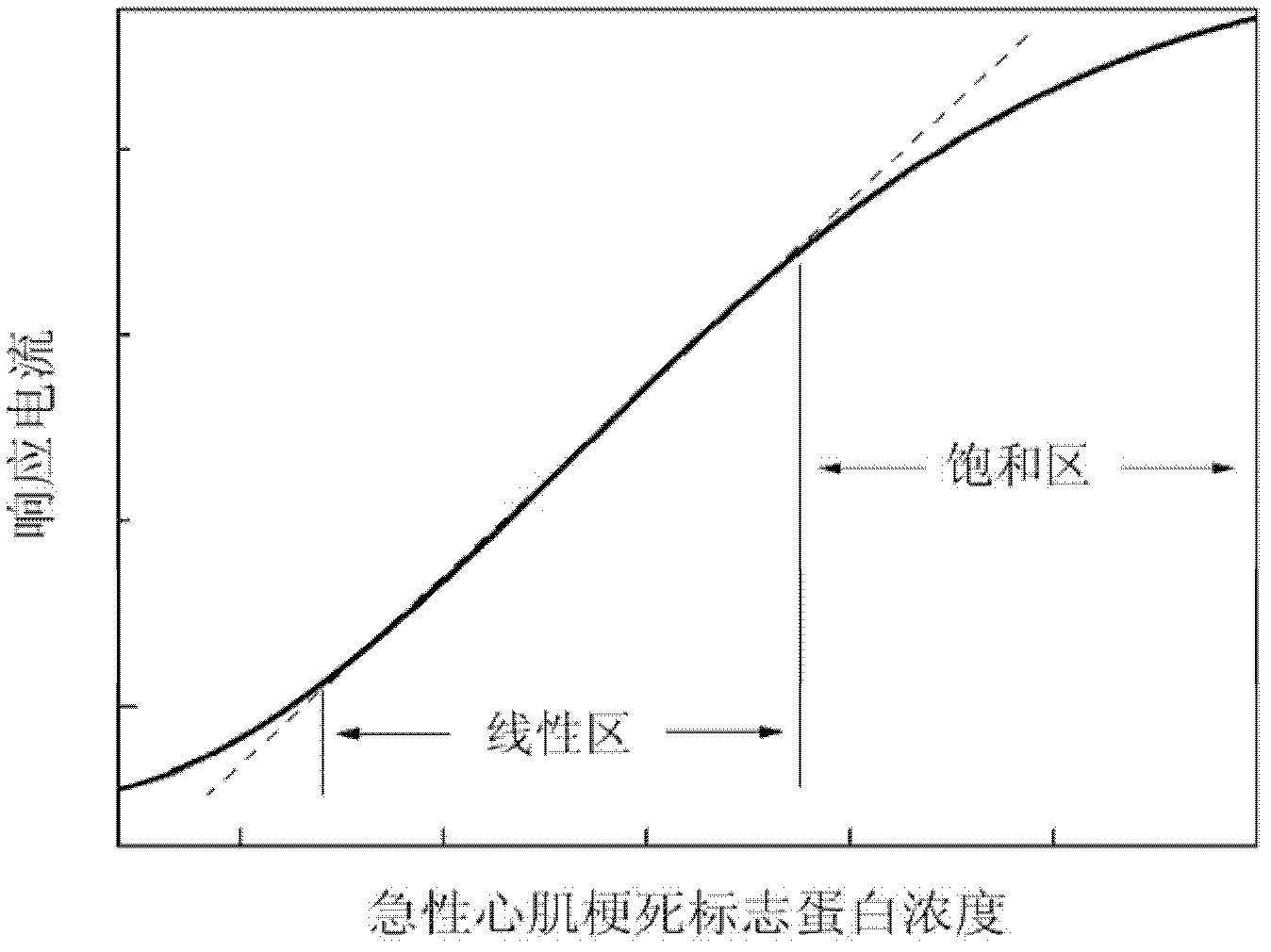
[0059] Determination of cardiac troponin I using a quasi-one-dimensional silicon nanowire field-effect tube biosensor:
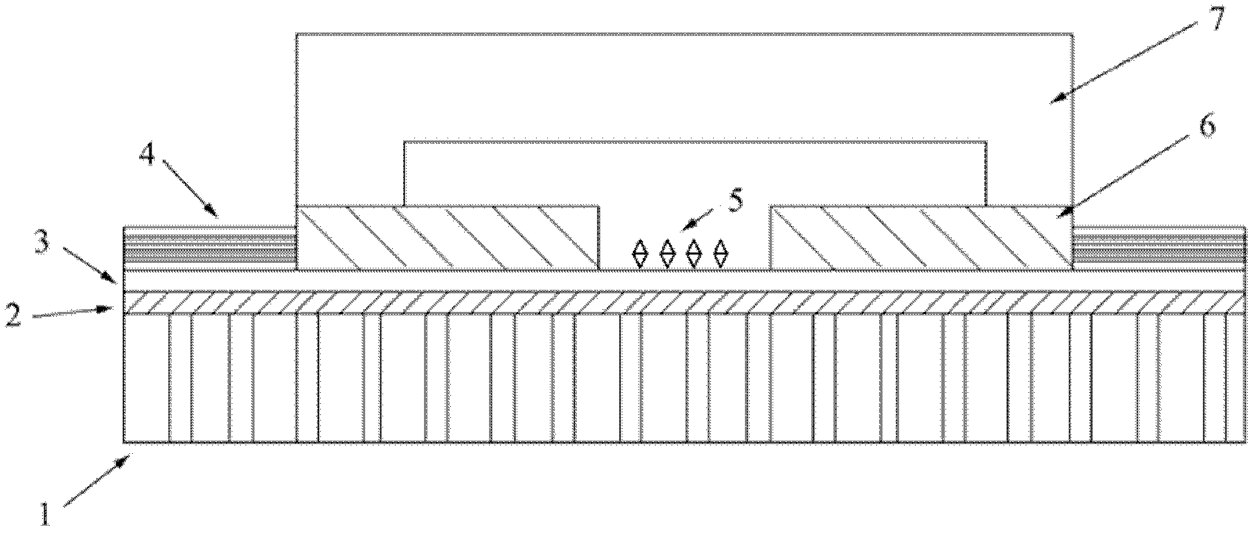
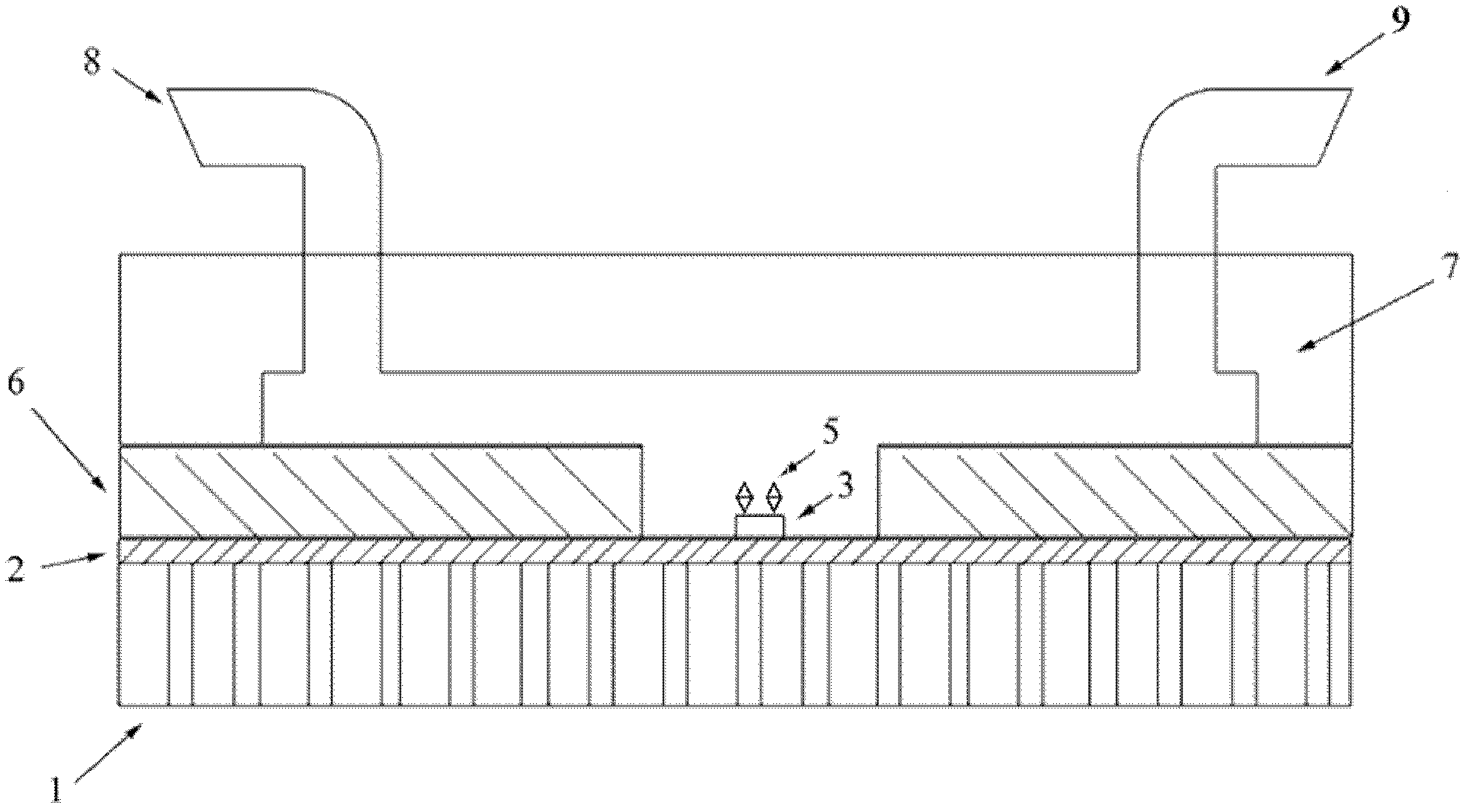
[0060] 1. Using the "top-down" method to prepare a field-effect transistor of a single quasi-one-dimensional silicon nanowire:
[0061] First, use electron beam lithography to process photoresist patterns on the surface of SOI silicon wafers, in which the width of the nanowire region is 50nm;
[0062] Then use ion beam etching (IBE) technology to etch the Si not covered by photoresist;
[0063] Then use ultraviolet lithography to prepare metal electrode patterns, and then use electron beam evaporation equipment to deposit nickel-gold electrodes;
[0064] Finally, the plasma-enhanced chemical vapor deposition (PECVD) technology is used to deposit silicon nitride / silicon oxide double-layer film on the surface of the device, in which the surface of the passivation layer is silicon nitride with a thickness of 100nm, and the silicon oxide film is at the bottom l...
Embodiment 2
[0070] Measurement of cardiac troponin T using a quasi-one-dimensional silicon nanowire field-effect tube biosensor:
[0071] 1. Using the "top-down" method to prepare a field-effect transistor of a single quasi-one-dimensional silicon nanowire:
[0072] First, use electron beam lithography to process photoresist patterns on the surface of SOI silicon wafers, in which the width of the nanowire region is 100nm;
[0073] Then use reactive ion etching (RIE) technology to etch the Si not covered by photoresist;
[0074] Then use ultraviolet lithography to prepare metal electrode patterns, and then use magnetron sputtering to deposit titanium-gold electrodes;
[0075] Finally, the plasma-enhanced chemical vapor deposition (PECVD) technology is used to deposit silicon nitride / silicon oxide double-layer film on the surface of the device, in which the surface of the passivation layer is silicon nitride with a thickness of 500nm, and the silicon oxide film is at the bottom layer with ...
Embodiment 3
[0081] Determination of Creatine Kinase Isoenzymes Using Quasi-One-Dimensional Silicon Nanowire Field-Effect Tube Biosensors:
[0082] 1. Using the "top-down" method to prepare a field-effect transistor of a single quasi-one-dimensional silicon nanowire:
[0083] First, use electron beam lithography to process photoresist patterns on the surface of SOI silicon wafers, in which the width of the nanowire region is 150nm;
[0084] Then use ion beam etching (IBE) technology to etch the Si not covered by photoresist;
[0085] Then use ultraviolet lithography to prepare metal electrode patterns, and then use thermal evaporation equipment to deposit nickel-gold electrodes;
[0086] Finally, the plasma-enhanced chemical vapor deposition (PECVD) technology is used to deposit silicon nitride / silicon oxide double-layer film on the surface of the device, in which the surface of the passivation layer is silicon nitride with a thickness of 300nm, and the silicon oxide film is at the bottom...
PUM
| Property | Measurement | Unit |
|---|---|---|
| Width | aaaaa | aaaaa |
| Thickness | aaaaa | aaaaa |
| Thickness | aaaaa | aaaaa |
Abstract
Description
Claims
Application Information
 - R&D
- Intellectual Property
- Life Sciences
- Materials
- Tech Scout
- Unparalleled Data Quality
- Higher Quality Content
- 60% Fewer Hallucinations
Browse by: Latest US Patents, China's latest patents, Technical Efficacy Thesaurus, Application Domain, Technology Topic, Popular Technical Reports.
© 2025 PatSnap. All rights reserved.Legal|Privacy policy|Modern Slavery Act Transparency Statement|Sitemap|About US| Contact US: help@patsnap.com