A preparing method of a small-intestine decellularized scaffold with a complete three-dimensional structure and a vascular network

A decellularized scaffold and three-dimensional structure technology, which is applied in the field of small intestine decellularized scaffold preparation, can solve the problems of reduced efficiency, insufficient decellularization and disinfection, and tearing of scaffold materials, so as to retain the complete three-dimensional structure, ensure the success rate and repair effect , the effect of maintaining integrity

Inactive Publication Date: 2015-09-02
SECOND MILITARY MEDICAL UNIV OF THE PEOPLES LIBERATION ARMY
View PDF1 Cites 6 Cited by
  • Summary
  • Abstract
  • Description
  • Claims
  • Application Information

AI Technical Summary

Problems solved by technology

[0004] At present, the preparation process of small intestine decellularized scaffolds is simple, and most of them are limited to single decellularization. Generally speaking, there will be problems of decellularization or insufficient disinfection, which leads to certain immunogenicity and antigenicity of the material. At the same time, the existing Most of the techniques used in combination of physical and chemical methods to treat the small intestine, in order to achieve the purpose of decellularization and antigen removal
It has been reported in the literature that the combination of physical and chemical methods to treat the small intestine can completely remove the cellular components in the small inte

Method used


Image

  • A preparing method of a small-intestine decellularized scaffold with a complete three-dimensional structure and a vascular network
  • A preparing method of a small-intestine decellularized scaffold with a complete three-dimensional structure and a vascular network
  • A preparing method of a small-intestine decellularized scaffold with a complete three-dimensional structure and a vascular network

Examples


Embodiment 1

[0038]Adult rats were taken, weighing 300-350g, fixed on the test table after weighing, anesthetized by intraperitoneal injection of 3% pentobarbital sodium, the dose was 1ml / kg, after the anesthesia was complete, intramuscularly injected low molecular weight heparin 500U for systemic anticoagulation deal with. After the abdomen is disinfected, a surgical towel is spread, and a segment of the jejunum with a rich blood supply is selected after the abdominal cavity is opened, and the arterial and venous blood vessels at the root of the selected jejunal mesentery are carefully separated, and the arterial and venous blood vessels are intubated and fixed in turn, and the proximal end is ligated and disconnected Blood vessel. Intestinal clamps are used to clamp both ends of the required intestinal tube, taking care to avoid branch vessels. Separate the two ends of the intestinal segment and intubate and fix it separately, separate the mesentery and remove the required intestinal tu...

Embodiment 2

[0040] The small intestine decellularized scaffold prepared by the method of Example 1 underwent a series of in vitro testing experiments, such as DNA concentration testing, HE staining, Masson's trichrome staining, scanning electron microscopy, and uniaxial mechanical property testing. The testing process and testing results are as follows:

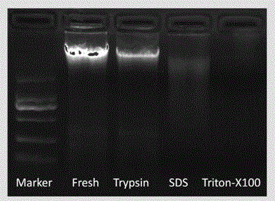
[0041] 2-1. Agarose gel electrophoresis detection experiment:

[0042] The DNA electrophoresis experiment of the small intestine decellularized scaffold prepared by the present invention and normal non-decellularized small intestine, the results are as follows figure 1 As shown in the figure, Fresh in the figure indicates normal small intestine samples that have not been decellularized, and Trypsin (trypsin), SDS (sodium dodecyl sulfate) and Triton (polyethylene glycol octylphenyl ether) respectively indicate that the corresponding reagents were passed through in sequence The DNA content of the scaffold material after treatment shows th...

Embodiment 3

[0072] 3-1. Partial defect repair experiment of abdominal wall:

[0073] The SD rats were intraperitoneally injected with 3% pentobarbital sodium to carry out the partial defect repair experiment of the abdominal wall. Method: After SD rats were anesthetized, they were fixed on the experimental table, the abdomen was shaved and the skin was prepared, the iodophor cotton ball was disinfected and the towel was spread, the skin was incised and the abdominal muscles were completely exposed, and a part about 2*2cm in size was cut off through the midline of the abdomen The abdominal wall muscles include the external oblique muscle and part of the rectus abdominus muscle. The canine small intestine decellularized scaffold prepared according to the method described in Example 1 was cut to cover the defect site with a size of about 1 cm larger than the abdominal wall defect, and was continuously sutured and fixed with sterile sutures. , align the cut skin edges, and suture. Put them i...


PUM


Abstract

A preparing method of a small-intestine decellularized scaffold with a complete three-dimensional structure and a vascular network is disclosed. The method is characterized by comprising following steps: a first step of selecting a healthy adult mammal, anesthetizing, injecting with an anticoagulant, opening the abdominal cavity, subjecting the small intestine section to cannula fixation for artery and vein vessels, and ligating at the proximal end to break the vessels; a second step of clipping two ends of the small intestine section, breaking the two ends of the small intestine section, respectively performing cannula fixation, separating a mesentery, taking down a complete intestinal tube and a complete mesenteric vessel which are needed, putting into a low-temperature environment, and repeatedly washing with normal saline; a third step of perfusing the artery, the vein and the intestinal tube with a phosphate buffer, a trypsin solution containing ethylenediaminetetraacetic acid, the phosphate buffer, a sodium dodecylsulfate solution, the phosphate buffer, a polyethylene glycol octylphenol ether solution and the phosphate buffer in order; and a fourth step of disinfecting after perfusion. The small-intestine decellularized scaffold prepared by the method can maintain a complete scaffold structure.

Description

technical field [0001] The invention relates to a method for preparing a small intestine decellularized scaffold with a complete three-dimensional structure and a vascular network, and belongs to the field of biotechnology. Background technique [0002] Tissue trauma or defect is a common clinical disease, such as partial defect of organ tissue caused by trauma, hernia, surface skin injury, burn, etc., which seriously affects the quality of life of patients, and is a problem that must be faced and urgently solved in medicine. At present, there are two main types of materials for tissue repair on the market: one is polymer synthetic materials such as polypropylene, polyester, and polytetrafluoroethylene, but because they are good adhesion carriers for bacteria, bacteria adhere to them. After being applied, a biofilm can be formed to protect it from the host immune defense mechanism and antibiotics, so that it can survive locally for a long time and cause chronic infection of ...

Claims


Application Information

Patent Timeline
IPC IPC(8): A61L27/36
Inventor 周海洋胡志前张剑张蓓蓓孙延平阮灿平张言言柴云笙
Owner SECOND MILITARY MEDICAL UNIV OF THE PEOPLES LIBERATION ARMY
Why Eureka
Try Eureka