3D tissue engineering product containing living cells, and preparation method thereof

A 3D, cell-based technology, applied in biochemical equipment and methods, fusion cells, tissue regeneration, etc., can solve the problems of unfavorable cells and complicated processes, and achieve the effect of simple manufacturing process and good biocompatibility

Inactive Publication Date: 2016-11-30
CHONGQING ACAD OF ANIMAL SCI
View PDF4 Cites 3 Cited by
  • Summary
  • Abstract
  • Description
  • Claims
  • Application Information

AI Technical Summary

Problems solved by technology

This process is complicated, and the chemical reagents left in t

Method used


Image

  • 3D tissue engineering product containing living cells, and preparation method thereof
  • 3D tissue engineering product containing living cells, and preparation method thereof
  • 3D tissue engineering product containing living cells, and preparation method thereof

Examples


Embodiment 1

[0039] The rapid construction of cell-containing 3D tissue engineering products includes the following steps:

[0040] 1. Preparation of SIS raw materials

[0041] (1) Take fresh porcine small intestine, remove the serosal layer and muscular layer of the small intestine by mechanical means, and rinse with normal saline repeatedly.

[0042] (2) Soak and stir in methanol and chloroform (1:1, V / V) degreasing solution for 12 to 24 hours (preferably 12 hours, repeat twice), and rinse with deionized water for 3 to 5 times to remove the organic solvent.

[0043] (3) Soak SIS in 0.05% trypsin / 0.05% ethylenediaminetetraacetic acid solution, shake and digest at room temperature for 12 hours, and wash repeatedly with physiological saline solution to remove trypsin.

[0044] (4) Shake and shake the SIS in 0.5% sodium dodecyl sulfate (SDS) saline for 4 hours, and rinse thoroughly with saline solution to remove the detergent.

[0045] (5) Soak SIS in 0.5% acetic acid solution overnight, c...

Embodiment 2

[0068] Semisolid Screening for Hybridoma Cells

[0069] (1) Prepare a SIS neutral solution with a concentration of 1%, add riboflavin at a final concentration of 0.1%, and mix well.

[0070] (2) Prepare 2X RPMI1640 cell culture medium (containing 20% ​​fetal bovine serum, 0.22g / L sodium pyruvate, 0.15% L-glutamine, 200IU / mL penicillin, 200ug / mL streptomycin, 2X HAT).

[0071] (3) Mix equal volumes of (1) and (2) evenly.

[0072] (4) Resuspend the conventionally fused hybridoma cells with the solution in (3), and mix well.

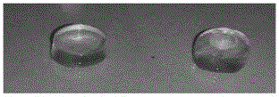
[0073] (5) Put 3 mL of the above cell suspension into a six-well plate, and irradiate it with a 200W incandescent bulb at a distance of 10-30 cm from the 96-well plate or mold for 3-5 minutes, and the solution undergoes a phase transition and forms a gel.

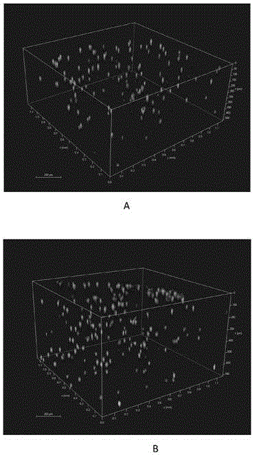
[0074] (6) 37°C, 5% CO 2 After 7-10 days of culture, white spots can be seen in the culture medium, and each spot is a hybridoma cell clone.


PUM


Abstract

The present invention provides a 3D tissue material, which adopts porcine small intestine mucosal decellularized matrix as a scaffold material, and is characterized by comprising cells, riboflavin or a derivative thereof. According to the present invention, the chemical components contained in the prepared 3D tissue material are highly close to the human extracellular matrix components, the prepared 3D tissue material is rich in growth factors, has good biocompatibility, and is favorable for cell growth; and the production process is simple, only the incandescent lamp is required to be adopted as the production equipment, the complex equipment is not required, the component harmful to the cell does not exist during the whole operation process, and the whole operation process cannot cause the environmental pollution.

Description

technical field [0001] The invention relates to a product and a manufacturing method of a 3D tissue engineering product containing living cells. Background technique [0002] In vitro construction of 3D tissue engineering products has broad application prospects in basic research of medical biology, in vitro drug screening and evaluation, and tissue regeneration. Various technologies including 3D printing are important processes for constructing 3D tissue engineering products. [0003] The construction of 3D tissue engineering products containing living cells requires good biocompatibility of the scaffold material, which can support cell growth. At the same time, a simple and easy processing technology is required to realize the phase transition of the scaffold material from liquid to solid state and fix the cells. In the scaffold material and form a product with a certain 3D structure. [0004] Commonly used scaffold materials include synthetic materials and natural sourc...

Claims


Application Information

Patent Timeline
IPC IPC(8): A61L27/38A61L27/36A61L27/52C12N5/20
CPCA61L27/38A61L27/3629A61L27/3687A61L27/52A61L2430/00C12N5/163C12N2500/38C12N2533/90
Inventor 葛良鹏刘作华
Owner CHONGQING ACAD OF ANIMAL SCI
Why Eureka
Try Eureka