Cell apoptosis double-color detection and imaging probe and application thereof

A technology of imaging probe and cytochrome, which is applied in the field of bioanalysis technology and nanomedicine to achieve the effect of good selectivity, high sensitivity and simple synthesis

Active Publication Date: 2017-11-24
MENGCHAO HEPATOBILIARY HOSPITAL OF FUJIAN MEDICAL UNIV
View PDF8 Cites 12 Cited by
  • Summary
  • Abstract
  • Description
  • Claims
  • Application Information

AI Technical Summary

Problems solved by technology

However, these methods are all aimed at the detection of a single target, such as the detection of cytochrome c or caspase-3 protease

Method used


Image

  • Cell apoptosis double-color detection and imaging probe and application thereof
  • Cell apoptosis double-color detection and imaging probe and application thereof
  • Cell apoptosis double-color detection and imaging probe and application thereof

Examples


Embodiment 1

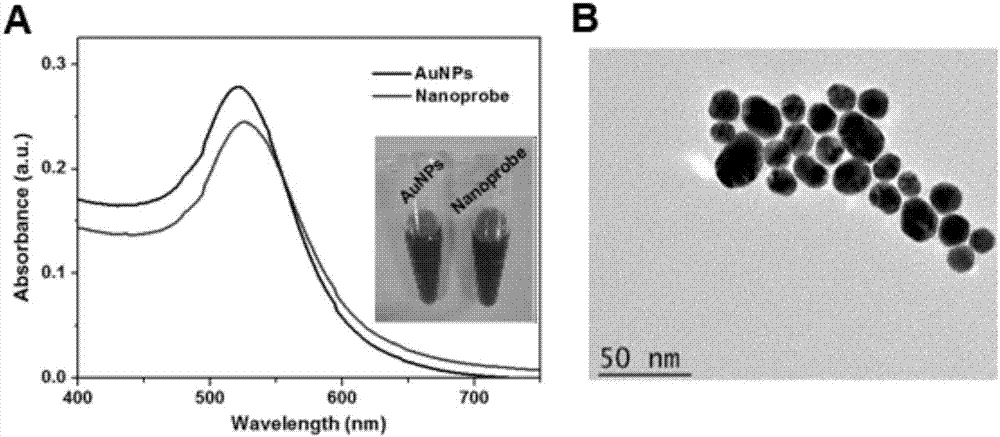
[0026] Synthesis of gold nanoparticles: The glassware used was soaked in aqua regia for 4 hours, washed thoroughly with secondary water and dried before use. After 50 ml of 0.01% wt chloroauric acid aqueous solution was heated to boiling, 1 ml of 1% wt trisodium citrate solution was quickly added under stirring, and the reaction was continued for 20 minutes until the color changed from colorless to wine red. Cool to room temperature, and store in a 4°C refrigerator away from light until use. figure 2 A shows the absorption spectrum of gold nanoparticles.

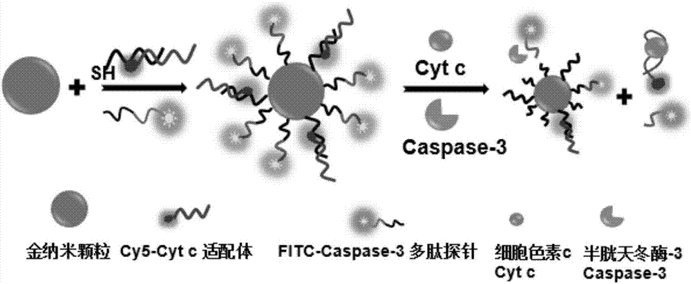
[0027] Cytochrome c aptamer hybridized with linked DNA: the aptamer sequence of cytochrome c was 5'-CCGTGTCTGGGGCCGACCGGCGCATTGGGTACGTTGTTGC-Cy5-3'; the aptamer linked DNA sequence was 5'-SH-TTTTTTTTTTGCAACAACGTA-3'. 24 μL 100 μM cytochrome c aptamer and 20 μL 100 μM linked DNA were reacted by DNA hybridization, mixed with hybridization solution 40 μL phosphate buffer saline (PBS) (10 mM PB solution, 137 mM NaCl, 2.7 mM KC...

Embodiment 2

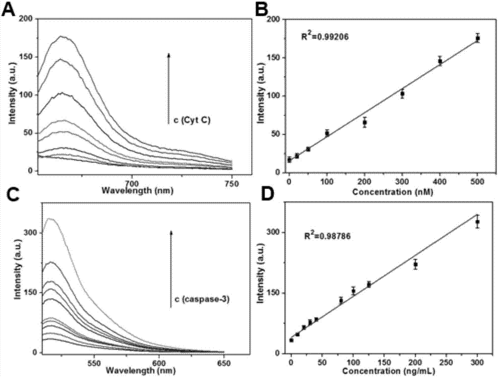
[0030] Fluorescence detection and caspase-3: When detecting cytochrome c, 1nM nanoprobes were reacted with different concentrations of cytochrome c in PBS at 37°C for 30 minutes to detect the fluorescence signal. The excitation wavelength is 635nm, and the detection wavelength is 665nm. When detecting caspase-3, 1nM nanoprobes were incubated with different concentrations of caspase-3 in 25mM HEPES buffer (pH 7.4, containing 100mM NaCl, 1mM EDTA, 10% sucrose, 0.1% CHAPS), and reacted at 37°C for 30 minutes Detection of fluorescent signal. The excitation wavelength is 480nm, and the detection wavelength is 520nm. The result is as image 3As shown, the detection linear range of cytochrome c is 0-500nM, and the detection limit is 10nM; the detection linear range of caspase-3 is 0-300ng / mL, and the detection limit is 5ng / mL.

[0031] Nanoprobe detection selectivity experiment: 1nM nanoprobes were reacted with different interfering substances at 37°C for 30 minutes to detect corr...

Embodiment 3

[0034] Nanoprobes for apoptosis imaging: HeLa cells were cultured in RPMI 1640 medium containing 10% fetal bovine serum, penicillin (100 U / mL) and streptomycin (100 μg / mL) at 37°C in high humidity containing 5% in a carbon dioxide incubator. HeLa cells were cultured in a confocal imaging culture dish, and when they grew to cover 60-70% of the culture dish, washed twice with PBS, added Opti-MEM culture solution containing 1nM nanoprobes, incubated for 2 hours, washed twice with PBS, Add culture medium containing different concentrations of STS and incubate for 2 hours. After washing twice with PBS, fluorescent confocal imaging was performed. The result is as Figure 5 As shown, it is proved that the nanoprobe can dual-color image apoptosis.

[0035] Nanoprobes for apoptosis drug evaluation: HeLa cells were cultured in RPMI 1640 medium containing 10% fetal bovine serum, penicillin (100 U / mL) and streptomycin (100 μg / mL) at 37°C in high humidity containing 5 % CO2 incubator. ...


PUM

PropertyMeasurementUnit
sizeaaaaaaaaaa

Abstract

The invention relates to a cell apoptosis double-color detection and imaging probe and application thereof. The probe adopts a fluorescently-labeled cytochrome c nucleic acid aptamer and a caspase-3 substrate sequence polypeptide sequence as recognition elements and golden nanoparticles as a fluorescent energy receptor, response molecules are subjected to covalent modification on the surface of the receptor, and then fluorescence of two fluorescent dyes is quenched off in the energy transfer process simultaneously. When no target is available, the dye fluorescence is quenched off. When target molecules are available, the cytochrome c is combined with the aptamer, a substrate polypeptide of the cytochrome c is hydrolyzed with caspase-3, then the fluorescent dye is far away from the surfaces of golden nanoparticles, a fluorescence signal is generated, and detection and imaging of cytochrome c and caspase-3 are achieved. The probe provided by the invention is simple and convenient to synthesize, high in sensitivity and good in selectivity; two biomarkers related to cell apoptosis can be detected at the same time; and the probe is effectively applied to cell apoptosis imaging detection and cell apoptosis medicine evaluation.

Description

(1) Technical field [0001] The invention relates to a cell apoptosis dual-color detection and imaging probe and application thereof, belonging to the fields of biological analysis technology and nanomedicine. (2) Background technology [0002] Apoptosis is a kind of programmed cell death, which plays an important role in the development, homeostasis and immunity of multicellular organisms. Usually, the imbalance of regulation of apoptosis will lead to the occurrence of various pathological processes, such as autoimmune disorders, degenerative diseases, cancer and so on. Therefore, real-time detection and monitoring of the apoptosis process of organisms is of great significance in the study of apoptosis signals, the evaluation of certain disease processes, and the development of apoptosis-related drugs. [0003] The biological changes of apoptosis mainly include annexin exposure of phosphatidylserine (PS), release of cytochrome c (Cyt c) from mitochondria, DNA fragmentation,...

Claims


Application Information

Patent Timeline
Patent Type & Authority Applications(China)
IPC IPC(8): G01N21/64C09K11/02
Inventor 刘景丰刘小龙张晓龙廖乃顺郑爱仙曾永毅
Owner MENGCHAO HEPATOBILIARY HOSPITAL OF FUJIAN MEDICAL UNIV
Why Eureka
Try Eureka