Fusion protein of equine influenza virus H3N8 subtype and preparation method, application and vaccine thereof
A fusion protein, H3N8 technology, applied in antiviral immunoglobulin, veterinary vaccines, biochemical equipment and methods, etc., to achieve good antigenicity, good immunogenicity, and a wide range of applications
- Summary
- Abstract
- Description
- Claims
- Application Information
AI Technical Summary
Problems solved by technology
Method used
Image
Examples
preparation example Construction
[0047] The present invention also provides a method for preparing the above-mentioned fusion protein, which can be performed by expressing the gene of the above-mentioned fusion protein in the host, for example, but not limited to, expression in E. coli expression system, yeast expression system, insect expression system, plant expression system, or mammalian expression system. Since the protein expressed by mammalian cells is translated and processed, its structure and biological characteristics are closer to natural proteins, so the present invention preferably uses a mammalian expression system to express the fusion The gene for the protein, more preferably the gene for expressing the fusion protein using a CHO cell expression system. CHO cells are Chinese hamster ovary cells. The CHO cell expression system has the following advantages:
[0048] (1) It has accurate post-translational folding and modification functions, and the expressed protein is closest to the natural pro...
Embodiment 1
[0068] Embodiment 1: HA-NA gene design and synthesis
[0069] HA-NA gene design and synthesis: After optimization, the HA gene (GenBank accession number KX815377.1) is designed to be 810bp in length and has the sequence shown in SEQ ID NO.1; the NA gene (GenBank accession number MG132049.1) is optimized The design length is 900bp, and it has the sequence shown in SEQ ID NO.2. The linker sequence is used to connect the HA gene and the NA gene, and the linker sequence has the sequence shown in SEQ ID NO.3. The full length of the HA-NA gene is 1740bp, and has the amino acid sequence shown in SEQ ID NO.4 and the nucleotide sequence shown in SEQ ID NO.5. HA-NA gene synthesis was completed by Shanghai Sangon Biological Company.
Embodiment 2
[0070] Embodiment 2: PEE6.4-HA-NA recombinant plasmid construction
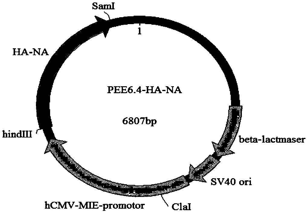
[0071] 2.1 Add restriction sites: Add restriction sites respectively upstream and downstream of the HA-NA gene sequence by PCR amplification: HindⅢ, SmaI, PCR amplification upstream primers are shown in SEQ ID NO.6, PCR amplification downstream Primers are shown in SEQ ID NO.7. PEE6.4-HA-NA plasmid map as figure 1 shown.
[0072] 2.2 Double digestion reaction of HA-NA gene and vector
[0073] 2.2.1 Construct a 50 μL reaction system, mix the components according to the table below, and then bathe in water at 37°C for 2 hours.
[0074]
[0075]
[0076] 2.2.2 Recovery of target DNA fragments: DNA gel recovery kit (purchased from Beijing Laibao Technology Co., Ltd.) was used to recover the enzyme-cut target fragments. The steps are as follows:
[0077] (1) After agarose gel electrophoresis, carefully cut the target DNA band from the agarose gel with a blade, put it into a 1.5mL EP tube, and weigh it. ...
PUM
 Abstract
Description
Claims
Application Information
 - R&D Engineer
- R&D Manager
- IP Professional
- Industry Leading Data Capabilities
- Powerful AI technology
- Patent DNA Extraction
Browse by: Latest US Patents, China's latest patents, Technical Efficacy Thesaurus, Application Domain, Technology Topic, Popular Technical Reports.
© 2024 PatSnap. All rights reserved.Legal|Privacy policy|Modern Slavery Act Transparency Statement|Sitemap|About US| Contact US: help@patsnap.com


