A kind of microneedle capable of extracting skin tissue fluid and its preparation method

An interstitial fluid and microneedle technology, which can be used in inoculation and ovulation diagnosis, medical science, surgery, etc., can solve the problems of complicated microneedle preparation process, high preparation cost, and brittle preparation materials, so as to achieve no risk of fracture, no additional devices, and rapid Extracted effects

Active Publication Date: 2021-07-27
HUAZHONG UNIV OF SCI & TECH +1
View PDF14 Cites 1 Cited by
  • Summary
  • Abstract
  • Description
  • Claims
  • Application Information

AI Technical Summary

Problems solved by technology

Hollow microneedles made of metal, glass, silicon, etc. are often used to extract skin tissue fluid, but the preparation process of these microneedles is complicated, the preparation cost is high, additional negative pressure devices are required, and the preparation materials are relatively brittle and prone to fracture. risk, which limits its application

Method used


Image

  • A kind of microneedle capable of extracting skin tissue fluid and its preparation method
  • A kind of microneedle capable of extracting skin tissue fluid and its preparation method
  • A kind of microneedle capable of extracting skin tissue fluid and its preparation method

Examples


Embodiment 1

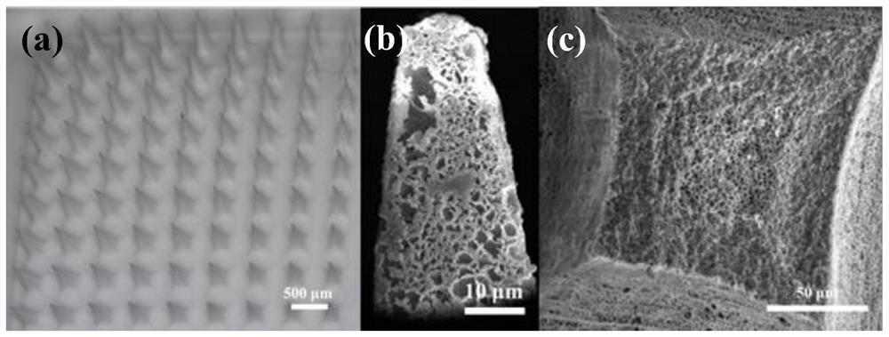
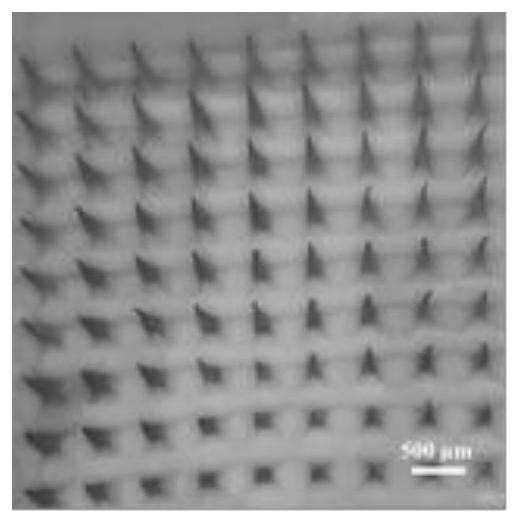
[0037] First, use the microneedle positive mold to obtain the polydimethylsiloxane (PDMS) negative mold by the method of reverse replication, then, the dimethyl cellulose acetate with a molecular weight of 10KDa and a mass fraction of 25% (w / w) The sulfoxide solution was added dropwise to the negative mold (the porogen is the solvent dimethyl sulfoxide), ultrasonicated for 1 hour and then placed in ultrapure water at 25°C to cause phase separation of the polymer, and then freeze-dried to obtain a pore size of 1500nm , a porous microneedle with a porosity of 57% (mercury porosimetry) (base diameter 210 μm, height 600 μm, 10*10 array, the following examples are all parameters). Then use the microneedle with O 2Ion treatment, and alternately soaked in 1mg / mL chitosan and hyaluronic acid solution to obtain a 20nm thick hydrophilic layer, washed with pure water and dried, the water extracted from the agarose hydrogel within 10min of the microneedle The mass of 0.8mg.

Embodiment 2-5

[0039] By adjusting the mass percentage of cellulose acetate in the dimethyl sulfoxide solution, the size of the pores in the microneedles and the porosity of the microneedles can be further regulated, and the hydrophilic ability of the microneedles can be adjusted by adjusting the thickness of the hydrophilic layer. In addition, other types of polymers can be used instead of cellulose acetate to form microneedles corresponding to the polymer material, and the pore size and porosity of the microneedles can also be adjusted by controlling the ratio of the polymer to the porogen.

[0040] Based on Example 1, Example 2-5 can be adjusted, and the microneedle parameters in Example 2-5 are shown in the following table:

[0041]

Embodiment 6

[0043] First, the PDMS negative mold was prepared by the micro-template method; then, polyethersulfone and calcium oxide with a size of 2 μm were added to N,N-dimethylformamide at a mass ratio of 3:7 to prepare a solution, and the above The solution was added dropwise onto the PDMS female mold, and ultrasound was used to promote the solution into the mold; N,N-dimethylformamide was heated and dried, and the solid polyethersulfone microneedles containing calcium oxide were peeled off; the obtained solid microneedles were placed in 10 -4 In mol / L dilute nitric acid solution, calcium oxide nanoparticles are removed to obtain porous polyethersulfone microneedles with a pore diameter of 2000 nm and a porosity of 65%. Then use the microneedle with O 2 Ion treatment, and alternately soaked in 1mg / mL chitosan and polyacrylic acid solution to obtain a 20nm thick hydrophilic layer, washed with pure water and dried, the microneedle within 10min from the water extracted from the agarose h...


PUM

PropertyMeasurementUnit
thicknessaaaaaaaaaa
pore sizeaaaaaaaaaa
pore sizeaaaaaaaaaa

Abstract

The invention discloses a microneedle capable of extracting skin tissue fluid and a preparation method thereof. The microneedle comprises a porous network framework interpenetrated and a hydrophilic polymer layer modified on the inner and outer surfaces of the framework. The preparation method is specifically to prepare a mixed solution of polymer and porogen first, then fill them into the microneedle mold, and then remove the porogen therein to obtain a porous network skeleton with interpenetration, and finally in the porous network skeleton The inner and outer surfaces are modified with a hydrophilic polymer layer. The present invention obtains the microneedle capable of extracting skin tissue fluid by improving the thickness of the hydrophilic polymer layer used in the microneedle, the porosity and aperture of the microneedle, and the overall process design of the corresponding preparation method. The microneedles provided by the invention have good biocompatibility, fast extraction of skin tissue fluid, no external negative pressure device, no obvious pain and skin infection, and no need for professional operation.

Description

technical field [0001] The invention belongs to the field of biomedical polymer materials, and more specifically relates to a microneedle capable of extracting skin tissue fluid and a preparation method thereof, which can be used for extracting skin tissue fluid. Background technique [0002] Timely diagnosis plays an important role in disease treatment and monitoring of physical status. Extracting body fluid samples is the first and critical step for timely diagnosis. Blood is often used for timely diagnosis due to its rich metabolite content. However, taking blood requires pricking the skin and penetrating the nerve cell-rich dermis, which can cause pain and skin irritation. [0003] The composition of skin tissue fluid is similar to that of blood, and the composition of skin tissue fluid changes with the composition of plasma. Therefore, skin tissue fluid can replace blood for sample extraction. Generally, skin tissue fluid can be extracted by pipette technology and mi...

Claims


Application Information

Patent Timeline
Patent Type & Authority Patents(China)
IPC IPC(8): A61B10/00
CPCA61B10/0045
Inventor 朱锦涛柳佩李钰策张连斌杜虹瑶王华陶娟
Owner HUAZHONG UNIV OF SCI & TECH
Why Eureka
Try Eureka