

Method for simultaneously detecting protein, RNA and exosome membrane protein in exosome
A technology of exosomes and internal proteins, which is applied in the field of exosome membrane proteins and simultaneously detects exosomes and internal proteins and RNA, can solve the problems that have not been reported, and achieve the effect of improving specificity and sensitivity
- Summary
- Abstract
- Description
- Claims
- Application Information
AI Technical Summary
Problems solved by technology
Method used
Image

Examples
Embodiment 1
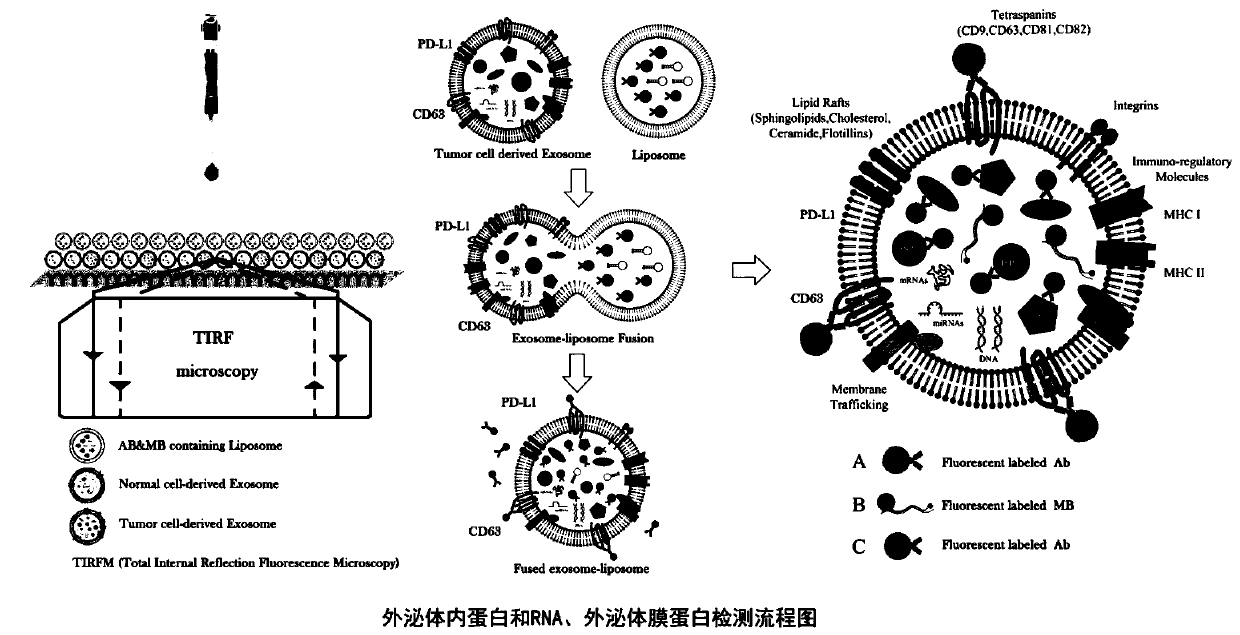
[0034] Example 1: Chip processing for in situ capture of exosomes
[0035] 1) Use ultra-thin (thickness 0.17-0.19mm), ultra-low fluorescence, ultra-high light transmittance, geometric mean roughness (RMS roughness) <100nm special glass, use high-purity electronic grade alcohol, at 24-200 After repeated cleaning under clean conditions at ℃, blow dry with high-purity nitrogen;
[0036] 2) After repeated washing with piranha solution, keep it clean and dry;
[0037] 3) The glass surface and organosiloxane with functional groups (Organosiloxane with functional groups) are placed in a vacuum container, the vacuum must be <10mmHg, and vapor deposition is carried out at 24-180°C for 6-24 hours;
[0038] 4) Mix the appropriate amount of avidin and biotin at a ratio of 1:7-1:30, mix at a low speed, let it stand for 30-60 minutes, and use PBS, BupH TM Phosphate Buffered Saline and ultrapure water (approximately 18 megohm) to prepare buffer, 0.01-1% Tween 20 repeatedly washed and place...
Embodiment 2
[0041] Example 2: Nanoparticle preparation and antibody coating
[0042] 1) DOTMA (trimethyl-2,3-dioleyloxypropyl ammonium chloride), DSPE-PEG-2000 (1,2-distearoyl-SN-glycerol-3-phosphoethanolamine- Polyethylene glycol 2000) and cholesterol are thoroughly mixed according to 3:1:4, ultrasonicated for 5 minutes, and left at room temperature for 2 hours to obtain stable cationic liposomes;
[0043] 2) Put an appropriate amount of molecular beacons, fluorescent monoclonal antibodies (RNA molecular beacons and antibodies can be mixed and encapsulated with nanoparticles at a molar ratio of 1:2 to 1:5 on the basis of excess) and the above cationic liposomes in PBS After fully blowing and mixing, ultrasonication for 5 minutes, the molecular beacon is that the 5' end stem and loop are completely complementary to the target gene, the 3' end stem is partially complementary to the 5' end stem, and the 5' end and 3' end are respectively separated by fluorescent groups. Group and quenching...
Embodiment 3
[0045] Example 3: A method for simultaneous detection of exosome internal protein and RNA, and exosome membrane protein
[0046] 1) Take the crude extracellular vesicles obtained by the precipitation method, and use the exosome in situ capture chip (prepared by the method in Example 1) under a DC electric field of 15-90V to directly capture the exosomes in the sample;
[0047] 2) Binding the target antibody to the exosome membrane captured in step 1), through the combination of the antigen and the antibody, a labeled type A fluorescent signal is emitted, and the corresponding membrane protein is detected;
[0048] 3) Take the RNA molecular beacon and fluorescent monoclonal antibody nanoparticles (prepared by the method in Example 2) that wrap the target to be detected;
[0049] 4) Fusion the encapsulated RNA molecular beacon and fluorescent monoclonal antibody nanoparticles obtained in step 3) with the exosomes captured in step 1), the RNA molecular beacon and fluorescent mono...
PUM
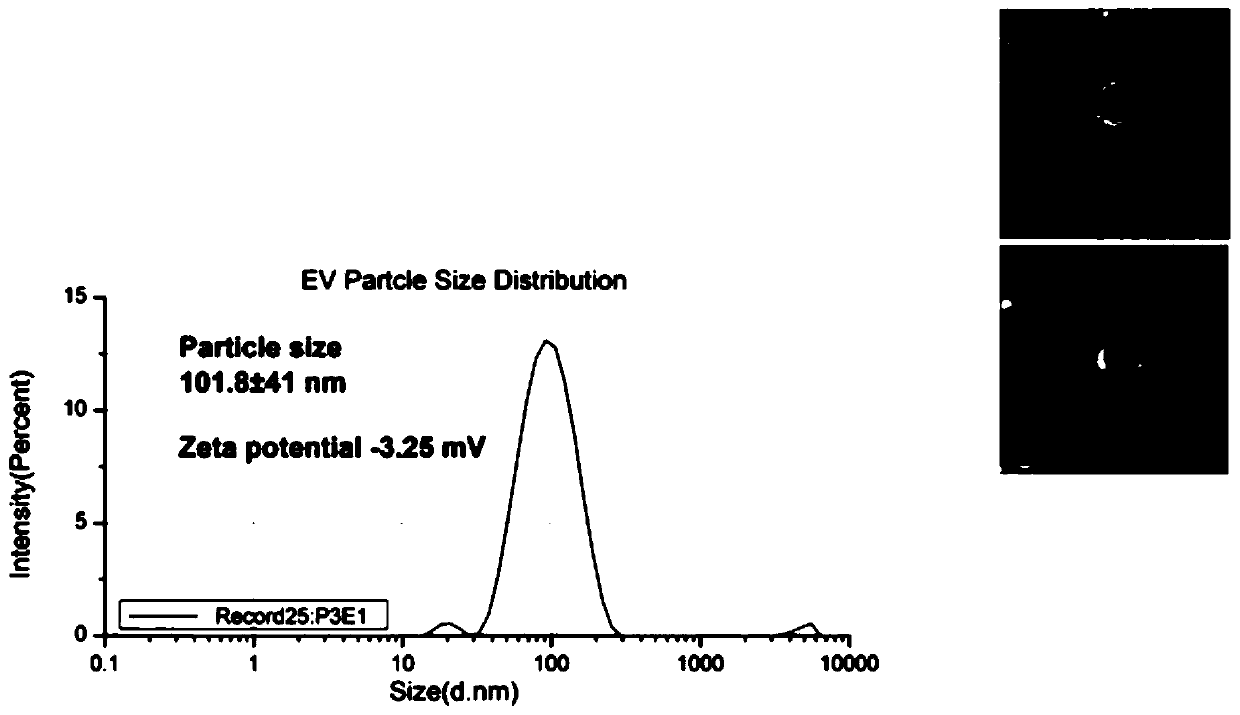
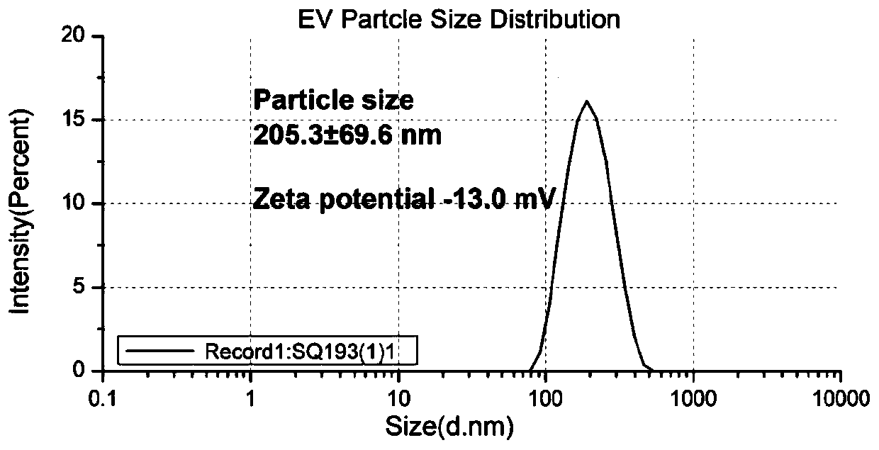
| Property | Measurement | Unit |
|---|---|---|
| diameter | aaaaa | aaaaa |
| thickness | aaaaa | aaaaa |
| particle diameter | aaaaa | aaaaa |
Abstract
Description
Claims
Application Information