Supercharge Your Innovation With Domain-Expert AI Agents!

A rare earth vanadate nano fluorescent labeling material and its preparation method and application

A rare earth vanadate and nano-fluorescence technology, applied in the direction of analyzing materials, material inspection products, instruments, etc., to achieve the effects of easy control, easy surface modification, and uniform size and morphology

Active Publication Date: 2022-02-18
FUJIAN INST OF RES ON THE STRUCTURE OF MATTER CHINESE ACAD OF SCI
View PDF15 Cites 0 Cited by
  • Summary
  • Abstract
  • Description
  • Claims
  • Application Information

AI Technical Summary

Problems solved by technology

However, bioluminescent labeling has requirements for nanomaterials with uniform shape and size and good dispersion

Method used


Image

  • A rare earth vanadate nano fluorescent labeling material and its preparation method and application
  • A rare earth vanadate nano fluorescent labeling material and its preparation method and application
  • A rare earth vanadate nano fluorescent labeling material and its preparation method and application

Examples


Embodiment 1

[0045] 1. Rare earth vanadate nano-fluorescence labeling material GdVO 4 : Preparation of 30%Eu:

[0046] In a 50 mL round bottom flask, add 20 mL of water, 0.35 mmol of gadolinium nitrate, 0.15 mmol of europium nitrate, 2 mmol of sodium citrate, 0.5 mmol of sodium orthovanadate, feed nitrogen, react at 85°C for 24 hours, centrifuge at 10,000 rpm, Wash 3 times with distilled water and dry to obtain water-soluble GdVO with good dispersibility 4 : 30% Eu nano fluorescent labeling material. Among them, GdVO 4 as matrix, Eu 3+ For doping ions, its mole percentage is 30%.

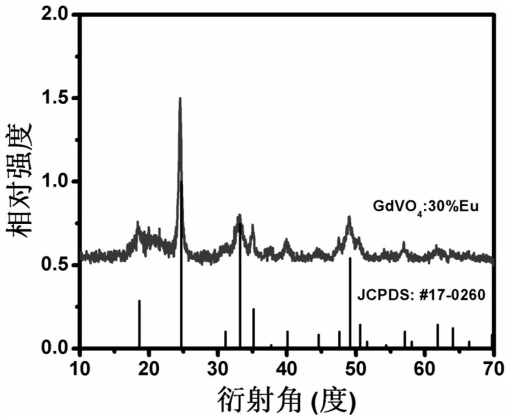
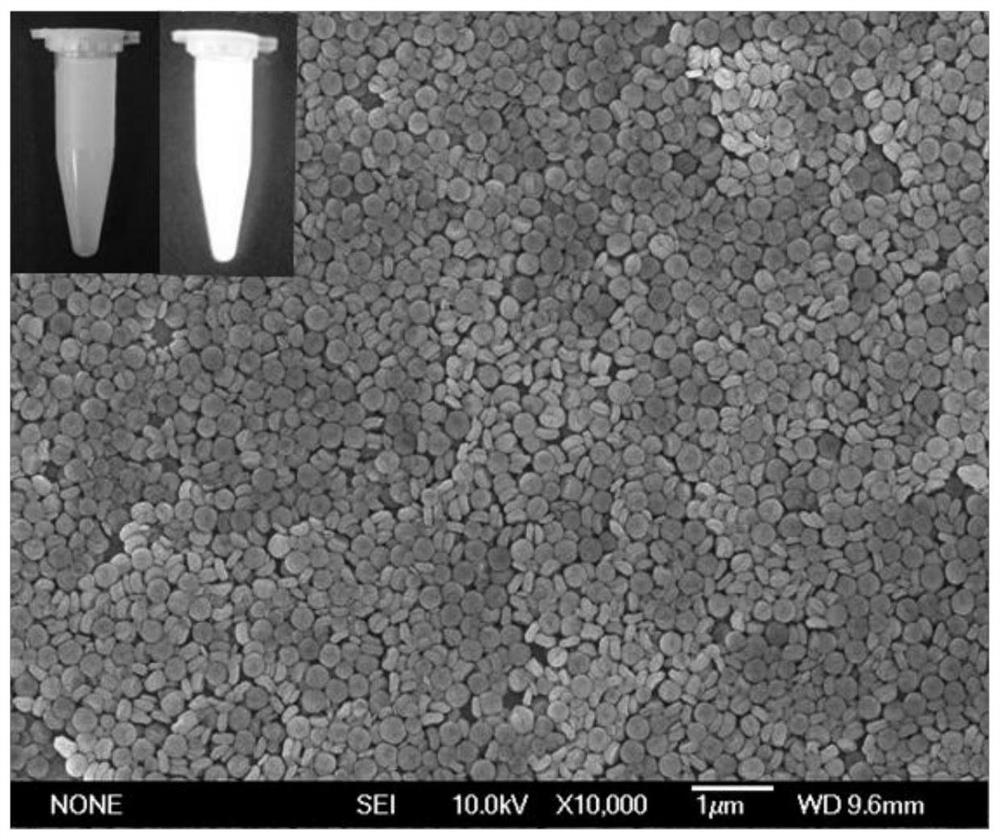
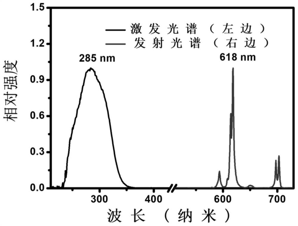
[0047] For the prepared GdVO 4 : 30% Eu nanoparticles were subjected to X-ray powder diffraction test (MiniFlex2, Rigaku), scanning electron microscope test (JSM6700, SEM), fluorescence excitation and emission test (FLS920, Edinburgh Instruments), luminescence decay characteristic test (FLS920, Edinburgh Instruments) .

[0048] figure 1 For the GdVO prepared in Example 1 4 : The X-ray powder diffraction...

Embodiment 2

[0058] Preparation of test strips

[0059] A. Sample binding pad 1 treatment: glass cellulose membrane with buffer solution containing surfactant (recipe: 100 mLPBS, pH 7.4, containing 2% NaCl, 2% BSA, 0.5% casein, 0.1% Tween-0.5% S9 and 5% sucrose) soaked for pre-blocking, dried overnight at 50°C, and sprayed GdVO through the airjet nozzle of the Biodot instrument 4 : 30% Eu-labeled cTnI monoclonal antibody 1 was ultrasonically sprayed at 8 μL / cm onto a glass cellulose membrane with a width of 1 cm, and dried overnight at 50°C to prepare a sample binding pad.

[0060] B. Preparation of nitrocellulose membrane 3: Take 50 mM pH 7.4 phosphate buffer solution to dilute another epitope cTnI monoclonal antibody 2 to 1 mg / mL for T line preparation; use 0.05 M MES Dilute the goat anti-mouse IgG antibody to 1 mg / mL in buffer solution for the preparation of C-line; draw the two diluted antibodies evenly and in parallel on the nitrocellulose membrane with a Biodot film drawer for coati...

Embodiment 3

[0062] Quantitative detection of embodiment 3 samples

[0063] 1. Establishment of standard curve

[0064] Detection method: Prepare standard solutions with cTnI concentrations of 500 pg / mL, 250 pg / mL, 125 pg / mL, 62.5 pg / mL, 31.25 pg / mL, and 15.625 pg / mL, and take calibrator solutions of each concentration 80 μL, respectively added to the test strips prepared in Example 2, after 15 min, use a time-resolved fluorescence quantitative analyzer to quantitatively detect the fluorescence values ​​of the T line and the C line, and test each concentration of the calibrator solution three times, and take the T / C The average value of the value, the specific results are shown in Table 1.

[0065] Table 1

[0066] cTnI (pg / mL) T / C log[cTnI] Log(T / C) 500 4.915 2.698 0.691 250 2.726 2.397 0.435 125 1.113 2.096 0.046 62.5 0.515 1.795 -0.288 31.25 0.226 1.494 -0.645 15.625 0.121 1.193 -0.917

[0067] According to the above test ...


PUM

PropertyMeasurementUnit
sizeaaaaaaaaaa
sizeaaaaaaaaaa
widthaaaaaaaaaa

Abstract

The invention provides a rare earth vanadate nano-fluorescent labeling material, a preparation method and application thereof. The preparation method has mild synthetic conditions and is easy to control. The prepared water-soluble rare earth vanadate nano-fluorescence marking material is tetragonal nano-particles, the size is about 100-500 nanometers, the shape is uniform, and the luminous performance is good. The rare earth vanadate nano fluorescent labeling material prepared by the present invention can be used to mark time-resolved fluorescence quantitative detection cTnI immunochromatographic test strips, because the rare earth vanadate nano material has the advantages of low background, strong fluorescent signal, and high signal-to-noise ratio. The test strip marked with the rare earth vanadate nano-fluorescence marking material of the present invention has high detection sensitivity, good precision, quickness and convenience, and has great significance in the field of biomedicine.

Description

technical field [0001] The invention belongs to the field of nano-luminescence technology, and in particular relates to a rare-earth vanadate nano-fluorescence labeling material and a preparation method and application thereof. Background technique [0002] Acute myocardial infarction (AMI) is ischemic necrosis of the myocardium, which is caused by sudden reduction or interruption of blood supply due to coronary artery disease. It often has complications such as arrhythmia, heart failure and cardiogenic shock, and is a serious type of coronary heart disease. AMI has become one of the main causes of human death due to the difficulty of early onset diagnosis, so early diagnosis of AMI is of great significance. Cardiac troponin I (cTnI) is a heart-specific antigen with high sensitivity and specificity to myocardial necrosis and injury. Since the content in the blood is extremely low, as long as there is a small amount of myocardial necrosis, the blood concentration will increa...

Claims


Application Information

Patent Timeline
Patent Type & Authority Patents(China)
IPC IPC(8): G01N33/53G01N33/533G01N33/577G01N33/558
CPCG01N33/53G01N33/533G01N33/577G01N33/558
Inventor 周山勇陈学元游文武李仁富涂大涛郑伟徐金
Owner FUJIAN INST OF RES ON THE STRUCTURE OF MATTER CHINESE ACAD OF SCI
Features
  • R&D
  • Intellectual Property
  • Life Sciences
  • Materials
  • Tech Scout
  • Unparalleled Data Quality
  • Higher Quality Content
  • 60% Fewer Hallucinations
Learn More