Attaching method of superthin slice for microscope obsservation onto mica surface based on interatomic force

An atomic force microscope and ultra-thin section technology, applied in the field of biomedical engineering, can solve the problems of cumbersome steps, high cost, sample damage and deformation, etc., and achieve the effect of simple steps and low cost

Inactive Publication Date: 2004-10-20
SHANGHAI JIAOTONG UNIV
View PDF0 Cites 0 Cited by
  • Summary
  • Abstract
  • Description
  • Claims
  • Application Information

AI Technical Summary

Problems solved by technology

However, while these methods have made breakthroughs, they also have their own defects: (1) Amako et al. only stated that AFM can observe ultra-thin sections with a flat and smooth surface cut with a diamond knife, but only the outline of Vibrio cholerae can be seen clearly and shape, unable to observe the structure inside the bacteria
(2) Ushiki et al. used AFM to observe biological tissue sections without embedding agents. They explained that this method was used because resin-embedded sections were difficult to give effective biological information.
However, the process of removing the embedding agent will destroy the original fine structure of the sample, especially for some soft samples, such as cultured cells
(3) The electron beam etching method used by Osada et al. does effectively improve the resolution, but if the slice is exposed to the electron flow for too long or the accelerating voltage is too high, it will cause the sample to deform and the fine structure will be destroyed.
Moreover, before preparing samples for AFM observation, it is necessary to go through an electron microscope sample preparation and observation procedure, which is not only cumbersome but also expensive.
[0004] In general, although these methods can improve the resolution of AFM to observe ultra-thin sections of biological tissues to a certain extent, they all have some shortcomings, such as easy to cause sample damage and deformation, cumbersome steps, high cost, etc.

Method used


Image

  • Attaching method of superthin slice for microscope obsservation onto mica surface based on interatomic force
  • Attaching method of superthin slice for microscope obsservation onto mica surface based on interatomic force
  • Attaching method of superthin slice for microscope obsservation onto mica surface based on interatomic force

Examples


Embodiment 1

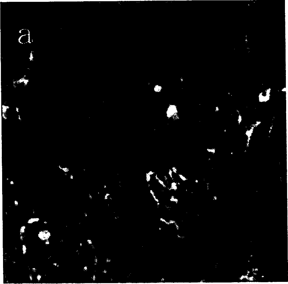
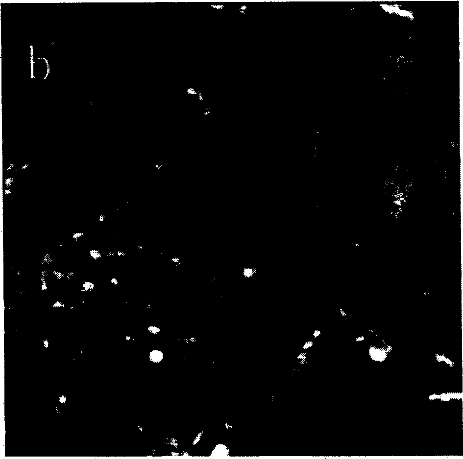
[0021] Human tongue squamous cell carcinoma tissue samples were taken from the oral and maxillofacial surgery of a certain hospital, fixed with 2% glutaraldehyde fixative solution at low temperature for 1 to 2 hours, or refrigerated overnight in the refrigerator. Rinse with PBS buffer, dehydrate with ethanol gradient, replace with ethylene oxide, infiltrate and embed with epoxy resin 618. LKB-2088 V-type ultra-microtome slices, and the glass knife used is newly prepared by LKB-7800 knife-making machine. Choose silvery white or lead white flakes, and the platinum ring is fished in the water drop placed on the mica. The temperature of the drying machine was adjusted to 60°C, and the mica was dried slowly. Observed in AFM tapping mode. The resulting image see figure 1 , figure 2 : Cell nuclei in mitotic phase can be seen in the field of vision, with active chromatin in the nucleus, relatively less cytoplasm, small nucleoplasmic ratio, and abnormal cell shape, which are typic...

Embodiment 2

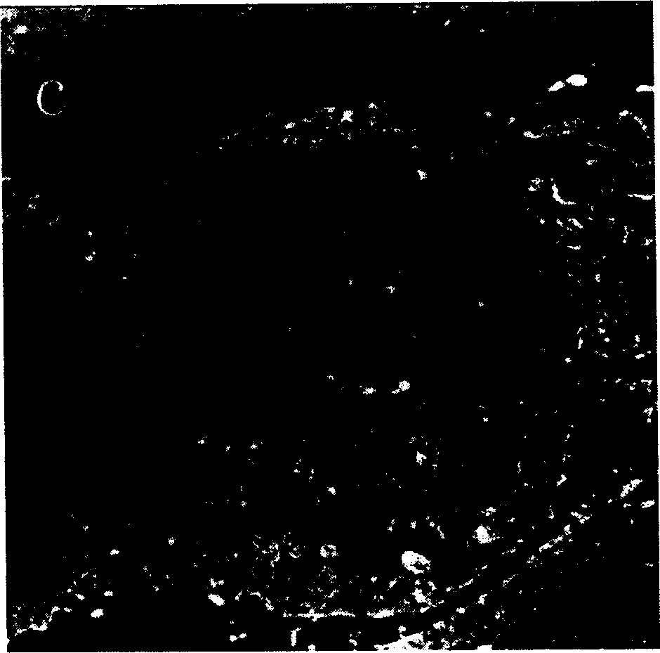
[0023] Tongue squamous cell carcinoma cultured cells PCA8113 were obtained from the Extraoral Tumor Biology Laboratory of an Institute of Stomatology, fixed with 2% glutaraldehyde, collected by centrifugation after dehydration, infiltrated with epoxy resin 618 and embedded. After slicing, the mica was collected and dried slowly on a drying machine at 50°C. Observed in AFM tapping mode. The resulting image see image 3 , Figure 4 .

[0024] image 3 What is shown is a tumor cell with abundant mitochondria in the cell, which suggests that the cell's metabolic activity is vigorous. Figure 4 Shown is a huge nucleus in a tumor cell with a clear nuclear membrane, two nucleoli can be seen, organelles are abundant in the perinuclear region, and the flat extracellular region is epoxy resin. image 3 Scanning range: 15μm×15μm, Figure 4 Scanning range: 20μm×20μm.


PUM


Abstract

The attaching method of superthin slice for microscope observation onto mica surface base on interatomic force includes preparing embedding lamp and superthin slice, selecting silvery white or plumbic white slice, gaining with platinum ring, dripping super pure water to split mica surface, soaking the platinum ring slightly in water drop to remove for the slice to float on the water surface and slowing stoving the mica in stoving machine to make the slice close to the surface of mica. The method is simple and low in cost, and has no damage to micro structure of the sample, can be used to obtain clear image and is suitable for preparing various biological sample.

Description

Technical field: [0001] The invention relates to a method for attaching ultra-thin slices, in particular to a method for attaching ultra-thin slices to mica surfaces for atomic force microscope observation, and belongs to the technical field of biomedical engineering. Background technique: [0002] Ultrathin sectioning is a method for preparing biological tissue samples for electron microscope (transmission electron microscope, TEM) observation. The general steps include: (1) fixation of biological samples, usually using glutaraldehyde and osmium tetroxide double fixation method; (2) dehydration, dehydration with ethanol or acetone gradient; (3) embedding, use domestic ring Oxygen resin 618 or imported Epon812 was infiltrated first and then embedded; (4) sliced, sliced ​​with an ultra-thin microtome, selected silvery white or leaded white slices, and dried with a copper net; (5) stained, usually with lead-uranium double Staining; (6) Transmission electron microscope observa...

Claims


Application Information

Patent Timeline
Patent Type & Authority Patents(China)
IPC IPC(8): G01N1/06G01N1/28G01Q60/24
Inventor 李鑫辉季彤胡钧胡晓芳张晓东孙洁林张陈平
Owner SHANGHAI JIAOTONG UNIV
Why Eureka
Try Eureka