
Compositions and methods for clearing a biological sample

a biological sample and composition technology, applied in the field of compositions and methods for clearing biological samples, can solve the problems of affecting the quality of tissue preparation, etc., and achieve the effect of reducing the time required for tissue preparation alone and reducing the difficulty of using traditional immunohistochemical (ihc) techniques to achieve large-scale results. desirable effects

Active Publication Date: 2020-02-25
WAKE FOREST UNIV HEALTH SCI INC
View PDF6 Cites 1 Cited by
  • Summary
  • Abstract
  • Description
  • Claims
  • Application Information

AI Technical Summary

Benefits of technology

The present disclosure provides improved methods for removing lipids from biological samples. The methods involve contacting the sample with a composition containing sodium dodecyl sulfate, 3-(N,N-dimethylmyristylammonio)propanesulfonate, Triton™ X-100, sodium deoxycholate, and a salt. The composition can be perfused into the sample for a short time to effectively remove lipids. The method can be used on fixed samples, such as embedded in fixative or contacted with an antibody. The patent also provides a phospholipase A2 and an imaging solution for further clarifying the sample. The method can be used on a variety of biological samples, such as plants, eukaryotic organisms, and cells. The imaging can provide three-dimensional information on the sample.

Problems solved by technology

One of the biggest issues with imaging the entirety of an organ, or even a relatively small sample of tissue (e.g., 1 mm×1 mm), is the difficulty of using traditional immunohistochemical (IHC) techniques to achieve the large scale results that are desirable.
The frozen blocks will retain a certain amount of fluorescence, however they will damage or destroy a lot of microstructures in the tissue.
This procedure would therefore take an unacceptable amount of time for the tissue preparation alone.
By cutting the tissue, damage to the edges of each tissue slice is inevitable.
This damage will create “gaps” and damaged areas in between each tissue slice when all of the slices are aligned and reconstructed.
Furthermore, the actual imaging of these 250 tissue slices would take an unacceptable amount of time.
Most oil-dipping objectives don't even have a working distance long enough to image through the entirety of a 4 μm thick tissue slice.
Even if all of the slices are fully imaged through the entirety of the slice thickness, the ability to align and accurately reconstruct the sample would be extremely difficult.
Due to the aforementioned difficulties that arise when performing tissue analysis on a larger (macro) scale, researchers have turned to using multiphoton imaging with a whole mounted, unsectioned specimen for deep tissue imaging.
Such a technique, however, carries with it a vast number of limitations and problems that arise when performing deep tissue imaging.
One of the most notable issues is the inability to limitlessly image into the depth of the tissue.
However, attaining such a result is extremely difficult and rare with all tissues, most notably the brain.
The inability to image deeper into the tissue arises from light absorption, and light scattering caused by lipids in the tissue.
While there has been some success with very small pieces of tissue, large samples cannot be used.
Sucrose causes a dehydration of the tissue, ultimately causing shrinkage of the actual tissue, most likely due to extreme osmolality changes.
The shrinkage of the tissue does not provide accurate information about the structure of the tissue, specifically the microstructure.
This technique does not permit the ability to label the tissue with antibodies.
Also, the sucrose solutions do not provide a good medium for maintaining endogenous fluorescence.
The light transmittance with sucrose-cleared tissues is also extremely poor.
True and complete clearing using sucrose clearing is unattainable.
The limitations of this technique are very similar to those mentioned in the sucrose technique.
Glycerol can only be used on extremely small and thin samples.
There is a smaller volume change in the sample when using glycerol; however, volume change does still occur.
Antibody labeling cannot be utilized with this technique.
Glycerol is also a very poor medium for endogenous fluorescence since it has been shown to quench a majority of the fluorescence.
True complete clearing using glycerol clearing is typically unachievable.
BABB causes the most drastic tissue shrinkage out of all the clearing techniques demonstrated in literature, thus creating the worst tissue structure representation.
Antibody labeling is also unachievable with this technique.
However, the major limitations are (i) the inability to do antibody labeling, (ii) the approximate 1.5× volume expansion of the tissue, and (iii) the amount of time that it takes to perform the technique.
While this technique has provided some advantages in the field of tissue clearing, it also possesses limitations.
For example, SCALE results in a denaturation of the majority of proteins from the sample, there is an inability to probe with antibodies in the SCALE solution, and the majority of fluorescent signal is lost in a time dependent manner.
SeeDB does not, however, allow antibody staining and the cleared sample can only be maintained in the final clearing solution for a maximum of 7 days, at which time the sample has to be washed free of fructose.
There have also been a lot of problems reported with autofluorescence and browning developing in the tissue due to a maillard reaction.
This technique has also not been proven to completely clear the entirety of an organ; in fact, the technique performs best with tissue slices that are about 1-2 millimeters in thickness.
The problems with CLARITY arise with the variety of tissues that are compatible with the technique.
While it has been shown that this works for nervous tissue, it is not as compatible with other organs / tissues in the body.
Attempts at using the CLARITY technique on other parts of the mouse were met with very little success.
Researchers have also reported difficulty even when attempting to clear the brain (see, e.g., the CLARITY Resources website).
The protocol itself is technically difficult to perform, while also being tremendously costly.
There is also an inability to perform the clearing technique on multiple samples if only one chamber is created.
It is therefore an ineffective technique if one is attempting a round of experiments that would require clearing of a dozen or more samples.
Another confounding factor is the mounting medium.
FocusClear™ has been shown to be a great product for tissue clearing; however, it is prohibitively costly.

Method used


Image

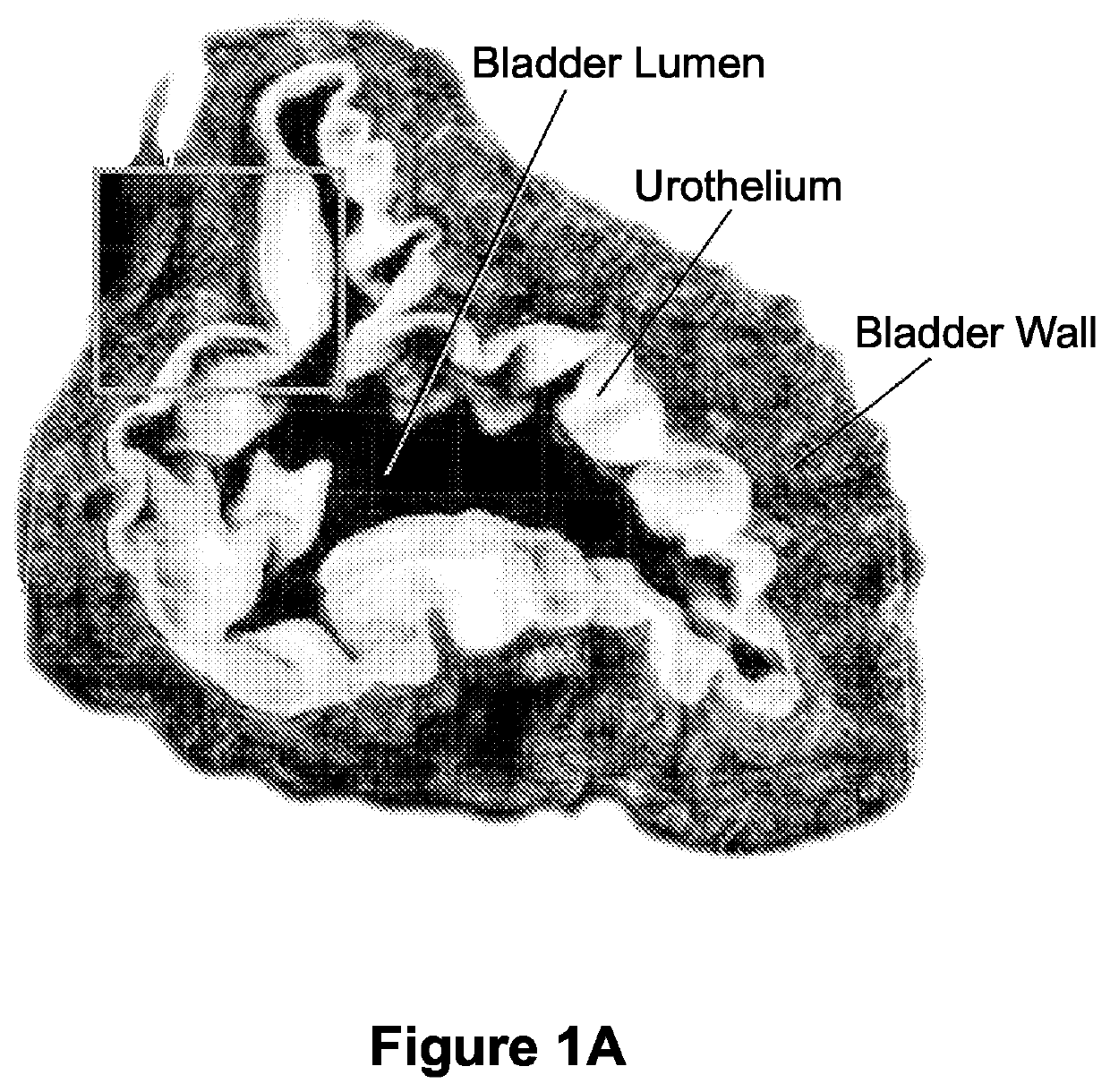
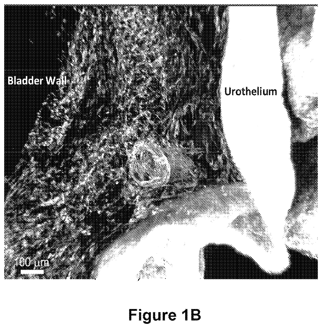
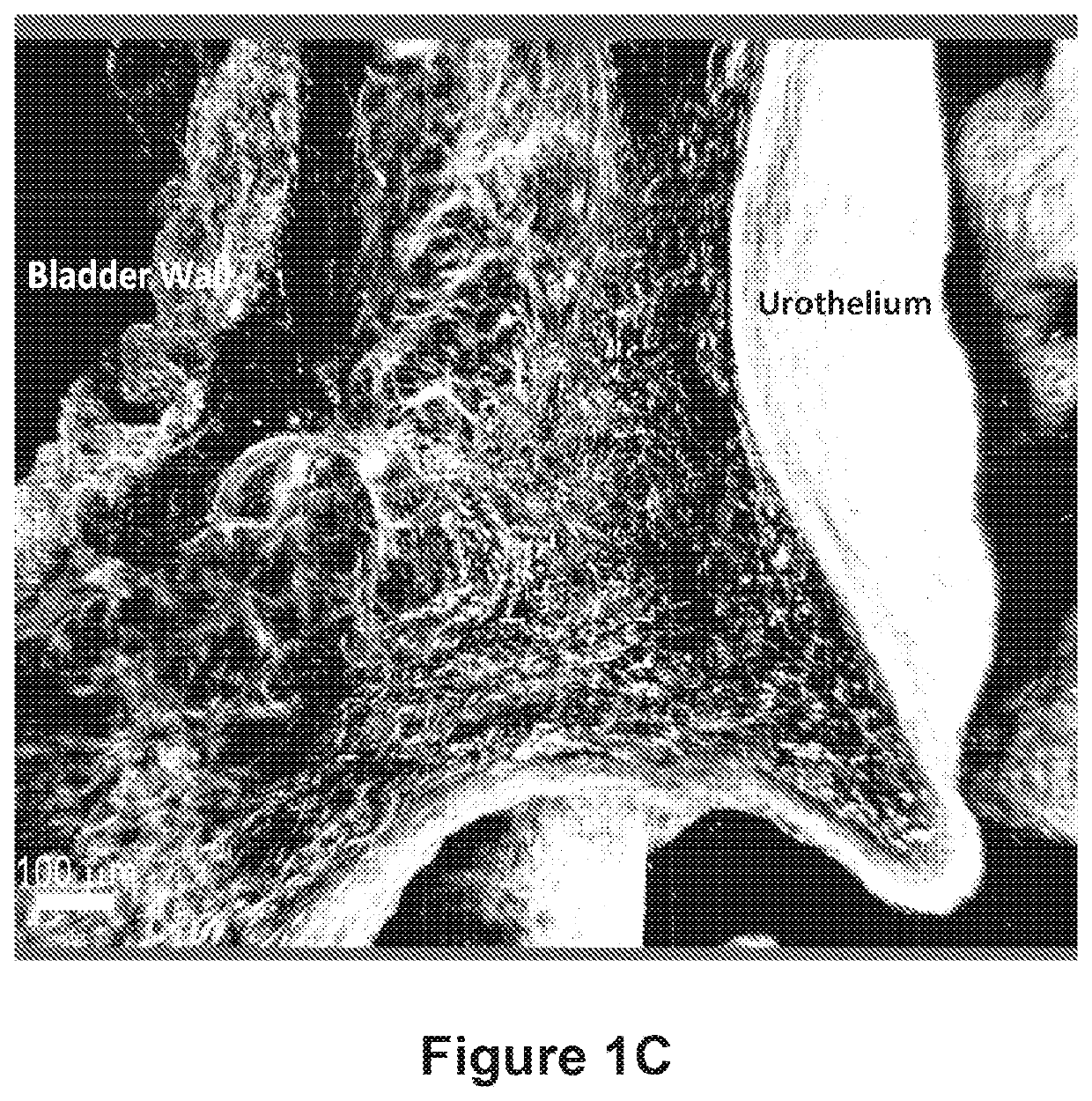
  • Compositions and methods for clearing a biological sample
  • Compositions and methods for clearing a biological sample
  • Compositions and methods for clearing a biological sample

Examples


example 1

This example discusses the tissue harvesting and embedding step of the protocol disclosed herein. First, an animal is euthanized according to approved protocol. Cervical dislocation is not performed as it will sever the vasculature to the brain.

Next, the animal is transcardially perfused with 20 mL of 1× ice-cold PBS followed by 20 mL of ice-cold gel / fixative solution. Perfusion can also be performed through the abdominal aorta if isolation of kidneys, liver, and spleen are desired. In addition, organs can be harvested first and then integrated with hydrogel.

Perfusion System

A 2″ PVC pipe is capped at both ends and retrofitted with ¼″ diameter nozzles at opposite ends. The tube is supported vertically using a chemistry stand and clamp, with the inlet of the tube at the top and the outlet at the bottom. One quarter inch tubing is then connected to both ends of the tube fittings and lead to their desired components of the perfusion system. The water circulator outlet is connected with ...

example 2

This example discusses the tissue clearing and sample preparation for imaging steps of the protocol disclosed herein. First, the gel samples are removed from the tubes and any excess gel is teased away from around the tissue. Then the samples are placed into covered 50 mL conical tubes with 50 mL of wash buffer for two 24-hour periods at 37° C. on an end-over-end rocker. This is to help remove any excess PFA or acrylamide, as well as to help prime the sample for perfusion with the clearing buffer.

The samples are then placed into individual immunohistochemistry (IHC) tissue cassettes, labeled using pencil with what tissue it is. The perfusion chamber and water circulator is then prepared by connecting the hoses and filling the circulator with the clearing buffer.

“Lipid Magnet” Clearing Buffer

This formulation is for 10 liters of a solution.

Amount to addComponentfor 10 Liter batch  5% SDS500g0.5% Sodium Deoxycholate50g  1% Triton X-100100mL  1% Tween-20100mL0.1% SB3-1410g150 mM NaCl or...


PUM

PropertyMeasurementUnit
depthaaaaaaaaaa
depthaaaaaaaaaa
depthaaaaaaaaaa

Abstract

The disclosure provides improved materials and methods for optically clearing biological tissue that is subsequently used for deep tissue imaging analysis. Also provided is a description of a microscopic image acquisition methodology in which imagery of intact tissues are acquired to rapidly acquire microscopy data on a whole-organ scale to maximize cost effectiveness for biological microscopy and minimize time spent performing such analysis.

Description

FIELD OF THE INVENTIONThe present disclosure relates to compositions and methods that are useful in optically clearing a biological sample so as to make the sample transparent.BACKGROUND OF THE INVENTIONAs imaging technology improves via advances including better optics, higher resolutions, and the ability to image deeper into tissue with three dimensional results, there is a higher demand for more advanced imaging protocols to image tissue on a large scale.One of the biggest issues with imaging the entirety of an organ, or even a relatively small sample of tissue (e.g., 1 mm×1 mm), is the difficulty of using traditional immunohistochemical (IHC) techniques to achieve the large scale results that are desirable. Typical IHC procedures require the embedding of the sample into paraffin or snap frozen into an optimum cutting temperature (OCT)-based block. Paraffin is useful for subsequent antibody labeling but will remove any endogenous fluorescence localized in the tissue. The frozen b...

Claims


Application Information

Patent Timeline
Patent Type & Authority Patents(United States)
IPC IPC(8): G01N1/30G01N1/36G01N1/40G01N33/483G06T7/00
CPCG01N1/36G01N1/4044G01N1/30G01N33/4833G01N2001/364G06T7/0012
Inventor MARINI, FRANKCHRIST, GEORGE
Owner WAKE FOREST UNIV HEALTH SCI INC
Learn More