
Concatameric immunoadhesion

a technology of immunoadhesion and catalytic molecules, applied in the field of catalytic immunoadhesion, can solve the problems of reducing the production yield of multimeric forms, unable to bind to light chains, and difficulty in purifying multimeric high molecular weight forms, etc., and achieves the effect of increasing efficacy and stability

Active Publication Date: 2003-10-16
MEDEXGEN
View PDF2 Cites 66 Cited by
  • Summary
  • Abstract
  • Description
  • Claims
  • Application Information

AI Technical Summary

Benefits of technology

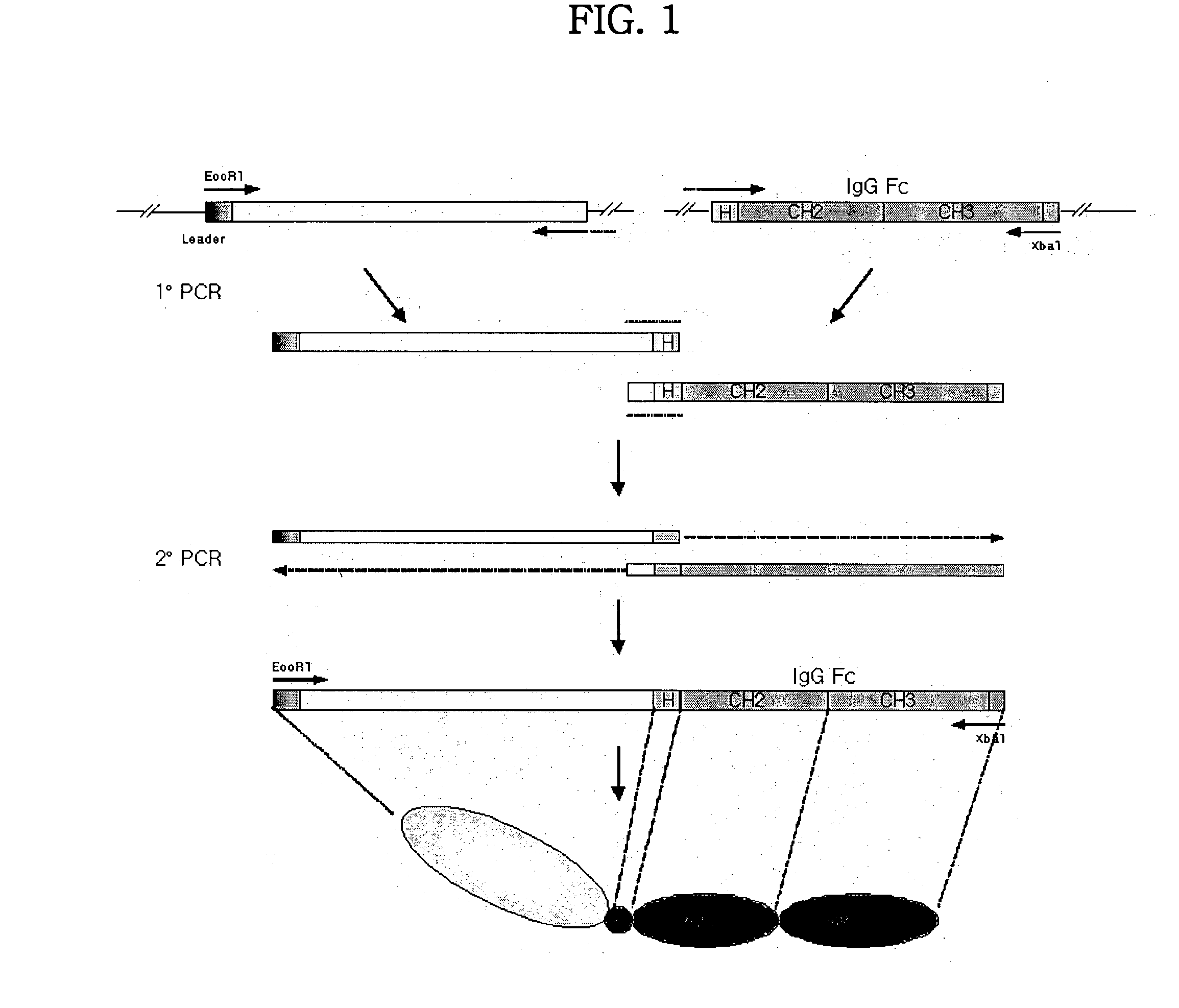
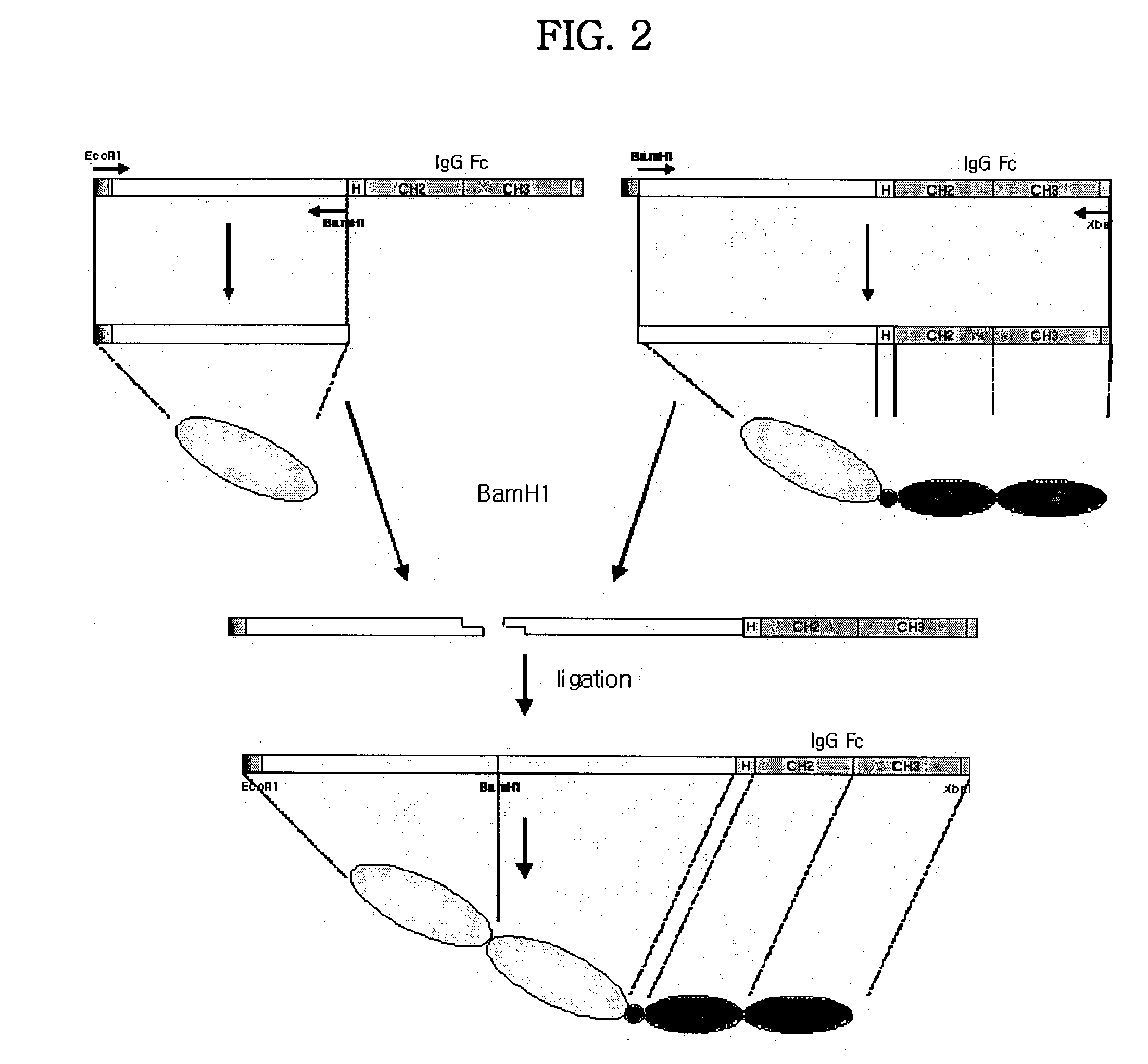
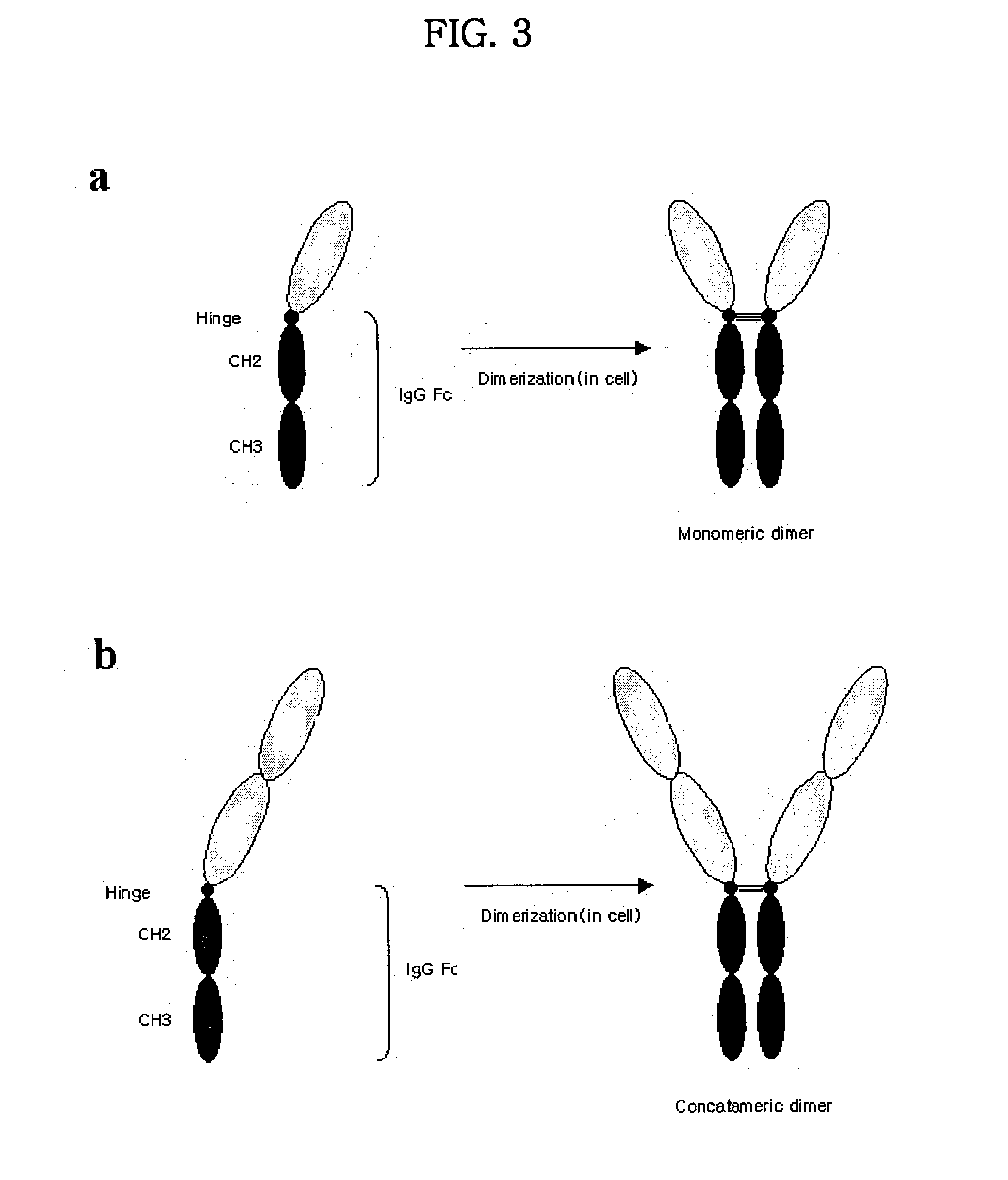
[0013] The present inventors have manufactured concatameric proteins by fusing the C-terminal end of soluble domain of biologically active protein to the N-terminal end of soluble domain of the same or other biologically active protein by using DNA recombination techniques. Also, the present inventors have dimerized this concatamers by linking it to the hinge region of Fc fragment of immunoglobulin and added more glycosylations by using DNA mutagenesis techniques. And the present inventors have found that concatamerized protein dimers and their glycosylated forms show increased efficacy and stability compared to conventional monomeric fusion proteins.
[0081] Host cells used in the present invention may be prokaryotic or eukaryotic. In addition, host cells having high introduction efficiency of foreign DNA and having high expression levels of an introduced gene may be typically used. Examples of the host cells useful in the present invention include prokaryotic and eukaryotic cells such as E. coli, Pseudomonas sp., Bacillus sp., Streptomyces sp., fungi or yeast, insect cells such as Spodoptera frugiperda (Sf9), animal cells such as Chinese hamster ovary cells (CHO) or mouse cells, African green monkey cells such as COS 1, COS 7, human embryonic kidney cells, BSC 1, BSC 40 or BMT 10, and tissue-cultured human cells. When cloning a DNA construct encoding the fusion protein according to the present invention, host cells are preferably animal cells. When using COS cells, since SV40 large T antigen is expressed in COS cells, a plasmid carrying a SV 40 replication origin may be present as a multicopy episome and thus allows high expression of an exogeneous gene. A DNA sequence introduced into a host cell may be homogeneous or heterogeneous to the host cell, or a hybrid DNA sequence containing a homogenous or heterogeneous DNA sequence.
[0085] The term "signal sequence", as used herein, means an amino acid sequence mediating transport of an expressed protein to the outside of the cell membrane, and is also called a "leader sequence". Cell surface proteins or secretory proteins, which are transported to the outside of the cell membrane, have an N-terminal sequence typically cut by signal peptidase in the cell membrane. Such a N-terminal sequence is called a signal sequence or signal peptide, or a leader sequence or leader peptide. Secretory (or transported) proteins or all proteins present outside of the cell membrane or in the extracellular environment have a specific signal sequence. There is no specific homology between such signal sequences and same proteins have different signal sequences according to their origin. Secondary structure or distribution of nonpolar and charged residues is more important for proper function of the signal sequences than primary structures thereof. Although not having specific homology, the signal sequences share several common features, as follows. The signal sequences contain an N domain at their N-termini, which is a hydrophilic region comprising one or more positively charged residues, and an H-domain follows the N domain, which is a somewhat long hydrophobic region. In the case of E. coli, the signal sequence comprises about 18-30 amino acids. The N domain contains many cationic amino acids such as Lys or Arg, and thus has a net positive charge. Many hydrophobic amino acids such as Ala or Leu are found in the H domain, and polar or charged amino acids such as Pro, Lys, Arg, Asn or Glu are rarely in the H domain. A large number of amino acids such as Ala and Leu residues form an .alpha.-helical structure to facilitate membrane penetration. A C domain is positioned between the H domain and an actually secreted portion of a protein. The C domain is less hydrophobic, and contains a sequence capable of being recognized by signal peptidase such as LebB or LspA. There have been no reports about an exact site cleaved by the signal peptidase, but the signal peptidase is typically known to mostly cleave behind the Ala-X-Ala sequence in the C domain. Preproteins containing the above-mentioned signal sequence arrive at the cell membrane through interaction with several proteins, and fold to their mature forms through cleavage of a specific region of a signal peptide. Such a signal sequence is very important in strategies to express a desired protein on the cell surface or in the extracellular environment. Foreign proteins and fusion proteins should be stably transported to the extracellular environment at high efficiency. Typically, cell surface proteins having excellent secretory ability are useful for cell surface expression of foreign proteins or fusion proteins, which typically have secretory signal sequences capable of offering excellent secretion efficiency.
[0092] The immunoglobulin fusion gene may further include a signal sequence to stimulate extracellular secretion of a protein encoded thereby. For example, the CTLA-4 molecule contains a unique leader sequence having highly hydrophilic redundancy at its N-terminus, and which is abnormally long and highly water-soluble (Harper, K. et al., J. Immunol. 147:1037-1044; and Brunet, J. F. Nature 328:267-270, 1987). Generally, most cell surface proteins or secretory proteins have a leader sequence comprising 20-24 highly hydrophobic amino acids at their N-termini. However, the CTLA-4 molecule used in the present invention comprises a total of 37 residues: 16 hydrophilic amino acids at its N-terminus, and 21 highly hydrophobic amino acids typical in its transmembrane regions. In the conventional method of preparing CTLA4Ig fusion proteins, the leader sequence of the CTLA-4 molecule was substituted with a leader sequence of oncostatin M (Linsley, P. S. et al., J. Exp. Med. 174:561-569, 1991) or IL-6 (Yamada, A, et al., Microbiol. Immunol. 40:513-518, 1996). The present inventors demonstrated that a CTLA-4 molecule containing a leader sequence having a "MRTWPCTLLFFIPVFCKA" sequence instead of the amino acid sequence consisting of 16 amino acids, "ACLGFQRHKAQKNLAA", is preferable, and the secretion of an expressed protein to the extracellular environment is easily achieved, as disclosed in International Pat. Publication No. WO98 / 31820.
[0096] Secretory proteins produced in eukaryotic cells as host cells are modified by glycosylation. Glycosylation is known to influence in vivo stability and functionality as well as physical properties of a protein. Therefore, a preferred aspect of the present invention includes facilitating production of a concatameric fusion dimeric protein of interest using recombinant DNA techniques and the above-mentioned animal cell lines as host cells, and linking additional sugar chains to a soluble extracellular domain of a protein involving immune response.
[0101] Tumor necrosis factor-alpha (TNF-.alpha.), which is known as the hormone cachectin, and tumor necrosis factor-beta (TNF-.beta.), which is also known as lymphotoxin, are multifunctional cytokines, inducing inflammation, cellular immune response, septicemia, cytotoxicity, cachexia, rheumatoid arthritis, inflammation-related diseases (Tartaglia, L. A. et al., Immunol. Today 13:151,1992), and antiviral reaction (Butler, P., Peptide Growth Factor II, 1990, Springer-Verlag, Berlin, pp.39-70). Such actions of TNF-.alpha. and TNF-.beta., including cytotoxic activity, originate from their binding to TNF receptors in a trimeric form (Eck, M. J. et al., J. Biol. Chem. 267:2119, 1992). As TNF receptors, 55 kDa-type I (TNFR1 or p55) and about 75 kDa-type II (TNFR2 or p75) are known (Smith, C. A. et al., Science 248:1019, 1990; Loetscher, H. et al., Cell 61:351, 1990; and Schall et al., Cell 61:361, 1990). The two receptors have similar affinity for TNF-.alpha. and TNF-.beta. (Schall et al., Cell 61:361, 1990). Immunoglobulin fusion proteins of such soluble receptors have effects of inhibiting the action of TNF-.alpha. and TNF-.beta. by inhibiting binding of TNF-.alpha. and TNF-.beta. to their receptors on the cell surface, which is known to be effective in reducing TNF-dependent inflammation.

Problems solved by technology

Most fusion proteins of receptor extracellular domain and immunoglobulin heavy chain are composed of heavy chain without CH1 domain, which result in dimers not binding to light chains.
However, this method had many difficulties for commercialization such as simultaneous expression of two different fusion genes in the same cell line, remarkably lower production yields of multimeric form; and difficulty in purifying multimeric high molecular weight forms.

Method used


Image

  • Concatameric immunoadhesion
  • Concatameric immunoadhesion
  • Concatameric immunoadhesion

Examples


example 2 and 3

CD2 and CTLA4

[0159] DNA fragments encoding soluble extracellular domain of CD2 and CTLA4 were constructed by PCR using a primer [CD2(the sequence of nucleotide of SEQ ID NO: 40), and CTLA4(the sequence of nucleotide of SEQ ID NO: 43)] with EcoRI restriction site and the coding sequence [CD2 (the sequence of nucleotide of SEQ ID NO: 13), and CTLA4 (the sequence of nucleotide of SEQ ID NO: 15)] encoding the leader sequence [CD2(the sequence of amino acid 1-24 of SEQ ID NO: 14), and CTLA4(the sequence of amino acid 1-21 of SEQ ID NO: 16)], and an antisense primer [CD2(the sequence of nucleotide of SEQ ID NO: 41), and CTLA4(the sequence of nucleotide of SEQ ID NO: 44)] with PstI restriction site and the sequence [CD2(the sequence of nucleotide of SEQ ID NO: 13), and CTLA4(the sequence of nucleotide of SEQ ID NO: 15)] encoding 3' end of the soluble extracellular domain of the proteins as described above. The template cDNA for this reaction was constructed by reverse transcription PCR (RT...

example 4

Expression and Purification of Simple / Concatameric Fusion Dimeric Protein of TNFR / Fc

[0171] In order to express the fusion proteins in CHO-K1 cell (ATCC CCL-61, Ovary, Chinese hamster, Cricetulus griseus), after pBluescript KS II (+) plasmid DNA including TNFR / Fc fusion gene was purified from transformed E. coli, an animal cell expression vectors were constructed as TNFR / Fc fragment produced by restriction using EcoRI and XbaI was inserted at EcoRI / XbaI site of an animal cell expression vector, pCR.TM.3 (Invitrogen, USA) plasmid. And these were designated plasmid pTR11-Top10' and plasmid pTR22-Top10', and deposited as accession numbers of KCCM 10288 and KCCM 10291, respectively, at Korean Culture Center of Microorganisms (KCCM) on Jul. 10. 2001.

[0172] Transfection was performed by mixing either the plasmid pTR11-Top10' or plasmid pTR22-Top10' DNA including TNFR / Fc fusion genes as described above with the reagent of Lipofectamin.TM. (Gibco BRL, USA). CHO-K1 cells with the concentratio...

example 5

SDS-PAGE of Purified TNFR1-TNFR1 / Fc and TNFR2-TNFR2 / Fc (FIG. 15)

[0176] Proteins purified using protein A column were electrophorized by the method of SDS-PAGE in reducing condition added by DTT, reducing reagent (which destroy disulfide bond), and in a non-reducing condition excluding DTT. The result of the estimation of molecular weight on SDS-PAGE is shown in Table 10. It was possible to confirm that TNFR / Fc proteins were the shape of a dimer in the cell. The molecular weight deduced from the amino acid sequence of TNFR1-TNFR1-Ig was about 70 kDa, and was estimated as about 102 kDa on SDS-PAGE. As this difference could be regarded as a general phenomenon which generate on the electrophoresis of glycoproteins, this feature seemed to occurr as the result from decrease in mobility on the electrophoresis by the site of glycosylation.

10TABLE 10 Molecular weight of TNFR-TNFR / Fc on the SDS-PAGE. Molecular weight (kDa) Proteins Reducing condition Non-reducing condition TNFR1-TNFR1 / Fc 102 ...


PUM

PropertyMeasurementUnit
temperatureaaaaaaaaaa
pHaaaaaaaaaa
pHaaaaaaaaaa

Abstract

Disclosed are concatameric proteins comprising two soluble domains, in which the C-terminus of a soluble domain of a biologically active protein is linked to the N-terminus of an identical soluble domain or a distinct soluble domain of a biologically active protein. Also, the present invention discloses dimeric proteins formed by formation of intermolecular disulfide bonds at the hinge region of two monomeric proteins formed by linkage of a concatamer of two identical soluble extracellular regions of proteins involving immune response to an Fc fragment of an immunoglobulin molecule, their glycosylated proteins, DNA constructs encoding the monomeric proteins, recombinant expression plasmids containing the DNA construct, host cells transformed or transfected with the recombinant expression plasmids, and a method of preparing the dimeric proteins by culturing the host cells. Further, the present invention discloses pharmaceutical or diagnostic compositions comprising the dimeric protein or its glycosylated form.

Description

[0001] The present invention relates to concatameric proteins, and more specifically, concatamerized structure of biologically active protein domains where C-terminal end of extracellular soluble domain of biologically active protein is fused to N-terminal end of the same or other extracellular soluble domain of biologically active protein, and dimerization of two concatamers by coupling to hinge region of Fc fragment of immunoglobulin, and glycosylated forms of the concatameric proteins.[0002] The activity of cytokine is associated with pathologic severity of inflammatory and / or immune response to various antigenic stimulations. Many antigen specific antibodies and soluble receptors which could recognize cytokines are currently in use to inhibit the function of cytokines for the therapeutic purposes (WO 93 / 016184, WO 96 / 02576, WO 96 / 023067, WO 1997 / 03682, and U.S. Pat. Nos. 5,434,131, 5,656,272, 5,977,318, 6,210,661, 6,225,117). Antibodies and soluble receptors inhibit cytokine sig...

Claims


Application Information

Patent Timeline
Patent Type & Authority Applications(United States)
IPC IPC(8): A61K38/16A61P19/02A61P29/00C12N15/09C07K14/47C07K14/475C07K14/52C07K14/705C07K14/715C07K16/46C07K19/00C12N1/15C12N1/19C12N1/21C12N5/10C12N9/12C12P21/02
CPCA61K2039/505C07K14/70507C07K2319/30C07K14/7151C07K2319/00C07K14/70521A61P19/02A61P29/00A61P37/00C07K16/46
Inventor CHUNG, YONG-HOONHAN, JI-WOONGLEE, HYE-JACHOI, EUN-YONGKIM, JIN-MI
Owner MEDEXGEN
Learn More