[0011] In a preferred embodiment, the expandable constrictor comprises an outer
conical shell and an inner
conical shell. Each shell has an apex and an open base to receive blood flow. One or a plurality of ports traverses the walls of the two conical shells. Blood flows through the open base and through the ports. The inner shell can be rotated relative to the outer shell so that the ports align or misalign with the ports in the outer shell to allow variable blood flow past the occluder, thereby providing adjustable and controlled flow. The inner shell is rotated by a rotating mechanism, e.g., a torque cable disposed within the elongate tube and coupled to the inner shell. The constrictor can be expanded by, e.g., a resilient pre-shaped ring, graduated rings, or a beveled lip formed at the base of the shell, and collapsed by, e.g., pull wires distally affixed to the occluder or a guide sheath.
[0013] In still another embodiment, the constrictor comprises a first cylindrical
balloon mounted to a distal end of the
catheter, and a second toroidal
balloon disposed about the cylindrical
balloon. The chamber of the first balloon communicates with an inflation lumen.
Blood flow occurs through the cylindrical balloon and through the center of the toroidal balloon. The toroidal balloon is expanded by inflation through a second and independent inflation lumen to reduce blood flow through the cylindrical balloon. In this manner, the first balloon provides an
inflatable sleeve and the second toroidal balloon provides variable control of blood flow through the sleeve. Other embodiments include an expandable sleeve (not a balloon) surrounded by a toroidal balloon for adjustably constricting the flow of blood through the cylindrical sleeve.
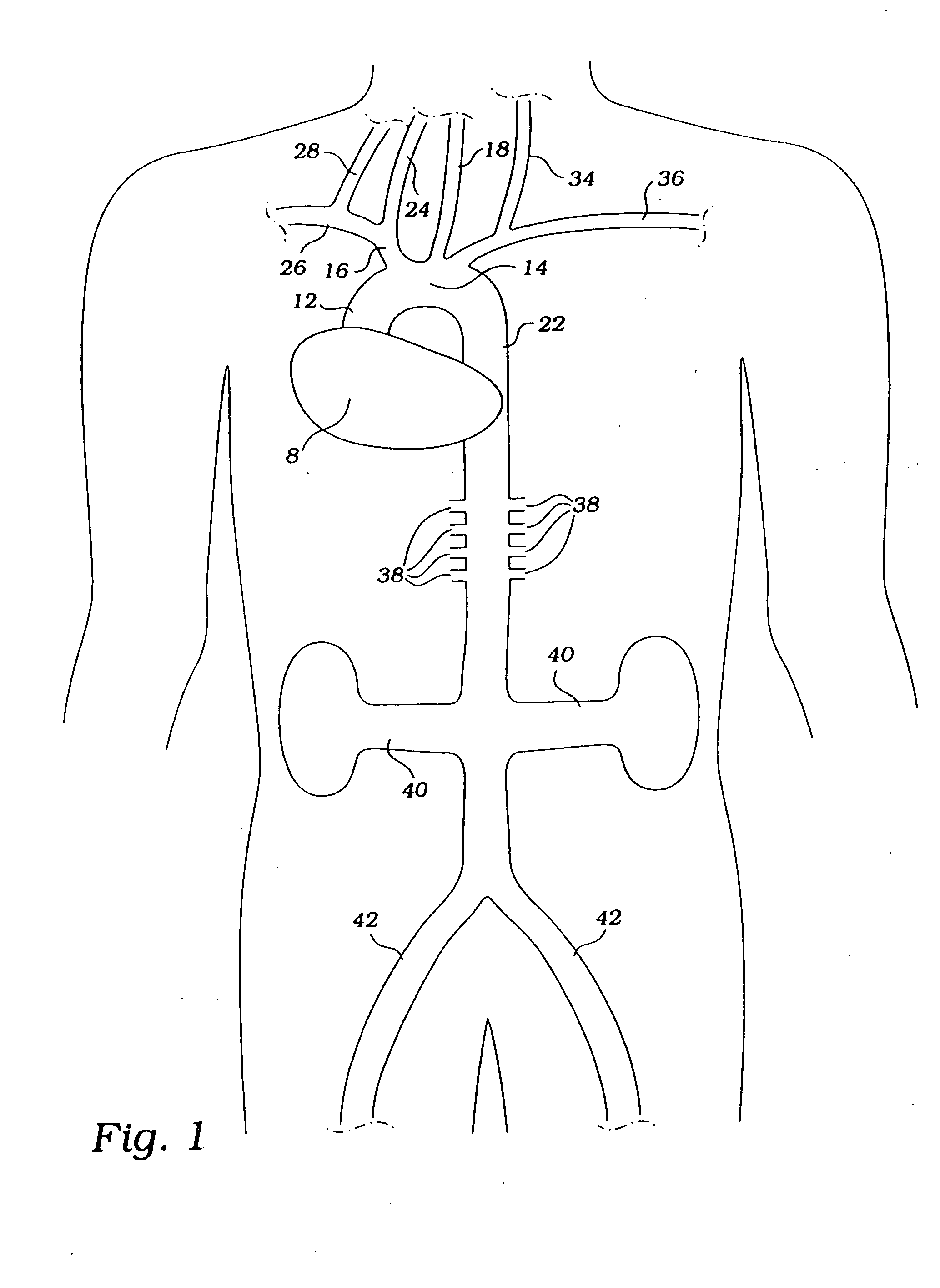
[0014] In a preferred method, the
occlusion devices described above are inserted into the
descending aorta through an incision on a
peripheral artery, such as the femoral, subclavian, axillary or
radial artery, in a patient suffering from global or focal cerebral ischemia, during
cardiac surgery (including any operation on the heart, with or without CPB), or during aortic
surgery (during circulatory arrest, as for
aortic arch surgery, repair of an
abdominal aortic aneurysm, or thoracic
aneurysm repair, to reduce perfusion and the amount of
blood loss in the operating field). The devices can be introduced over a guide wire. With assistance of transesophageal echocardiography (TEE), transthoracic echocardiography (TTE),
intravascular ultrasound (IVUS),
aortic arch cutaneous
ultrasound, or angiogram, the constrictor is positioned downstream from the
takeoff of the brachiocephalic
artery and upstream from the renal arteries. The constrictor is expanded to partially occlude blood flow in the
aorta and maintained during
systole, during
diastole, or during
systole and
diastole. The constrictor preferably achieves continuous
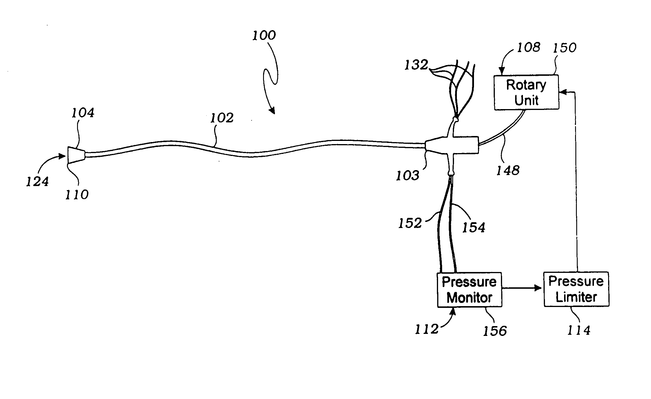
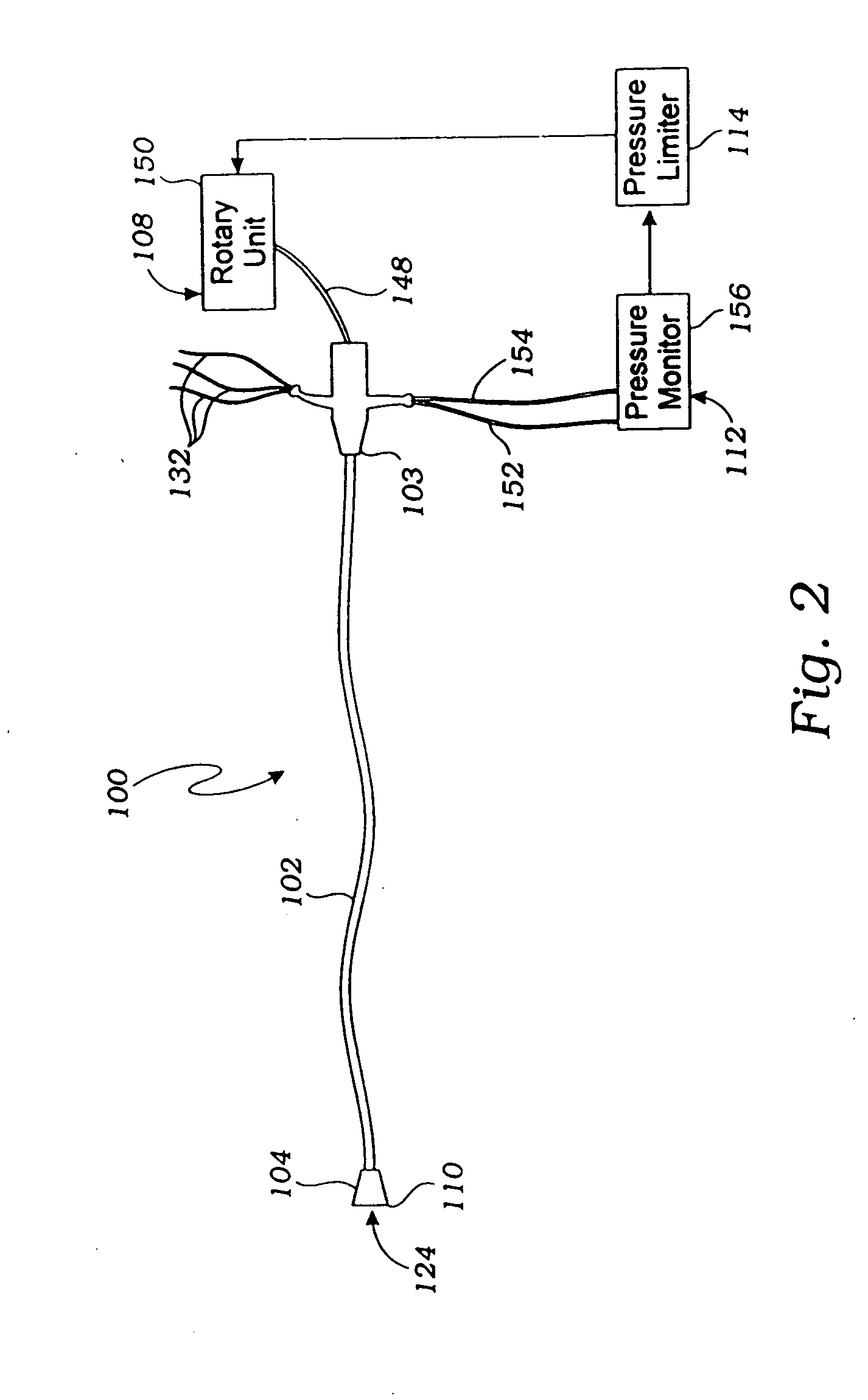
apposition to the wall of the vessel, resulting in fewer emboli dislodgment. The pressure
limiter, connected to the rotary unit and the pressure monitor, prevents the upstream and downstream
blood pressure from exceeding, respectively, a set maximum and minimum pressure differential.
[0015] Flow rates can be varied within one
cardiac cycle (e.g., 80% during systole, 20% during diastole, or 70% during systole, 30% during diastole), and every few cycles or seconds (e.g., 80% for 6 cycles, 20% for 2 cycles, or 70% for 5 cycles, 10% for 1 cycle). In certain cases it may be preferred to cycle to cycle between lesser and greater occlusion so that the brain does not autoregulate. This ensures constant and continued increased cerebral perfusion. In this manner, blood in the
descending aorta is diverted to the cerebral vasculature, thereby increasing cerebral perfusion and minimizing neurological deficits. By selectively increasing
cerebral blood flow, the use of systemically administered vasoconstrictors or inotropic agents to treat shock may be reduced or eliminated.
[0017] Prolonged hypertension often causes ischemic damage to the kidneys. In still another method, the
partial occlusion devices are introduced peripherally and positioned in the renal arteries to reduce
blood pressure to the renal vasculature, thereby minimizing damage to the kidneys that might otherwise result from hypertension.
[0018] It will be understood that there are many advantages in using the partial aortic occlusion devices and methods disclosed herein. For example, the devices can be used (1) to provide variable
partial occlusion of a vessel; (2) to augment and maintain cerebral perfusion in patients suffering from global or focal ischemia; (3) to condition the brain or
spinal cord to secrete neuroprotective agents prior to a major
surgery which will necessitate reduced cerebral or spinal perfusion; (4) to prolong the
therapeutic window in global or focal ischemia; (5) to accommodate other medical devices, such as an
atherectomy catheter; (6) prophylactically by an interventional radiologist, neuroradiologist, or cardiologist in an angiogram or
fluoroscopy suite; (7) for prevention of cerebral ischemia in patients undergoing procedures, such as coronary catheterization or surgery, where
cardiac output might fall as a result of arrhythmia,
myocardial infarction or failure; (8) to treat shock, thereby eliminating or reducing the use of systemic vasoconstrictors; and (8) to prevent
renal damage in hypertensives.

