Method of full-color optical coherence tomography

a tomography and color technology, applied in the field of full-color optical coherence tomography, can solve the problems of compromising the lateral resolution to a few m, placing a fundamental limit on the image acquisition speed, and low light efficiency or image contrast, and achieves high frame rate

US20050185192A1Active Publication Date: 2005-08-25UNIV OF SOUTH FLORIDA
3 Cites 57 Cited by

Patent Information

Authority / Receiving Office
US · United States
Current Assignee / Owner
Publication Date
2005-08-25

Smart Images

  • Figure 1
    Figure 1
  • Figure 2
    Figure 2
  • Figure 3
    Figure 3
Patent Text Reader

Abstract

Two-dimensional and three-dimensional optical coherence tomography is obtained by differential imaging of full-frame interference images using a white light source. Full-color tomographic imaging is also possible by processing the three-color channels of the interference images. A technique is described to obtain two-dimensional OCT images with full natural color representation. In a particular embodiment, the interference image is acquired using a color camera and the three-color channels are processed separately, recomposing the final image. In an additional embodiment, the interference images are acquired using separate red, blue and green light sources and the three color channels are combined to recompose the final image.
Need to check novelty before this filing date? Find Prior Art

Description

CROSS-REFERENCE TO RELATED DISCLOSURES

[0001] This disclosure claims priority from a provisional application filed Feb. 20, 2004, entitled: “Method and Apparatus for Full-Color Two-Dimensional Optical Coherence Tomography,” bearing application No. 60 / 546,408. Priority is also claimed to a second provisional application filed Dec. 10, 2004 entitled: “Full-Color Three-Dimensional Microscopy by Wide-Field Optical Coherence Tomography” bearing application No. 60 / 635,162.BACKGROUND OF INVENTION

[0002] In the field of biomedical imaging, various methods of optical imaging of biomedical tissues known in the art offer significant advantages over other biomedical imaging approaches. These advantages include non-ionizing radiation, wide range of resolution, numerous and effective contrast mechanisms, and relatively compact and inexpensive instrumentation. Optical imaging can be applied to a wide range of biological systems, from cells and subcellular organelles, in vivo and ex vivo tissues, t...

Examples

Embodiment Construction

[0030] In the following detailed description of the preferred embodiments, reference is made to the accompanying drawings, which form a part hereof, and within which are shown by way of illustration specific embodiments by which the invention may be practiced. It is to be understood that other embodiments may be utilized and structural changes may be made without departing from the scope of the invention.

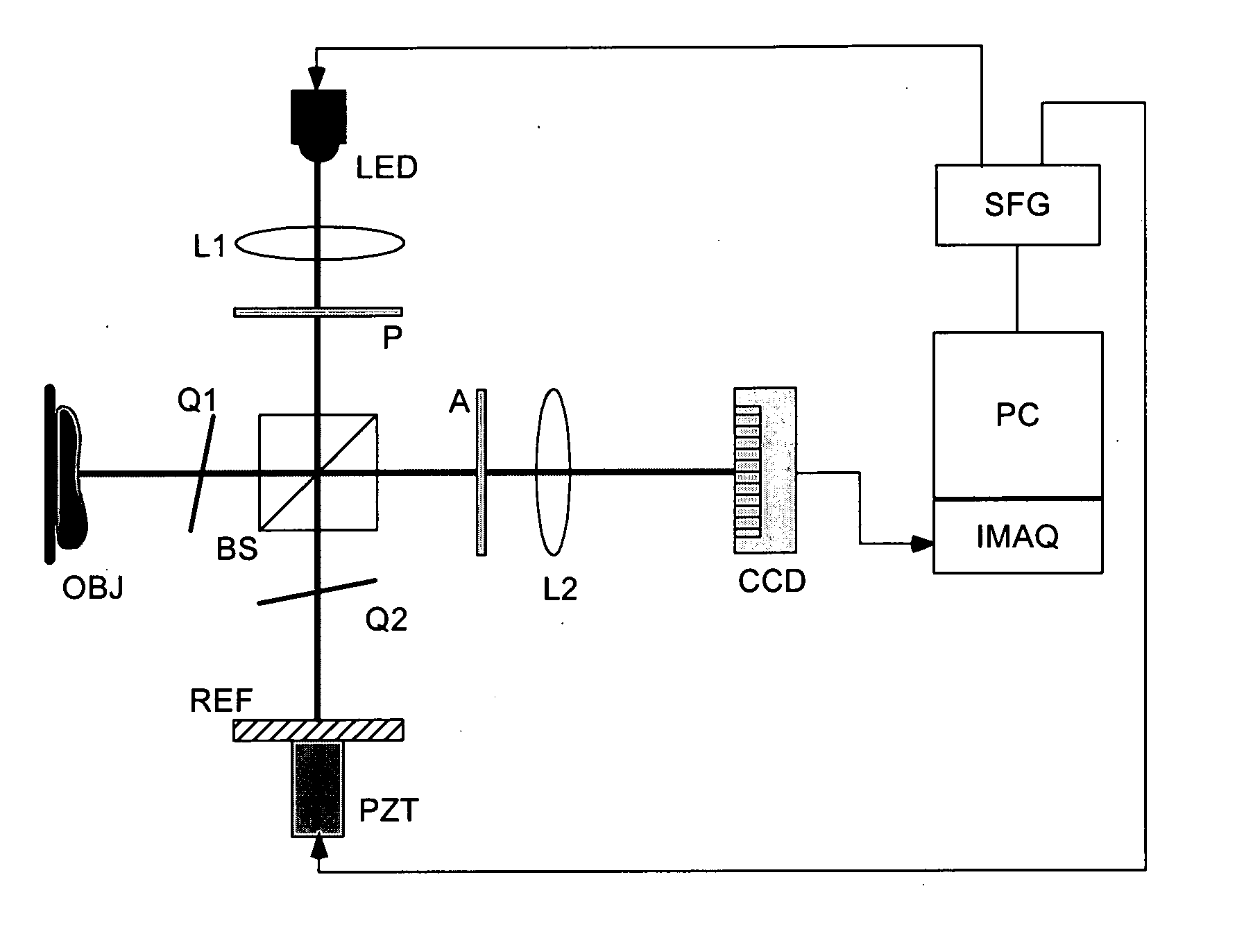
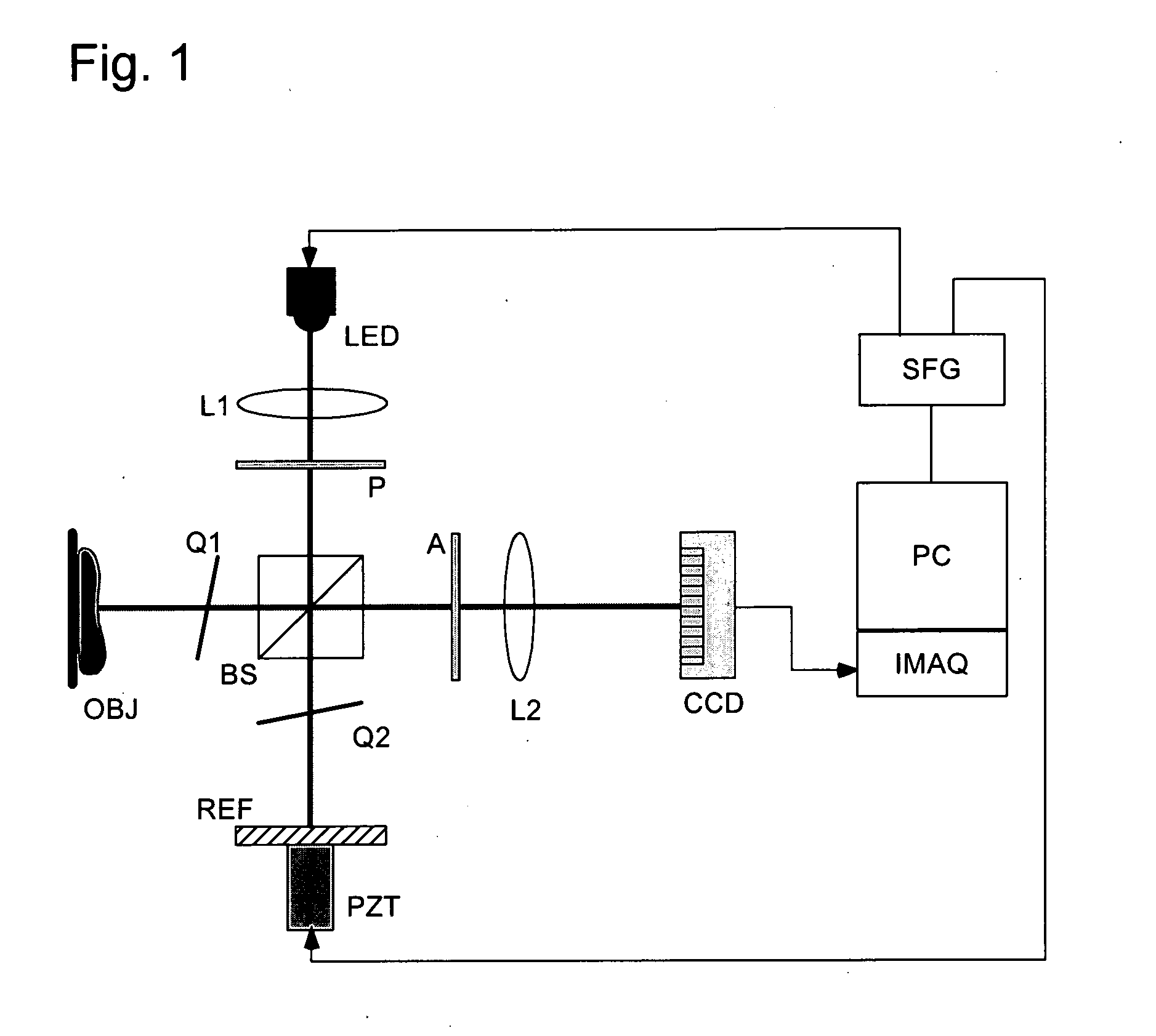
[0031] The principle of color 3D microscopy by wide-field optical coherence tomography (WFOCT) is described referring to the diagram of the apparatus illustrated in FIG. 1. A high-brightness LED (˜30 lumens) illuminates the Michelson interferometer through a collimating lens L1, a polarizer P, and the broadband polarizing beam splitter BS. The quarterwave plates Q1 and Q2 in the object or reference arms change the orthogonal polarization states, so that all of the reflections from the object or the reference mirror are steered toward the monochrome CCD camera through the imaging le...