
Cell separation matrix

a cell and matrix technology, applied in the field of cells-separation matrix, can solve the problems of not having a cell viability reference, complex sample processing, and not solving cells that existed in clusters

Inactive Publication Date: 2005-12-08
CHEN WEN TIEN
View PDF9 Cites 55 Cited by
  • Summary
  • Abstract
  • Description
  • Claims
  • Application Information

AI Technical Summary

Benefits of technology

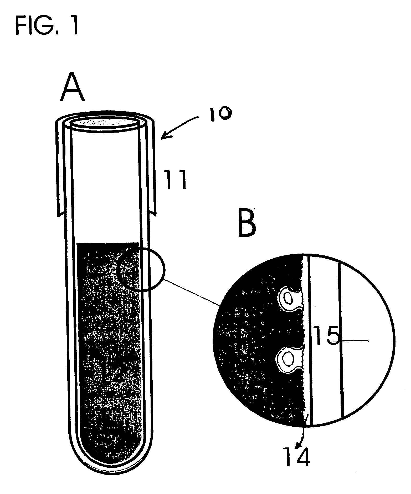
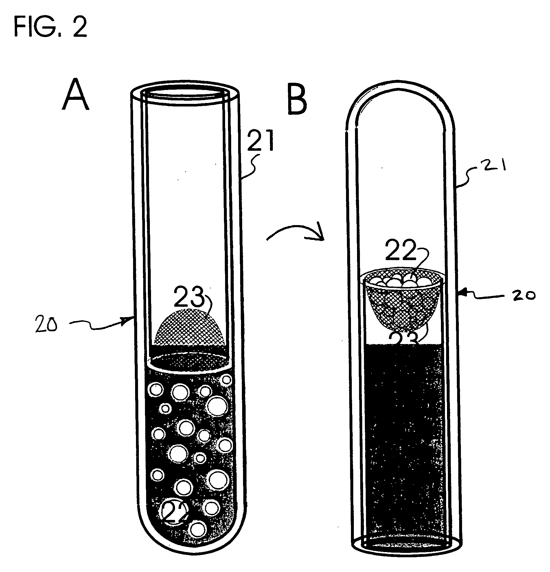
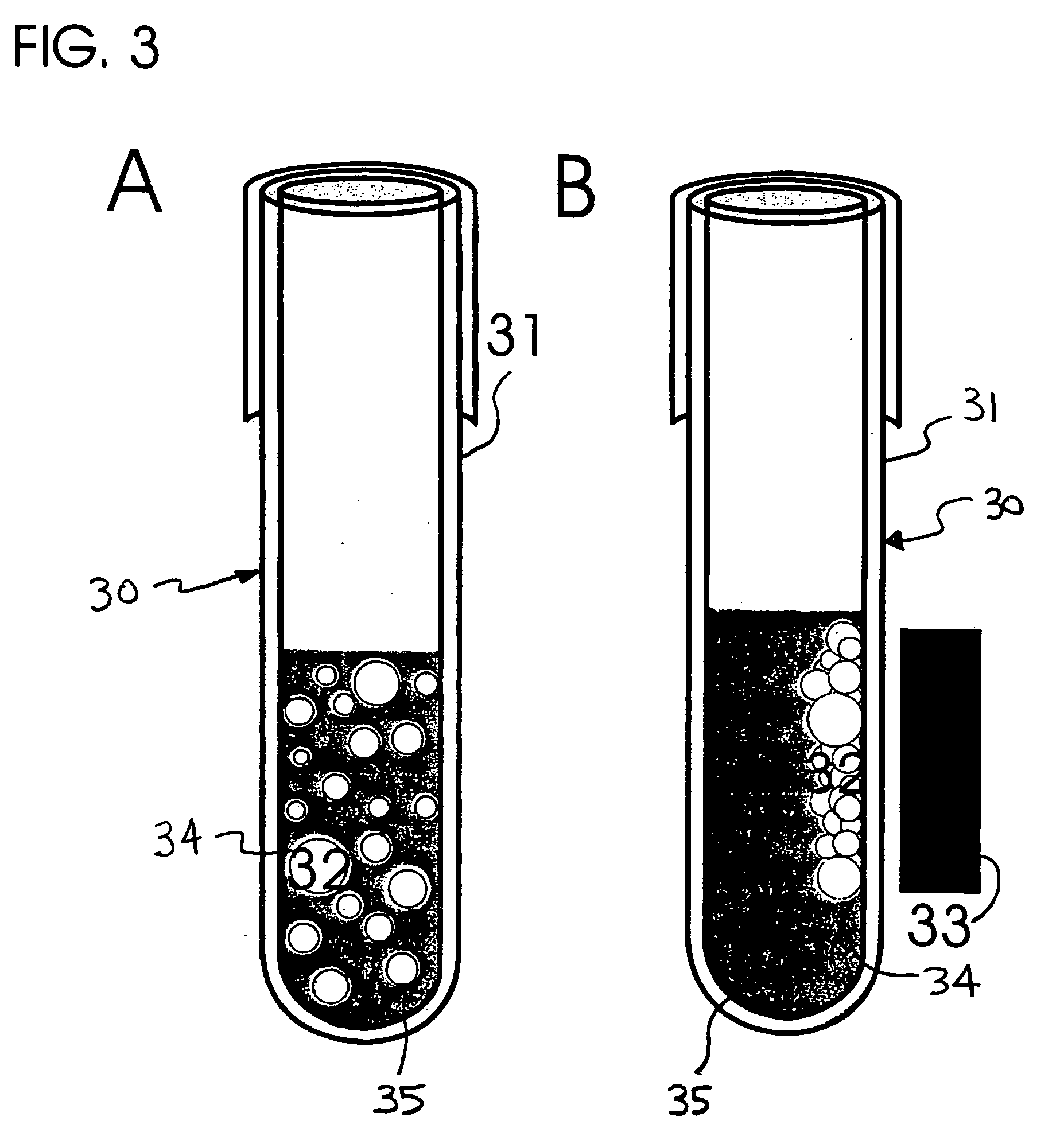
[0016] The present invention provides a cell-separation matrix modified from that described in co-pending PCT Patent Application PCT / US01 / 26735 (claiming priority to U.S. Provisional Patent Application No. 60 / 231,517) which provides an improved matrix for separating cells in a manner to isolate and detect metastatic cells, and a small fraction of hematopoietic cells associated with metastasis, from blood and tissues of patients inflicted with metastasic cancer. The modified-matrix provides a “cancer cell trap” that allows for the efficient removal of viable cancer cells from the tissue fluids. The modified-matrix is useful for separating over 99% of blood cells from such metastatic, and associated-metastatic, cells. Metastatic cells may be characterized by in vitro assays including the local collagen or fibronectin degradation and internalization, cell proliferation, pathological and immunocytochemical identification, and apoptotic and cytolytic assays.

Problems solved by technology

Two major problems have been identified with respect to such cancer cell separation proposal: (1) the proposed method must isolate specifically viable cancer and related tissue cells but leave alone unrelated or damaged cells (Karczewski et al., 1994), and (2) that the proposed method must achieve the specificity in cell separation of one cell from over one million nucleate cells, or over one billion cells in whole blood.
These methods have several disadvantages, particularly with respect to complicated sample processing, no reference for cell viability, and false-positive results.
However, it could not resolve cells that existed in clusters, which may be the case in some cancers.
There are numerous disadvantages associated with antibody-based cell separation methods, including flow cytometry and magnetic cell separation.
There is also frequently significant non-specific antibody binding to damaged cells, with such techniques often including no reference for cell viability.
Overall such antibody-based cell separation methods have a higher than desired false-positive rate.
Furthermore, these cell separation methods are time consuming and cost intensive.

Method used


Image

  • Cell separation matrix
  • Cell separation matrix
  • Cell separation matrix

Examples


example 1

Preparation of Crosslinked Gelatin Films.

[0108] The following method may be followed to prepare crosslinked gelatin films useful in respect of preparing a modified matrix embodiment of the present invention: [0109] (a) gelatin is isolated from connective tissues of human or other animals [0110] Type I collagen is purified from connective tissues of rat tails or human placenta and heat-denatured by boiling for 5 minutes. The gelatin solution is then allowed to dry at 100° C. in an oven under vacuum. Gelatin powders include these produced by acid- or heat-extraction and these from commercial sources including, but not limited to, heat-denatured bovine type I collagen type A derived from porcine skin, Sigma Chemical Co., St. Louis, Mo., USA. [0111] (b) Core materials are coated with gelatin [0112] Gelatin powders are washed with chill distilled water three times by stirring and centrifugation of the gelatin particles. The gelatin solution, containing 2.5% gelatin w / v and 2.5% sucrose...

example 2

Blood Cell Separation Using the Modified Matrix Film.

[0121] (a) Blood or buffy coat are prepared as sources of cells [0122] Five to ten ml of blood are drawn from control subjects or patients with a diagnosis of the presence of primary tumor or metastatic cancer into a blood collection tube (Vacutainer, Becton Dickinson, green top, each tube holds 7-ml) containing lithium heparin as an anticoagulant. Blood or cells collected from an in vivo source are subjected to cell isolation within a relatively short time after their collection because the cells may lose their viability. In order to maintain the optimal isolation of cancer cells, it is preferred that blood or tissue samples are stored at 4° C. and used within 24 hours after their collection, most preferably, within four hours. [0123] Buffy coat is processed from blood by conventional density gradient centrifugation using Ficoll-Paque (Pharmacia) that removes the majority of red cells leaving a thin layer of nucleate cells, cal...

example 3

Identification of Viable Cancer Cells

[0130] (a) Colony Formation

[0131] A portion of enriched nucleate cells, i.e., equivalent to 0.1-ml blood volume per well, are seeded onto a 16-well microtiter plate-glass slide (in 96-well microtiter plate format; Lab-Tek, Rochester, N.Y.) comprising tissue culture medium containing 10% heat-inactivated human plasma (complement-inactivated human sera) or plasma. Cells are allowed to propagate for four days to two weeks thereby allowing the cancer cells to form colonies. It was estimated that, among approximately 100 putative metastatic cells isolated from the blood of patients with metastatic diseases, there was only one colony of carcinoma cells formed after one week of culture. The efficiency of colony growth in culture appears to be 10,000 folds higher than what was observed in vivo, suggesting that, free of host immunity, cultured cancer cells increase their capability to grow.

[0132] (b) Apoptosis and Cytolysis

[0133] Cells are cultured f...


PUM

PropertyMeasurementUnit
diameteraaaaaaaaaa
mesh-opening widthsaaaaaaaaaa
widthsaaaaaaaaaa

Abstract

A novel modified matrix system, mimicking a metastatic environment, that can be used to capture and detect viable cancer and normal cells from tissue fluid samples derived from cancer subjects and which provides effective cell separation for diagnostic and therapeutic applications in treating patients with metastatic diseases.

Description

RELATED APPLICATIONS [0001] This application claims priority from U.S. Provisional Patent Application No. 60 / 332,408, filed Nov. 16, 2001, and is a continuation-in-part application of parent application PCT / US01 / 26735, filed Aug. 28, 2001, and U.S. Provisional Patent Application No. 60 / 231,517, filed Sep. 9, 2000.BACKGROUND OF THE INVENTION [0002] 1. Field of the Invention [0003] The present invention generally relates to a matrix for separating cells. More particularly, the present invention relates to a cell-separation matrix that may be used to selectively isolate cells with metastatic potential. The cell-separation matrix may be used in the diagnosis of metastatic cancers and in the treatment of cancer by reducing circulating metastatic cells. [0004] 2. Description of the Related Art [0005] Primary cancers frequently shed neoplastic cells into the circulation at an early stage of metastases formation (Fidler I J, 1973, European Journal of Cancer 9:223-227; Liotta L A et al., 197...

Claims


Application Information

Patent Timeline
Patent Type & Authority Applications(United States)
IPC IPC(8): G01N33/569G01N33/574
CPCG01N33/56966G01N33/57488G01N33/574
Inventor CHEN, WEN-TIEN
Owner CHEN WEN TIEN
Learn More