Hydrogel implants for replacing hyaline cartilage, with charged surfaces and improved anchoring

a technology of hydrogel implants and hyaline cartilage, applied in the field of surgical implants, can solve the problems of limiting the utility and effectiveness of hydrogel implants to replace injured or diseased cartilage in human surgery, affecting the quality of cartilage replacement, so as to increase the strength and durability of composite hydrogel implants, improve the interaction

Inactive Publication Date: 2005-12-29
MANSMANN KEVIN A
View PDF0 Cites 192 Cited by
  • Summary
  • Abstract
  • Description
  • Claims
  • Application Information

AI Technical Summary

Benefits of technology

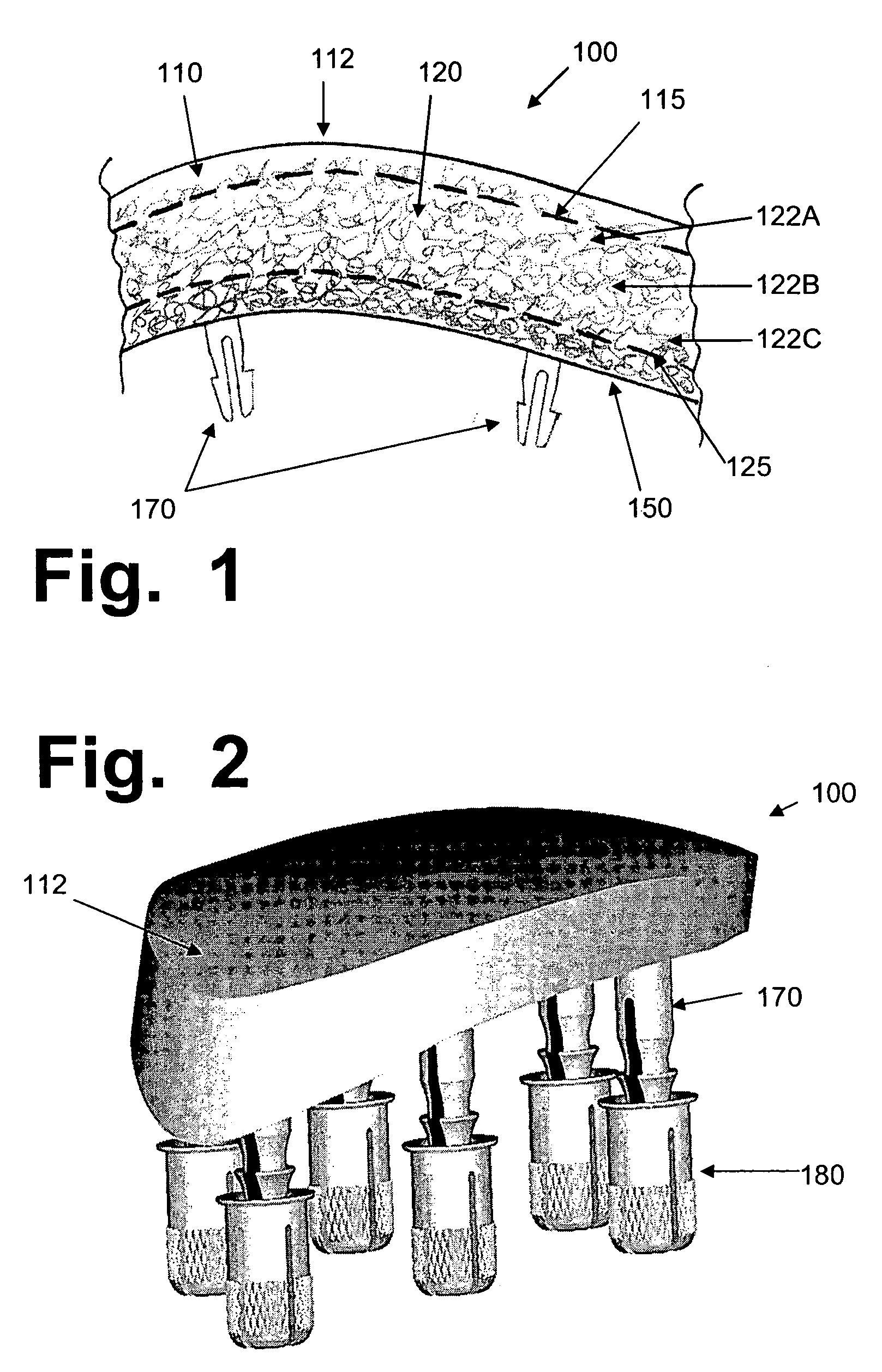
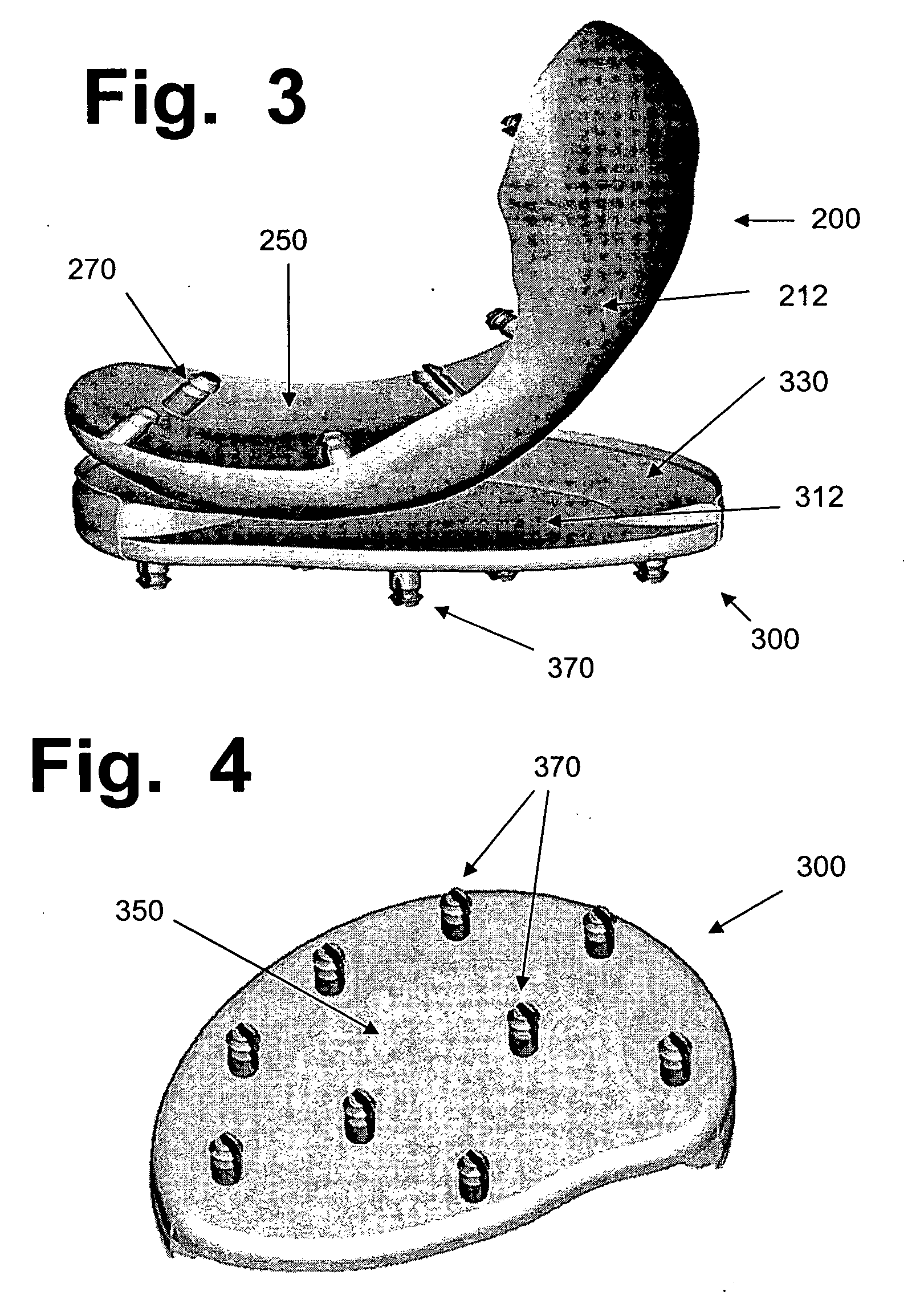
[0047] A hydrogel device for surgical implantation to replace damaged hyaline or meniscal cartilage in a mammalian joint is disclosed, with a combination of enhancements and improvements over previous proposed implants. One improvement comprises a hydrogel surface that has been chemically treated, by sulfonation or other means, to give it a negative electrical charge that emulates natural cartilage and improves its interactions with certain components of synovial fluid. Another improvement comprises a porous anchoring surface provided with anchoring pegs that will lock in place when pressed into receptacles that have been set and anchored in hard bone, prior to insertion of the implant. A thi

Problems solved by technology

While these materials have numerous laboratory uses, the use of hydrogel implants to replace injured or diseased cartilage, in surgery on humans, has been very limited, for a number of reasons.
That approach suffers from shortcomings that limit its utility and effectiveness, notably including problems involving minor edges and noncomformities that lead to potentially abrasive surfaces around the periphery of any such inserted plug surrounded by cartilage.
Also, those implants are believed to be made of polyvinyl alcohol (PVA), which is not as strong or durable as other known hydrogel materials.
However, collagen hydrogels suffer from problems and limitations, if used in implants for replacing cartilage.
Those problems include: (1) the risk that a foreign protein will provoke a tissue rejection, especially if the protein is from a non-human source such as cowhide (the source of most collagen available for testing and use); (2) collagen fibers are typically digested, resorbed, and replaced within a span of months, as part of natural tissue regeneration processes; (3) toxic chemicals are usually needed to crosslink collagen fibers in ways that will create matrices; and, (4) collagen has less strength and durability than various types of known synthetic polymers.
Although certain synthetic hydrogels (such as polyhydroxy-ethyl-methacrylate) are used for contact lenses and slow-release drug carriers, they are not strong or durable enough to replace hyaline cartilage.
However, none of those are being used to replace hyaline cartilage in load-bearing joints, such as knees or hips.
These operations inflict severe damage on the muscles, tendons, ligaments, bones, and blood vessels, in and around a knee or hip.
They cause severe pain, and since they inflict so much damage on tissues and vasculature, many elderly people never fully recover from these surgeries.
The problems that render hydrogels too weak and fragile for use in knee or hip replacements have arisen because, in a typical hydrogel, the fibers that hold the gel together take up only a small portion of the volume (usually less than about 10%, and many hydrogels contain less than 5% fiber volume).
Since water molecules cannot contribute any significant strength to a hydrogel, large loads must be imposed on the fibers that form the matrix and hold the water molecules together.
The problems of low strength and durability are also aggravated by the lack of a crystalline structure in a hydrogel.
If a hydrogel material had a crystalline structure, with repeating units in regular rows and columns that could reinforce the matrix, it would be stronger, but i

Method used


Image

  • Hydrogel implants for replacing hyaline cartilage, with charged surfaces and improved anchoring
  • Hydrogel implants for replacing hyaline cartilage, with charged surfaces and improved anchoring
  • Hydrogel implants for replacing hyaline cartilage, with charged surfaces and improved anchoring

Examples


example 1

PVA and PVA / PVP Samples

[0125] Granular PVA (grade 71-30, with an average molecular weight of about 140 kilodalton) was supplied at no cost by DuPont. PVP (average molecular weight about 40 kd) was obtained from Sigma Chemical. When PVA / PVP copolymers were tested, they contained a ratio of 99% PVA and 1% PVP, by weight. In either case, a total polymer weight of 10% w / v in distilled and deionized water was used. The mixture was stirred for 20 minutes, by which time the solution appeared to be completely uniform and consistent. It was heated to 85° C. overnight, then cooled to room temperature, and stirred again for 20 minutes.

[0126] An aliquot of this solution was poured into a shallow flat mold, which was then kept in a warm ventilated incubator at 37° C. until essentially all water had been removed, leaving a polymeric sheet with a thickness of about 1.75 to 2 mm. This usually took about 4 to 5 days.

[0127] A punch was used to remove circular samples, usually with 0.67, 1.5, or 1....

example 2

Polyacrylonitrile Samples

[0133] Sample sheets of polyacrylonitrile, 2.55 to 2.6 mm thick, were provided by the PragTech company (Flemington, N.J.). These sheets were of a type designated as “Qpan” by Pragtech. The exact details of the process use to manufacture the “Qpan” class of PAN are proprietary, and may be covered by one or more currently pending patent applications (including U.S. application Ser. Nos. 09 / 383,020 and 10 / 193,578, both by Stoy et al and accessible on the U.S. Patent Office website). Methods for manufacturing polyacrylonitrile are disclosed in various patents that can be located by searching for “Stoy” as the inventor, in the U.S. patent database (www.uspto.gov). Such US patents range from U.S. Pat. No. 4,107,121 (“Ionogenic hydrophilic water-insoluble gels from partially hydrolyzed acrylonitrile polymers . . . ”) to U.S. Pat. No. 6,593,451 (“Method of processing polyacrylonitrile”), and include 14 additional patents in between those two. U.S. Pat. Nos. 3,895,1...

example 3

Standardizing Tests on Tribometer

[0135] Before the tribometer (made by AMTI, www.amtiweb.com, and connected to a desktop computer using AMTI software) could be used for testing hydrogels, it had to be standardized, which is comparable to calibrating it. This is done using the procedures set forth in ASTM protocol F732 (“Standard Test Method for Wear Testing of Polymer Materials Used in Total Joint Prostheses”).

[0136] Briefly, the tribometer machine is used to rub pins having smooth, flat-faced surfaces made of a known type of plastic, called “ultrahigh-molecular-weight polyethylene” (UHMWPE), against smooth disks made of a very hard cobalt-chromium alloy (supplied by Biomet Inc., www.biomet.com). Prior to the tests, the pins (having 0.5 inch diameters for the standardizing tests) were pre-soaked in distilled water for a month, to minimize fluid absorption during the test. A load of 253 newtons was applied to the pins, to generate an average contact stress of 3.54 megapascals (Mpa)...


PUM

PropertyMeasurementUnit
Densityaaaaaaaaaa
Flexibilityaaaaaaaaaa
Stiffnessaaaaaaaaaa

Abstract

Hydrogel devices for surgical implantation to replace damaged cartilage in a mammalian joint (such as a knee, hip, shoulder, etc.) are disclosed, with one or more of the following enhancements: (1) articulating surfaces that have been given negative surface charge densities that emulate natural cartilage and that interact with positively charged components of synovial fluid; (2) anchoring systems with affixed pegs that will lock into accommodating receptacles, which will be anchored into hard bone before the implant is inserted into a joint; (3) a three-dimensional reinforcing mesh made of strong but flexible fibers, embedded within at least a portion of the hydrogel.

Description

PRIORITY CLAIM [0001] This application claims the benefit under 35 USC 119(e) of provisional application 60 / 562,176, filed Apr. 14, 2004.BACKGROUND OF THE INVENTION [0002] This invention relates to surgical implants for replacing or repairing hyaline cartilage, in joints such as knees, hips, shoulders, etc. As used herein, all references to implants, surgery, etc., refer to surgical (which includes arthroscopic) implantation of a device into a mammalian joint. [0003] As known in the art, hydrogels are materials that are somewhat flexible and pliable, and do not have rigid or crystalline structures. In hydrated form, they contain water molecules, which can permeate through a matrix (i.e., three-dimensional network) of flexible crosslinked fibers. In animals, nearly all types of soft tissues are hydrogels, with matrices made of collagen (a bundled protein that provides tensile strength) and proteoglycan filaments (extremely thin protein strands surrounded by hyaluronate, a natural pol...

Claims


Application Information

Patent Timeline
IPC IPC(8): A61F2/00A61F2/02A61F2/08A61F2/30A61F2/32A61F2/38A61F2/40A61F2/42A61F2/46
CPCA61F2/30749A61L2430/24A61F2/30965A61F2/32A61F2/40A61F2/4241A61F2/4261A61F2/468A61F2002/30006A61F2002/30062A61F2002/30382A61F2002/30489A61F2002/30594A61F2002/30751A61F2002/30892A61F2002/30894A61F2002/3895A61F2002/4631A61F2210/0004A61F2220/0008A61F2220/0025A61F2220/0033A61F2250/0015A61L27/14A61L27/50A61L27/52A61L2400/10A61L2400/18A61F2/30756A61L2430/06A61F2002/30487A61F2002/30331
Inventor MANSMANN, KEVIN A.
Owner MANSMANN KEVIN A
Why Eureka
Try Eureka