
Light induced immobilisation

a technology of immobilisation and light, applied in the direction of immobilised enzymes, enzymes, non-active ingredients of pharmaceuticals, etc., can solve the problems of increased detection limits, increased chemical reactions, and significant risk of reducing biological activity

Inactive Publication Date: 2006-07-13
BIONANOPHOTONICS
View PDF4 Cites 2 Cited by
  • Summary
  • Abstract
  • Description
  • Claims
  • Application Information

AI Technical Summary

Benefits of technology

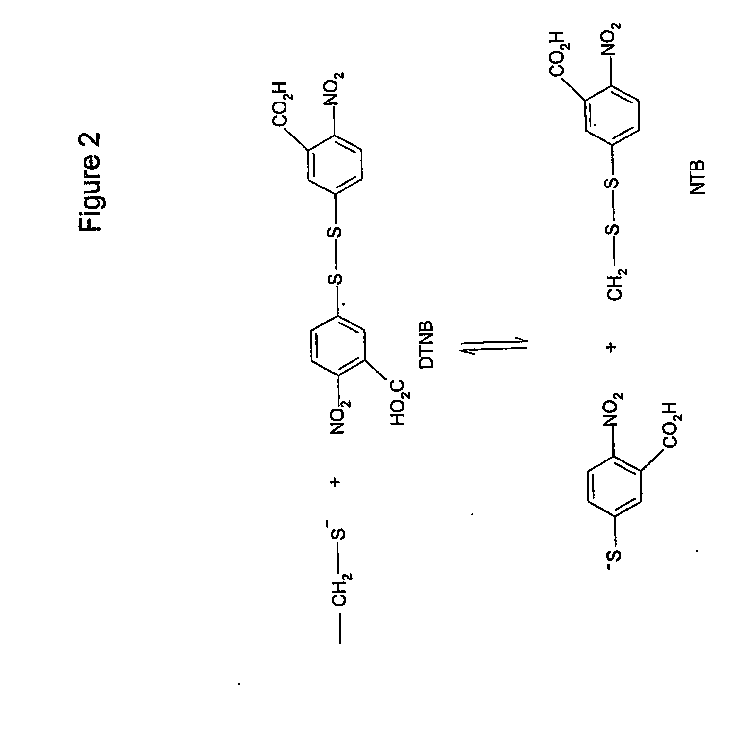
[0058] This method of light induced thiol coupling can also be used to immobilise a protein on a support. The most common types of bonds that are formed during coupling to a support are disulfide bonds and sulfur-metal bonds (primarily sulfur-gold) where a self-assembled layer is formed. As both types of bonds are stable, extensive washing after immobilization will not displace the protein. The density of proteins on a support can be controlled by varying the protein concentration, or the intensity and duration of UV-irradiation, and subsequently blocking the remaining activated thiol groups on the surface with reagents such as L-cysteine, (2-(2-pyridinyidithio)ethaneamine hydrochloride (PDEA) or with a thiol-lipid bilayer (Hong Q., et. al., 2001, Biochemical Society Transactions 29(4):587-582). The support, with evenly distributed immobilized proteins, is therefore blocked to prevent non-specific binding. According to the present invention the method of immobilization does not involve any chemical steps, since the thiol-activated proteins formed by UV radiation, spontaneously self-assemble on the support. The described thiol and disulfide exchange reactions are an effective and rapid way to bind molecules to supports. A particular advantage of the present invention is its avoidance of the several disadvantages associated with the chemical generation of free thiols in a protein. Some proteins, e.g., cutinase, are inactivated by the reducing agents (DTT or beta-mercaptoethanol) used to generate free thiols, as shown in Example 10. If a reducing agent comprising a thiol group, is used to chemically generate free thiols in a protein, it must be removed before immobilisation by can be performed, during which step the disulfide bonds can reform. Alternative reducing agents, such as 2-carboxyethyl)phosphine, lacking a thiol group, have the disadvantage that they are reactive to other groups. The use of reducing agents to disrupt disulfide bonds has the additional disadvantage that they are pH dependent, both with regards their chemical stability and their reducing activity.
is its avoidance of the several disadvantages associated with the chemical generation of free thiols in a protein. Some proteins, e.g., cutinase, are inactivated by the reducing agents (DTT or beta-mercaptoethanol) used to generate free thiols, as shown in

Problems solved by technology

This contrasts with traditional coupling methods for protein immobilisation, which typically involve several, chemical reactions, which can be costly, time-consuming as well as deleterious to the structure / function of the bound protein.
In comparison, the majority of known protein coupling methods lead to a random orientation of the proteins immobilised on a carrier, with the significant risk of lower biological activity and raised detection limits.

Method used


Image

  • Light induced immobilisation
  • Light induced immobilisation
  • Light induced immobilisation

Examples


example 1

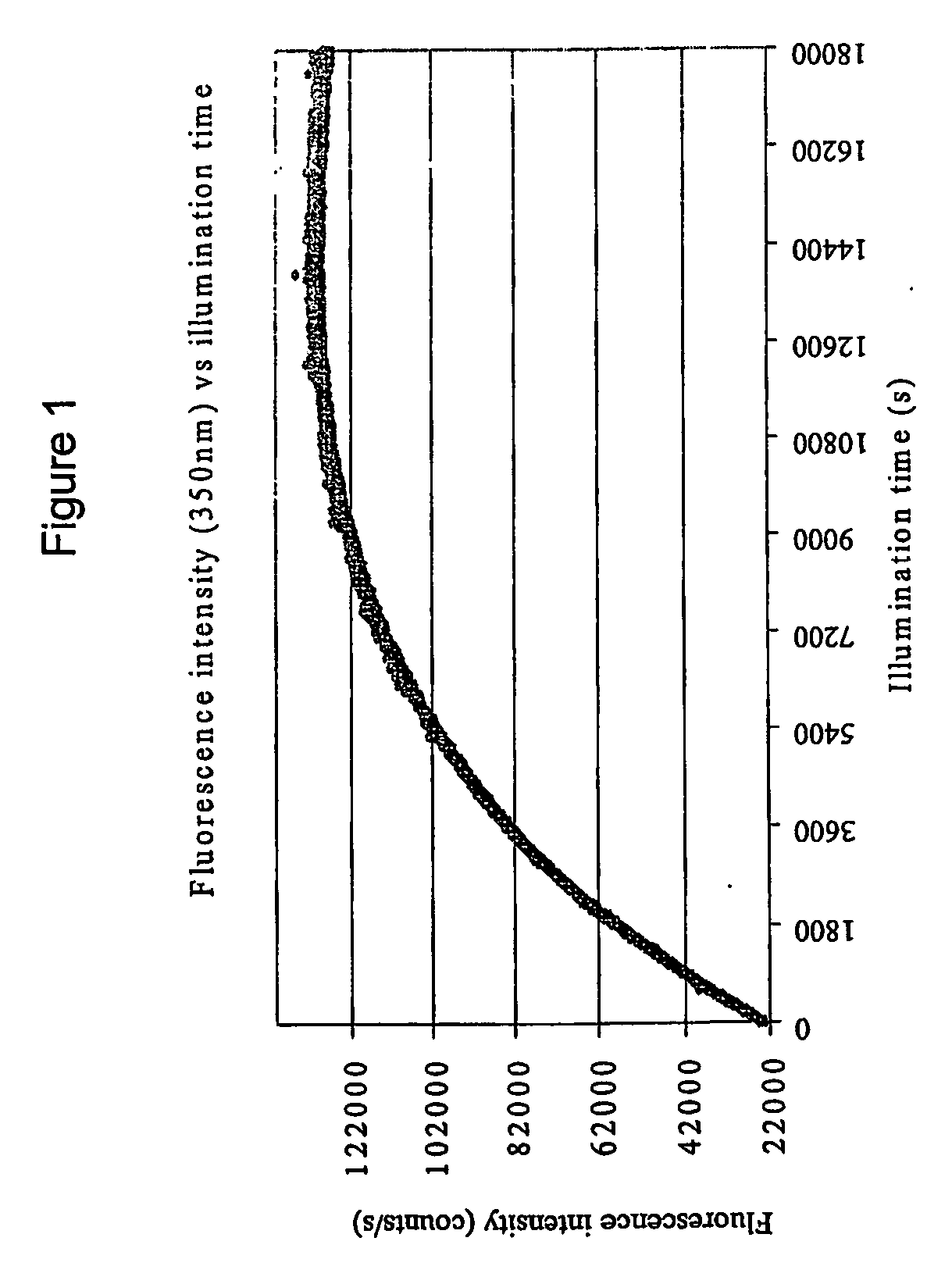
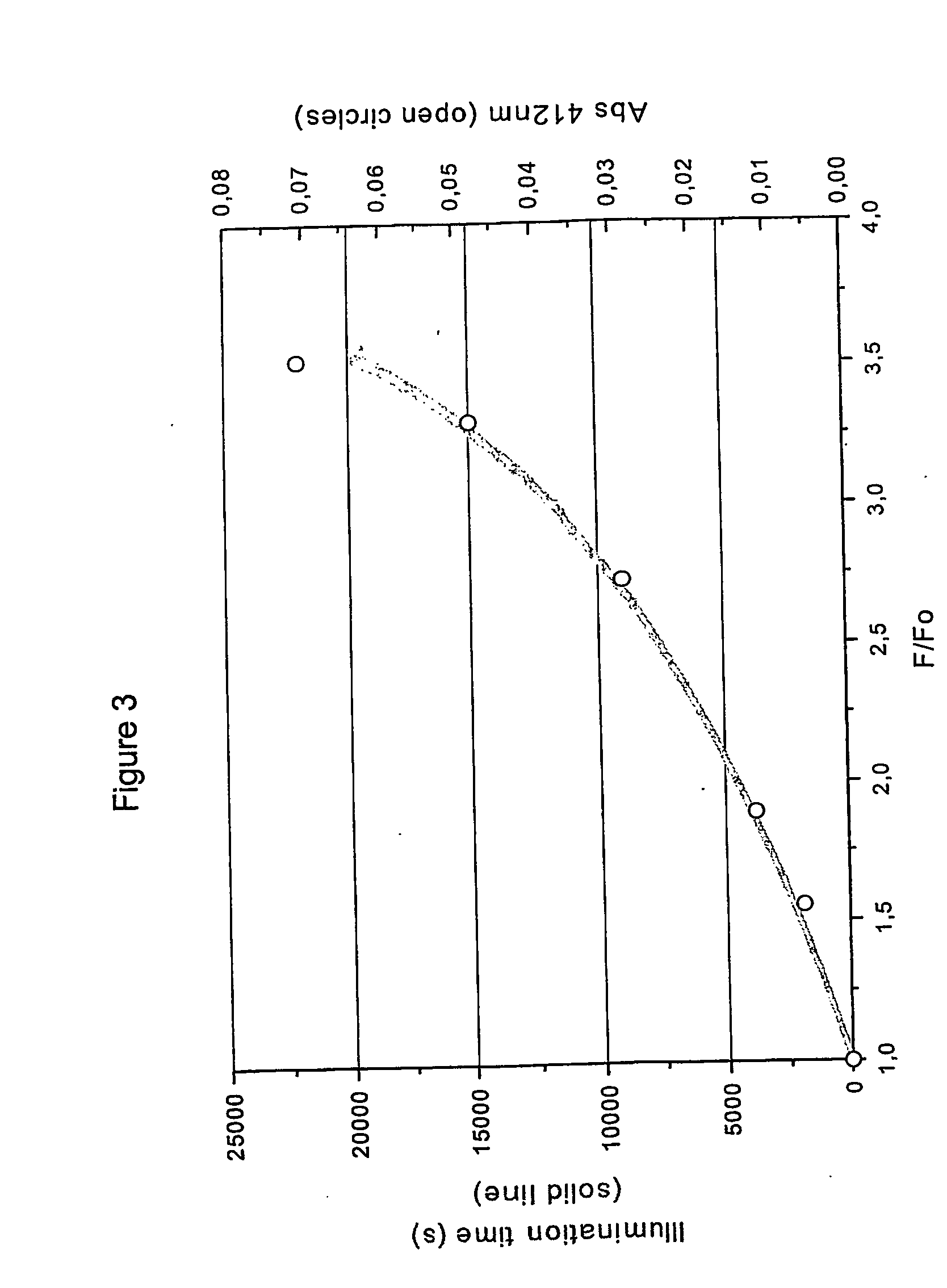
Formation of Free Thiol Groups in Cutinase Upon UV Illumination

[0063] The disruption of disulfide bridges in a protein following UV illumination was examined using a cutinase, with lipase / esterase properties isolated from Fusarium solani pisi.

Steady-Sate Fluorescence Emission Intensity of Cutinase

[0064] Cutinase preparations were subjected to UV irradiation at 295 nm under the following conditions in order to follow its steady-state fluorescence emission intensity of continuous illumination: Three ml of 2 μM stock solution of cutinase was continuously illuminated at 295 nm for increasing periods of time (Oh, 1h, 2h, 3h, 4h, and 5h) in a quartz macro-cuvette (1 cm path length). Light excitation at 295 nm was supplied by a Xenon Arc Lamp coupled to a monochromator provided by a RTC 2000 PTI spectrometer. The cuvette was mounted in a thermostated cuvette, kept at a constant temperature of 25° C. The cutinase sample was maintained as a homogeneous solution by continuous stirring at ...

example 2

Irradiation-Induced Disruption of a Disulfide Bridge in Native Cutinase is Dependent on the Presence of a Trp.Residue as Spatial Neighbour

[0069] In order to demonstrate the role of a tryptophan residue in cutinase in the irradiation induced disruption of a disulfide bridge, the Trp fluorescence emission intensity of the native protein was compared to that of reduced cutinase in which all disulfide bridges were chemically disrupted. The cutinase protein was partially denatured by heat in order to facilitate reduction of all its disulfide bridges. The Trp fluorescence emission intensity of the native and denatured cutinase, following irradiation, was used to demonstrate the importance of a Trp residue being a spatial neighbour of a disulfide bridge, for the transfer of excitation energy from Trp to the disulfide bridge.

Reduction of Native Cutinase with DTT

[0070] In native cutinase, the disulfide bridges within the folded protein are inaccessible to solvent, and they cannot be dire...

example 3

Specificity of Light-Induced Disulphide Bridge Disruption in Proteins

[0075] The cutinase gene of Fusarium solani pisi has been mutated to encode a mutant cutinase polypeptide where the single tryptophan residue is replaced by alanine, a non-fluorescent amino acid (W69A). The absence of light-inducible disulfide bridge disruption in the mutant in comparison to native cutinase, demonstrated below, further confirms the requirement for an aromatic amino acid (e.g. trp) in close spatial proximity to the disulfide bridge. The mutant cutinase (W69A) was expressed recombinantly. Three ml of a 2 μM solution of the mutant or native protein, in 50 mM Tris-HCl pH 7.0, was transferred to a cuvette, thermostated at 25° C., with magnetic stirring at 700 rpm, and illuminated at 296 nm using a 75 W xenon lamp from an RTC 2000 PTI spectrophotomoter coupled to a monochromator, with an excitation and emission slit of 10 and 2 nm, respectively. As can be seen from the absorption (A) and emission spectr...


PUM

PropertyMeasurementUnit
wavelengthaaaaaaaaaa

Abstract

The present invention involves a method of coupling disulfide bridge containing proteins to a carrier by inducing the formation of thiol groups on a protein with irradiation, and coupling the protein to the carrier. The formation of thiol group(s) in the protein is a consequence of the disruption of disulfide bridges following electronic excitation of aromatic amino acid residues located in close spacial proximity to the disulfide bridge. The light-induced coupling method facilitates the orientated immobilization of a protein on a carrier.

Description

FIELD OF THE INVENTION [0001] The present invention relates to a method of cross-linking or immobilising proteins on a carrier. BACKGROUND OF THE INVENTION [0002] Molecules can be immobilised on a carrier or solid surface either passively through hydrophobic or ionic interactions, or covalently by attachment to activated surface groups. In response to the enormous importance of immobilisation for solid phase chemistry and biological screening, the analytical uses of the technology have been widely explored. The technology has found broad application in many different areas of biotechnology, e.g. diagnostics, biosensors, affinity chromatography and immobilisation of molecules in ELISA assays. The value of immobilisation technology is demonstrated by the recent development of DNA microarrays, where multiple oligonucleotide or cDNA samples are immobilised on a solid surface in a spacially addressable manner. These arrays have revolutionised genetic studies by facilitating the global an...

Claims


Application Information

Patent Timeline
Patent Type & Authority Applications(United States)
IPC IPC(8): A61K9/14C12N11/16C07K16/46C07K17/00G01N33/68
CPCA61K47/48246C07K17/00C12N9/18C12N11/14C12Y301/01074G01N33/6803G01N33/6842A61K47/64
Inventor PETERSEN, STEFFEN BJORNDA CRUZ AUGUSTO NEVES PETERSEN, MARIA TERESA
Owner BIONANOPHOTONICS
Learn More