
Method and device for sanitation using bacteriophages

a technology of bacteriophage and sanitizer, which is applied in the field of bacteriophage, can solve the problems of untreatable strains of vre, and untreatable strains, and achieve the effects of preventing colonization and invasion of laying hens, reducing salmonella contamination levels, and protecting against salmonella colonization

Inactive Publication Date: 2007-10-25
INTRALYTIX
View PDF23 Cites 11 Cited by
  • Summary
  • Abstract
  • Description
  • Claims
  • Application Information

AI Technical Summary

Benefits of technology

The present invention provides a method for sanitation using bacteriophages to clean surfaces and reduce poultry contamination with Salmonella. The bacteriophages are applied to the surface using a dispersing mechanism, such as a pressurized container or a fogging device. The bacteriophages can be poured, brushed, wiped, or painted onto the surface. The areas that can be sanitized include livestock pens, live stock feeding areas, livestock slaughter areas, livestock waste areas, knives, shovels, rakes, saws, and other medical equipment. The bacteriophages can be used in poultry processing to decontaminate surfaces and prevent Salmonella infections. The invention also provides a sanitation device containing bacteriophages for poultry processing. The use of bacteriophages for foodstuff packaging is also disclosed.

Problems solved by technology

A particular problem in this regard has been vancomycin-resistant enterococci (VRE), which are not treatable with standard classes of antibiotics.
Use of antibiotics has been clearly shown to increase the density, or level of colonization, in an individual patient (Donskey CJ et al, “Effects of antibiotic therapy on the density of vancomycin-resistant enterococci in the stool of colonized patients.” N Engl J Med 2000;343: 1925-32): this, in turn, would appear to increase the risk of subsequent infection, and the risk of transmission of the organism to other patients.
Given the virulence of S. aureus, the emergence of such untreatable strains would be devastating and have a major impact on the way in which medicine is practiced in this country.
Persons who are nasally colonized with MRSA have an increased risk of developing serious systemic infections with this microorganism, and, in particular, colonization or prior infection with MDRSA significantly increases the risk of subsequent bacteremia with MDRSA (Roghmann MC, “Predicting methicillin resistance and the effect of inadequate empiric therapy on survival in patients with Staphylococcus aureus bacteremia.
Many of these strains are resistant to all major antibiotic classes, presenting substantive difficulties in management of infected patients.
In this setting, colonization with multi-drug resistant Pseudomonas represents a potentially serious risk factor for development of multi-drug resistant Pseudomonas pneumonia.
However the results from early studies to evaluate bacteriophage as antimicrobial agents were variable due to the uncontrolled study design and the inability to standardize reagents.
This initial failure of phage as antibacterial agents may have been due to the failure to select for phage that demonstrated high in vitro lytic activity prior to in vivo use.
With the advent of antibiotics, the therapeutic use of phage gradually fell out of favor in the U.S. and Western Europe and little subsequent research was conducted.
However, this literature does not describe, in any way anticipate, or otherwise suggest the use of bacteriophage to modify the composition of colonizing bacterial flora in humans, thereby reducing the risk of subsequent development of active infections.
Antibiotic therapy of diarrheal illness is not effective, and may actually prolong intestinal carriage.
Bacteremia is, obviously, treated with antibiotics, although the emergence of highly resistant strains such as DT104 has begun to create problems in patient management.
There is currently no effective means of limiting or eradicating carriage of the organism in the intestinal tract.
Contamination may result from rupture of the intestinal tract during slaughter.
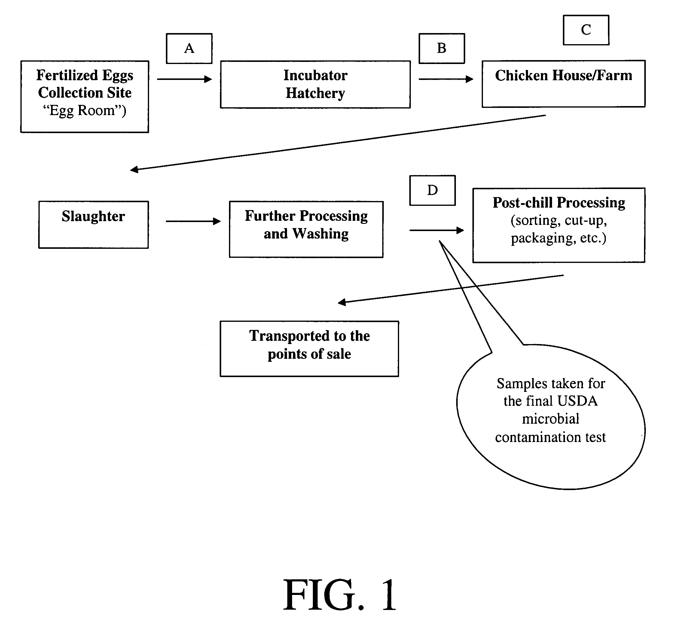
However, with current slaughter techniques, removal of the viscera seldom results in intestinal rupture and carcass contamination—and, when it does occur, the carcass is immediately tagged for “reprocessing.” The more common source of Salmonella is the skin of the animal itself, with the feather follicles serving as a sanctuary for bacteria.
The close quarters in chicken houses, and the piling of chicken crates on trucks on the way to slaughterhouses, results in frequent contamination of feathers by feces.
If members of a flock have high levels of intestinal colonization with Salmonella, there are multiple opportunities for contamination of feathers and feather follicles with the microorganism, and, in turn, for Salmonella contamination of the final product.
Plants must meet specific standards for percentage of product contaminated, based on national averages; failure to meet these standards results in plant closure.
Concerns about Salmonella contamination have also become a major issue in international trade, with Russia and other countries having embargoed millions of dollars worth lots of chickens because of identification of Salmonella in the product.
Irradiation of raw product (i.e., chicken carcasses) is efficacious, but expensive, and is limited by the small number of irradiation facilities and by consumer acceptance.
Antibiotics (in contrast to phage) generally have activity against multiple bacterial species; their administration can result in serious perturbations in the microbial ecology of the animal's intestinal tract, with accompanying loss of “colonization resistance” and overgrowth of microorganisms that are resistant to the antimicrobial agent used.
There is now a commercially available competitive exclusion product, PreEmpt (produced by MS Bioscience), that consists of 27 different bacteria strains- In preliminary testing, it appears to be effective in limiting Salmonella colonization, but its usage is hampered by the cost.

Method used


Image

  • Method and device for sanitation using bacteriophages

Examples


example 1

Obtaining VRE Isolates

[0158] Isolation of VRE

[0159] VRE were isolated by standard methods from patients in the surgical intensive care and intermediate care units of the University of Maryland Medical Center in Baltimore. Trypticase Soy Agar supplemented with 5% sheep blood (BBL, Cockeysville MD) was used to isolate enterococci from urine, wounds and sterile body fluids. VRE were isolated from stool specimens on Colistin Nalidixic Acid (CNA) agar (Difco labs, Detroit, Mich.) supplemented with defibrinated sheep blood (5%), vancomycin (10 μg / ml ) and amphotericin (1 μg / ml). See Facklam, R. R., and D. F. Sahm. 1995. Enterococcus. In: Manual of Clinical Microbiology, 6th edition, American Society for Microbiology, Washington, D.C., pp. 308-312.

[0160] Identification of VRE

[0161]Enterococci were identified by esculin hydrolysis and growth in 6.5% NaCl at 45° C. Identification to the species level was done using conventional testing as indicated in Facklam and Collins (Facklam, et al....

example 2

Isolation of VRE Phage

[0166] 500 ml of raw sewage from the University of Maryland is mixed with 100 ml of 10 times concentrated LB broth (Difco Laboratories). This sewage-broth mixture is inoculated with a 18-24 hour LB broth culture (1 ml) of a VRE strain and incubated at 37° C. for 24 hours to enrich the mixture for bacteriophage which can infect the VRE strain added. After incubation, the mixture is centrifuged at 5000 g for 15 minutes to eliminate matter which may interfere with subsequent filtration. The supernatant is filtered through a 0.45 μm Millipore filter. Filtrate is assayed using the Streak Plate Method and / or Appelman Tube Turbidity Test to detect lytic activity against different strains of VRE.

[0167] Method for Testing Phage Against VRE Isolates

[0168] Three methods are employed: Plaque Assay; Streak Plate Method; and Tube Turbidity Method, and the procedures for each follow.

[0169] Plaque Assay:

[0170] A 18-24 hour nutrient broth culture of the VILE strain (0.1 ml...

example 3

A Phage Strain is Active Against Over 200 VRE Isolates

[0178] A collection of 234 VRE isolates; 187 E. faecium of which 3 strains are from ATCC, 41 E. faecalis strains, and 6 E. gallinarium strains as well as 6 E. faecium strains which are vancomycin sensitive were tested for susceptibility of infection by 7 monophages isolated as described in Example 2. Susceptibility of infection was determined by the 3 techniques described. The majority of VRE strains in this collection were isolated from patients at the University of Maryland and Baltimore VA Medical Centers as indicated in Example 1. Such VRE isolates were determined to be distinct and genetically diverse by pulsed field gel electrophoresis typing. Of the 7 monophages, VRE / E2 and VRE / E3 have a relatively narrow host range compared to other VRE phages, but are able to infect the small proportion of VRE strains which were resistant to other phages collected. A phage cocktail containing the above 7 VRE monophages lysed 95% of the ...


PUM

PropertyMeasurementUnit
timeaaaaaaaaaa
temperaturesaaaaaaaaaa
minimum inhibitory concentrationaaaaaaaaaa

Abstract

Methods and devices for sanitation using bacteriophage are disclosed. According to one embodiment of the present invention, a method for sanitation using at least one bacteriophage includes the steps of (1) storing the at least one bacteriophage in a container; and (2) applying the at least one bacteriophage to a surface to be sanitized with a dispersing mechanism. According to another embodiment of the present invention, a sanitation device that dispenses at least one bacteriophage includes a container, at least one bacteriophage stored in the container, and a dispersing mechanism that disperses the at least one bacteriophage from the container.

Description

CROSS REFERENCE TO RELATED APPLICATIONS [0001] This application is a continuation of U.S. application Ser. No. 10 / 733,064, filed on Dec. 11, 2003, which is a divisional of U.S. application Ser. No. 09 / 757,687, now U.S. Pat. No. 6,699,701, filed on Jan. 11, 2001, which claims priority to U.S. Provisional Patent Application No. 60 / 175,377 filed Jan. 11, 2000, U.S. Provisional Patent Application No. 60 / 175,415 filed Jan. 11, 2000, U.S. Provisional Patent Application No. 60 / 175,416 filed Jan. 11, 2000, entitled “Method and Device for Sanitation Using a Bacteriophage,” and U.S. Provisional Patent Application No. 60 / 205,240 filed May 19, 2000, entitled “Method and Device for Sanitation Using a Bacteriophage.” The disclosures of these applications are incorporated herein, by reference, in their entireties. [0002] In addition, the present application is related to the following U.S. Provisional Patent Applications: U.S. Provisional Patent Application No. 60 / 175,377 filed Jan. 11, 2000, enti...

Claims


Application Information

Patent Timeline
Patent Type & Authority Applications(United States)
IPC IPC(8): A23L3/3463A23B4/22A23B5/16A23B7/155A23B9/28A01N63/40A23G9/30A23L3/3571A61K9/70A61K45/00A61L2/00A61L2/18A61L2/22A61L15/22A61L15/26A61L15/38A61L15/44A61L26/00C08G69/44C08L77/12
CPCA01N63/00A23B4/20C08L77/12C08G69/44A61L2300/404A61L26/0066A61L26/0052A61L26/0019A61L15/44A23B4/22A23B5/16A23B7/154A23B7/155A23G9/30A23L3/3463A23L3/34635A23L3/3472A23L3/3571A61K9/7007A61L2/00A61L2/0005A61L2/18A61L2/22A61L15/225A61L15/26A61L15/38Y02A50/30A01N63/40
Inventor SULAKVELIDZE, ALEXANDERMORRIS, J. GLENN JR.ALAVIDZE, ZEMPHIRAPASTERNACK, GARY R.BROWN, TORREY C.
Owner INTRALYTIX
Learn More