Controlled release microparticles

a microparticle and controlled release technology, applied in the field of drug delivery, can solve the problems of small implant size, inability to deliver large, inconvenient intraocular injection, etc., and achieve the effects of reducing burst, increasing duration, and increasing stability

Active Publication Date: 2007-12-20
EYETECH
View PDF5 Cites 38 Cited by
  • Summary
  • Abstract
  • Description
  • Claims
  • Application Information

AI Technical Summary

Benefits of technology

[0023] According to another embodiment, microparticles comprising a biologically active agent are suspended in a solution comprising the biologically active agent and administered by syringe to the eye. Utilizing the vitreal residence time of the biologically active agent as a polymer clearance window allows the polymeric metabolites to be cleared while sustai...

Problems solved by technology

While these implants are effective for the delivery of steroids, the small size of the implants preclude long-term (>30 days) delivery of large, water-soluble compounds.
In addition, formulation conditions for most polymeric delivery systems are not compatible with proteins, antibodies, and other biotherapeutics (S. P. Schwendeman et al., “Peptide, protein,...

Method used


Image

  • Controlled release microparticles
  • Controlled release microparticles
  • Controlled release microparticles

Examples


example 1

Preparation of Microparticles (Oil-in-Water)

[0136] Formulations were prepared via an oil-in-water solvent extraction / evaporation method. Macugen®; pegaptanib sodium ((OSI) Eyetech, Inc., NY, N.Y.) and PLGA were dissolved in methylene chloride and an emulsion was formed according to the process disclosed in PCT publication No. WO 2005 / 003180, which is incorporated herein by reference in its entirety. Following solvent extraction from the emulsion particles, the hardened microparticles were sieved through a 45 μm screen. Microparticles≦45 μm were collected by centrifugation and dried by lyophilization.

example 2

Preparation of Pegaptanib Microparticles (Water-in-Oil-in-Water).

[0137] A batch size of 200 milligrams dry microspheres containing pegaptanib was prepared according to the following procedure:

Step 1. Preparation of Primary Aqueous Phase

[0138] a. 30 mg Pegaptanib [0139] b. 300 μL water [0140] c. The mixture was vortexed to dissolve components

Step 2. Preparation of Organic Phase [0141] a. 200 mg PLGA (i.e. 50:50 lactide:glycolide, IV=0.37 dL / g) [0142] b. 7 ml methylene chloride (CH2Cl2) [0143] c. The mixture was vortexed to dissolve components

Step 3. Preparation of Secondary Aqueous Phase / Quench Solution [0144] b. 10.2 g polyvinyl alcohol [0145] c. 104 g sucrose [0146] d. 1.25 mL 1M Tris, pH 8.0 [0147] e. 1 mL 0.5M EDTA, pH 8.0 [0148] f. All components were dissolved in ˜800 mL water. The pH was adjusted to 7.4 and the final volume to was brought to 1 L.

Step 4.

[0149] The organic solution was homogenized at 20000 RPM for a total of 2 minutes using a Virtis homogenizer. While...

example 3

Morphology Analysis of Microparticles

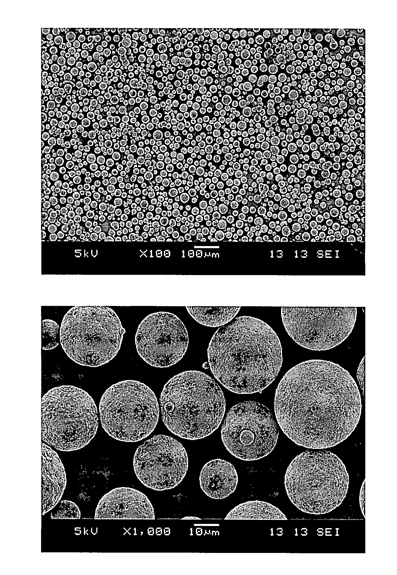
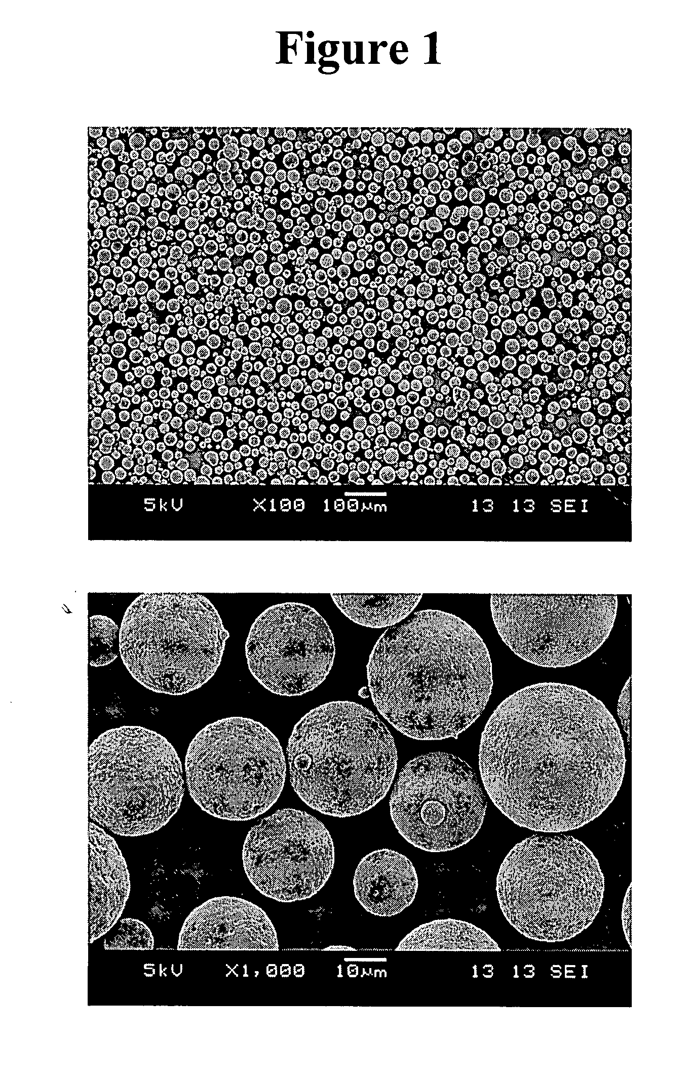
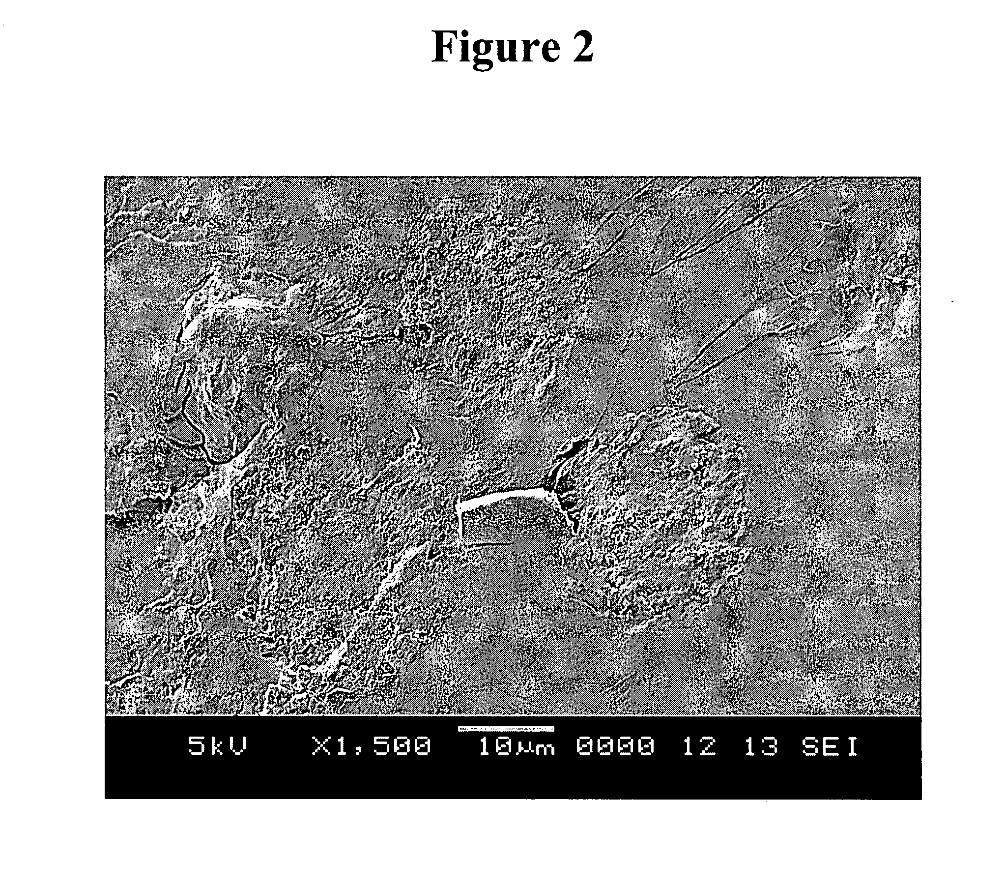
[0156] Microparticle morphology was observed by Scanning Electron Microscopy (SEM) analysis. Microparticles were sputter coated with gold using an Anatech LTD Hummer 6.2 system. Scanning electron microscopic images were taken using a JEOL JSM-5600 scanning electron microscope and accompanying software at an accelerating voltage of 5-10 keV. For selected samples, SEM analysis of the internal microsphere structure was made after embedding microparticles in L.R. White Resin and then splitting the preparation after the resin hardened.

[0157] Scanning electron micrograph images of microparticles formed by the process as set forth in Example 1 are shown in FIGS. 1 and 2. The images of FIG. 1 show that the microparticles have a smooth external morphology. The image of FIG. 2 shows that the microparticles have a monolithic internal morphology.

[0158] A scanning electron micrograph image of microparticles formed by the water-in-oil-in water process as se...


PUM

PropertyMeasurementUnit
Fractionaaaaaaaaaa
Fractionaaaaaaaaaa
Fractionaaaaaaaaaa

Abstract

Formulations for controlled, sustained release of biologically active agents for the treatment of ocular disorders have been developed. These formulations are based on solid microparticles formed of the combination of biodegradable, synthetic polymers such as poly(lactic acid) (PLA), poly(glycolic acid) (PGA), and copolymers thereof. The microparticles are characterized by low burst levels and efficient drug loading and sustained release.

Description

RELATED APPLICATIONS [0001] This application claims priority to U.S. Provisional Application Ser. No. 60 / 741,741, filed Dec. 2, 2005, and U.S. Provisional Application Ser. No. 60 / 780,760, filed Mar. 9, 2006, and U.S. Provisional Application Ser. No. 60 / 796,071, filed Apr. 28, 2006, all of which are hereby incorporated in its entirety by reference.FIELD OF THE INVENTION [0002] The invention relates to drug delivery. In particular, the invention relates to compositions and methods for the sustained delivery of therapeutic agents using microparticles. More particularly, the invention relates to sustained release microparticle compositions and methods of use for ophthalmic administration. BACKGROUND OF THE INVENTION [0003] As new treatment modalities for ophthalmic diseases become available, the number of intravitreous injections administered is expected to increase dramatically. For example, intravitreous injection of the vascular endothelial growth factor (VEGF) inhibitor, Macugen® ((...

Claims


Application Information

Patent Timeline
IPC IPC(8): A61K9/52A61P27/02
CPCA61K9/0048A61K9/1647A61K38/1866A61K9/0019A61K31/00A61P27/02
Inventor CAMPBELL, KATHLEEN M.CALIAS, PERICLESCOOK, GARY P.GANLEY, MARY A.
Owner EYETECH
Why Eureka
Try Eureka