
Cultured Melanocytes on Bioploymer Membranes for Treatment of Hyper and Hypopigmentation Disorders

a melanocyte and membrane technology, applied in the field of skin melanocyte delivery system, can solve the problems of cell genetic coding or dna mutation, cell function, aging skin,

Inactive Publication Date: 2008-04-17
RELIANCE LIFE SCI PVT
View PDF9 Cites 6 Cited by
  • Summary
  • Abstract
  • Description
  • Claims
  • Application Information

AI Technical Summary

Benefits of technology

[0087] The present invention has provided a process for co-culturing and its use in transplantation in skin conditions such as vitiligo. The advantage of co-delivering keratinocytes with melanocytes is that it will enhance the rate of epithelialization of the affected skin and, by secreting growth factors, will accelerate pigmentation.

Problems solved by technology

Without protection, UV radiation can cause the cell's genetic coding or DNA to mutate.
This can significantly alter cell functions, resulting in changes associated with rosacea, aging skin, compromised immune function and cancer.
In addition, inflammation resulting from rosacea, laser therapies, UV exposure, or other factors, can affect the functioning of the melanocytes, resulting in an abnormal increase in the production of melanin.
The change in appearance caused by vitiligo can affect a person's emotional and psychological well-being and may create difficulty getting or keeping a job.
Adolescents, who are often particularly concerned about their appearance, can be devastated by widespread vitiligo.
Some people who have vitiligo feel embarrassed, ashamed, depressed, or worried about how others will react.
Current therapy for vitiligo often takes a long time, for example, 6 to 18 months.
There is no universally effective medical or surgical treatment for vitiligo; although a number approaches are known to be effective.
There is also a risk of side effects such as skin shrinkage and skin striae (streaks or lines on the skin) for which the doctor has to closely monitor.
Patients must minimize exposure to sunlight at other times. However, it is time-consuming and care must be taken to avoid side effects, which can sometimes be severe.
There are two major potential side effects of topical PUVA therapy: (1) severe sunburn and blistering and (2) too much re-pigmentation or darkening of the treated patches or the normal skin surrounding the vitiligo (hyperpigmentation).
Other known side effects of oral psoralen include nausea and vomiting, itching, abnormal hair growth, and an increased skin cancer risk.
The major side effect of de-pigmentation therapy is inflammation (redness and swelling) of the skin.
Patients may also experience itching, dry skin or abnormal darkening of the membrane that covers the white of the eye.
De-pigmentation is permanent and cannot be reversed.
In addition, a person who undergoes de-pigmentation will always be abnormally sensitive to sunlight.
The risks of blister grafting include the development of a cobblestone appearance, scarring, and lack of re-pigmentation.
This technique, although effective, leads to a cobblestone appearance and scarring.
Tattooing works best for the lip area, particularly in people with dark skin; however, it is difficult for the doctor to match perfectly the color of the skin of the surrounding area.
In addition, tattooing of the lips may lead to episodes of blister outbreaks caused by the herpes simplex virus.
The disadvantages of such methods include:
(c) Area coverage: If the initial biopsy taken is small compared to the de-pigmented area, then the number of melanocytes administered to the de-pigmented site would not be sufficient to produce satisfactory levels of pigmentation.
This delivery method is inefficient, with cells not being correctly delivered to the wound bed or becoming trapped in the gauze applied over the treated skin.
Although strides have been made into understanding the pathogenesis, and treatment of vitiligo, many of the commercially available products are only marginally effective and rarely achieve an even-looking skin tone.
To date, it has not been possible to conduct transplantation of melanocytes remotely, due to the inability to safely and reliably transport the melanocyte cultures over distances.

Method used


Image

  • Cultured Melanocytes on Bioploymer Membranes for Treatment of Hyper and Hypopigmentation Disorders
  • Cultured Melanocytes on Bioploymer Membranes for Treatment of Hyper and Hypopigmentation Disorders
  • Cultured Melanocytes on Bioploymer Membranes for Treatment of Hyper and Hypopigmentation Disorders

Examples


example 1

Preparation of Cultured Melanocyte Graft on Biopolymer

Collection of the Skin

[0101] A punch biopsy was collected from the person who needs to be grafted hereto referred to as patient. Patient's blood (5 ml) was collected for infectious disease testing to determine the ID status of the patient to ensure safety to the operator as well as the patient at the time of transplantation. Skin biopsy collection vials containing DMEM, Iscove medium (Invirtrogen USA), 10% fetal bovine serum (FBS) (Hyclone, USA), antibiotic-antimycotic solution (Sigma USA) and gentamycin (Invitrogen, USA) were transported to hospitals in insulated boxes containing ice-packs. The insulated boxes were made of EPS (expanded polystyrene) and maintained at a temperature of 8-25° C. for 72 hours. The vials were stored in the refrigerator for a maximum of one week till use. The biopsies were collected in these transport vials and shipped back in insulated boxes for further processing within 48 hours of sample collect...

example 2

Co-Culture Techniques

[0116] The application of keratinocytes to skin resulted in enhanced rate of epithelialization and improved the rate of healing of the wound that is produced following debridement of the de-pigmented skin.

[0117] Keratinocytes were obtained either from the patient undergoing melanocyte treatment or from another donor. FIG. 10 shows the co-cultured cells on a biopolymer. When using cultured keratinocytes obtained from the patient directly, keratinocytes will continue to survive for a longer time and form part of the reconstituted epithelia. The application of allogeneic cultured keratinocytes will enhance the rate of healing by stimulating the body to regenerate even if these cells only survive temporarily, however.

example 3

Transport of the Graft of Cultured Melanocyte Biopolymer

[0118] There are no previous reports of transportation of melanocytes beyond a hospital. The present invention demonstrates how such portability may be achieved.

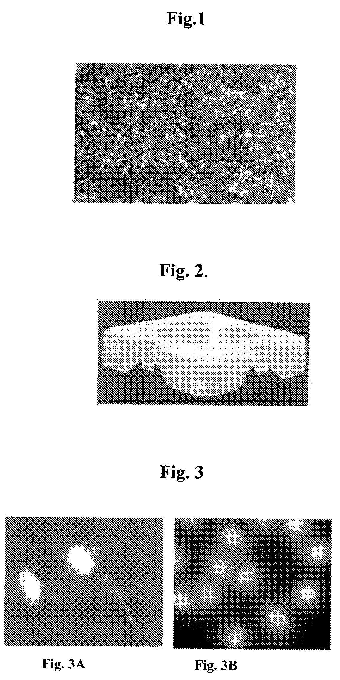
[0119] An example design of a transport container, its assembly, and use in transport of cultured cells is presented in Indian patent application 60 / MUM / 2006, which is incorporated herein by reference. The design of the container is shown in FIG. 2. Sterilized PLA films were placed in these transport dishes and soaked in PBS for 1 hour. The film in each dish was held in place with polycarbonate ring. The cells were seeded with 3-5×10⁴ cells / cm2 and cultured for 2 days before transport. Before dispatching to hospitals, the media in the dishes were replaced with CO2 enriched culture media using a flow meter. Sterilized PLA films were placed in specially designed transport dishes (Ampson, India) and soaked in PBS for 1 h. The film in each dish was held in place with poly...


PUM

PropertyMeasurementUnit
molecular weightaaaaaaaaaa
timeaaaaaaaaaa
melting pointaaaaaaaaaa

Abstract

The present invention relates to a graft wherein cultured autologous melanocytes are delivered using a biopolymer. The present invention describes the composition, method of preparation and its properties relating to safety and efficacy. The graft of the present invention has a potential use in treating hyper- and hypopigmentation disorders.

Description

CROSS-REFERENCE TO RELATED PATENT APPLICATIONS [0001] This application claims the benefit of Indian provisional application No. 1697 / MUM / 2006, filed on Oct. 13, 2006, the disclosure of which is incorporated by reference in its entirety. FIELD OF THE INVENTION [0002] The present invention relates to a delivery system of skin melanocytes on a suitable carrier system, and its method of preparation. In one embodiment, the invention relates to grafts of autologous melanocytes, its methods of preparation, transportation to hospitals and their therapeutic applications including vitiligo. BACKGROUND OF THE INVENTION Skin and Physiological Role of Melanocytes [0003] The skin is the largest organ of the body and is composed of an external epithelial component called the epidermis separated from an underlying connective tissue component, the dermis, by a basement membrane. The portion of the dermis adjacent to the epidermis is called the reticular dermis and is composed primarily of collagen ...

Claims


Application Information

Patent Timeline
Patent Type & Authority Applications(United States)
IPC IPC(8): A61K45/00A61P17/00C12N5/08C12N5/071
CPCA61L27/3813A61L27/60C12N5/0626C12N5/0698C12N2533/40C12N2502/094C12N2502/1323C12N2533/30C12N2502/091A61P17/00
Inventor GHOSH, DEEPASHENOY, SUDHEERKUCHROO, PUSHPASHAH, VIRU
Owner RELIANCE LIFE SCI PVT
Learn More