Method of Detecting Carcinogenesis Caused by Hepatitis B Virus

Inactive Publication Date: 2008-07-03
OTSUKA PHARM CO LTD
View PDF0 Cites 4 Cited by
  • Summary
  • Abstract
  • Description
  • Claims
  • Application Information

AI Technical Summary

Benefits of technology

[0011]An object of the present invention is to provide a method and a kit using same, that can effectively detect, for example, liver cancer by identifying, in liver tissue s

Problems solved by technology

Acute HBV infection results in serious illness in some cases, and about 0.5% of patients die of fulminant hepatitis.
However, oncogenesis by HBV integration at these sites has not been reproduced in samples from other hepatocellular carcinoma patients.
Notwithstanding th

Method used


Image

  • Method of Detecting Carcinogenesis Caused by Hepatitis B Virus
  • Method of Detecting Carcinogenesis Caused by Hepatitis B Virus
  • Method of Detecting Carcinogenesis Caused by Hepatitis B Virus

Examples


example 1

(a) Histological and Serological Examinations of the Hepatocyte Samples

[0153]The following tests were carried out using resected liver tumor tissue (hepatocellular carcinoma or HCC) from 26 Japanese patients obtained at the Second Department of Surgery, Chiba University Hospital, between 1987 and 2003. These patients had not undergone preoperative treatment.

[0154]Clinical, histological, and serological examinations were carried out on the patients providing the samples. In the histological examination, the tumor size, degree of differentiation of the tumor cells (three stages), and type of liver disease (CPH: chronic persistent hepatitis; CAH: chronic acute hepatitis; LC: liver cirrhosis) were investigated. In the serological examination, hepatitis B surface antigen (HBsAg) detection was carried out using an enzyme immunoassay (EIA) kit from Dinabot Co., Ltd., and antibody against the hepatitis B surface antigen (anti-HBs antibody) and antibody against the hepatitis B core antigen (...

example 2

Detection of HBx / MLL4 Fusion Transcription Products by RT-PCR

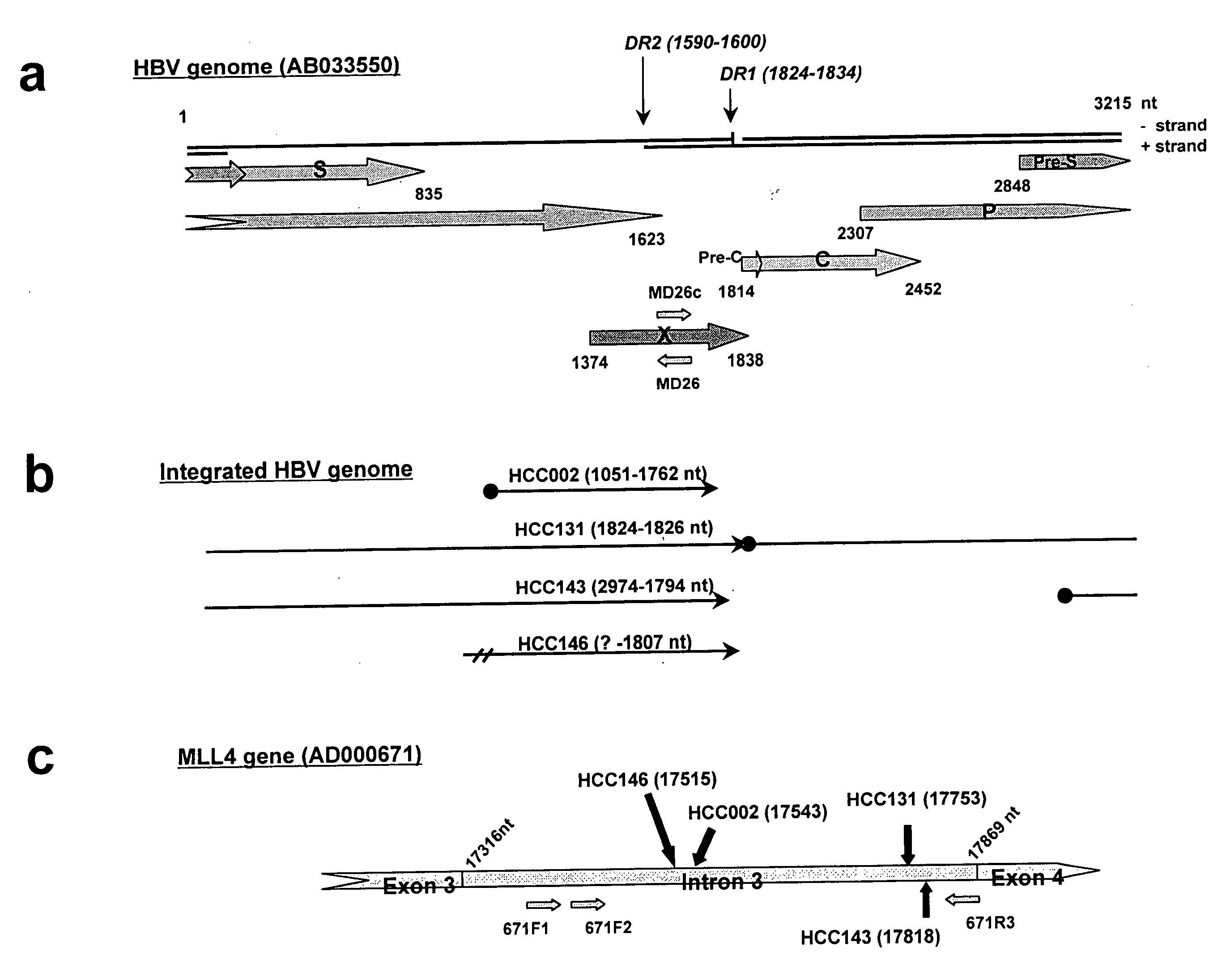
[0181]The coding region for HBx protein excluding the C-terminal and the promoter region of HBx (X gene of HBV-DNA) were present in all 4 cases in which MLL4 / HBV-DNA integration had been confirmed. The presence of HBx / MLL4 fusion transcription products in these 4 samples was predicted on this basis. The total RNA was extracted from HCC131, HCC143, HCC146, and HCC002 and the transcription products obtained by RT-PCR were investigated.

[0182]The details of the test procedure are given below.

[0183]The RNA was extracted using Trizol (Invitrogen) according to the instructions for use. The extracted RNA was confirmed by electrophoresis on 1.2% agarose gel. RT-PCR was carried out as follows using a Superscript One-Step RT-PCR system (Invitrogen) according to the instructions for use; gene-specific primers designed on exon 5 or 6 of MLL4 (E5-1: SEQ ID NO. 15; E5-2: SEQ ID NO. 16; and E6-1: SEQ ID NO. 17) were used as the reverse tr...

example 3

Detection of HBx / MLL4 Fusion Proteins by Immunoblotting Analysis

[0188]In order to confirm the actual translation of these fusion transcription products into protein, a comparison was carried out by immunological test procedures using liver tumor cells and cells adjacent thereto for HCC131, HCC143, and HCC002.

[0189]The details of the test procedures are as follows. The complete protein was prepared by lysing 100 to 200 mg of cells with a buffer that contained 0.1% SDS, 0.5% deoxycholate, 1% NP-40, 150 mM NaCl, 50 mM Tris-HCl (pH 8.0), 1 mM APMSF, and other protease inhibitors (complete protease inhibitor tablets, Roche) followed by centrifugation at 12,500 rpm for 30 minutes at 4° C. Optionally, 500 μg of the obtained total protein was incubated with anti-HBx (hepatitis B virus X protein) monoclonal antibody (Advanced Immuno Chemical, Inc.), and immunoprecipitation was performed by a standard method.

[0190]The total protein and the immunoprecipitated product prepared by the preceding ...


PUM

PropertyMeasurementUnit
Volumeaaaaaaaaaa
Sizeaaaaaaaaaa
Sizeaaaaaaaaaa

Abstract

A method for detecting, in liver tissue isolated from a human subject and suspected of being cancerous, integration of HBV-DNA into the MLL4 gene, this integration of HBV-DNA indicating the cancerous nature of a tissue. In a typical case, this integration can be detected by carrying out PCR using a primer specific to the region containing intron 3 of the MLL4 gene and a primer specific to the X gene region of HBV.

Description

TECHNICAL FIELD[0001]The present invention relates to a method and kit for detecting the integration of HBV-DNA into the MLL4 gene, which indicates that tissue from diseased tissue such as, for example, chronic hepatitis or liver cirrhosis, is cancerous, or to a method and kit for detecting fusion products thereof. The present invention further relates to a method and kit for detecting chromosomal translocation between intron 3 of the MLL4 gene on the long arm of chromosome 19 and the short arm of chromosome 17, which indicates that a tissue is cancerous.BACKGROUND ART[0002]The human hepatitis B virus (HBV), which is in the hepadnavirus family, exhibits a very restricted host range and shows a strong tropism for liver parenchymal cells. Acute HBV infection results in serious illness in some cases, and about 0.5% of patients die of fulminant hepatitis. Chronic infection, on the other hand, causes moderate to severe liver diseases, such as, for example, chronic hepatitis, liver cirrho...

Claims


Application Information

Patent Timeline
IPC IPC(8): C12Q1/68C12Q1/02G01N33/53C12N15/09C12Q1/70G01N33/574G01N33/576
CPCC12Q1/6886G01N2333/02C12Q1/706C12Q2600/156
Inventor INOUE, ITUROSAIGO, KENICHI
Owner OTSUKA PHARM CO LTD
Why Eureka
Try Eureka