
Novel method of using triacetin and auxiliary agent for ultrasonic diagnostic examination

Inactive Publication Date: 2009-08-27
SHINSHU UNIVERSITY +2
View PDF7 Cites 4 Cited by
  • Summary
  • Abstract
  • Description
  • Claims
  • Application Information

AI Technical Summary

Benefits of technology

[0039]According to the present invention, the pylorus is temporarily closed and a liquid is allowed to stay in the stomach, whereby the head and tail of the pancreas the images of which are particularly difficult to acquire can be easily imaged by observation through the stomach (FIGS. 5, 6, and 7).
[0040]Further, by means of observation through the stomach according to the present invention, it also becomes possible to obtain image information from new angles for the edge of the outward area of the left lobe of the liver, the left side of an adrenal, the superior pole and the area inward region of the left kidney, and the upper portion of the spleen (under the dome of the left diaphragm), whereupon these areas can be observed with ease.
[0041]As a further advantage, it becomes possible to reduce the mental and physical burdens that may be imposed on the person under examination by treatments such as preliminary ones that precede a radiographic or endoscopic examination of the stomach.

Problems solved by technology

However, ultrasonic examination has one weak feature in that it tends to be influenced by the physical conditions of the person under test such as the gas in the digestive tract and obesity.
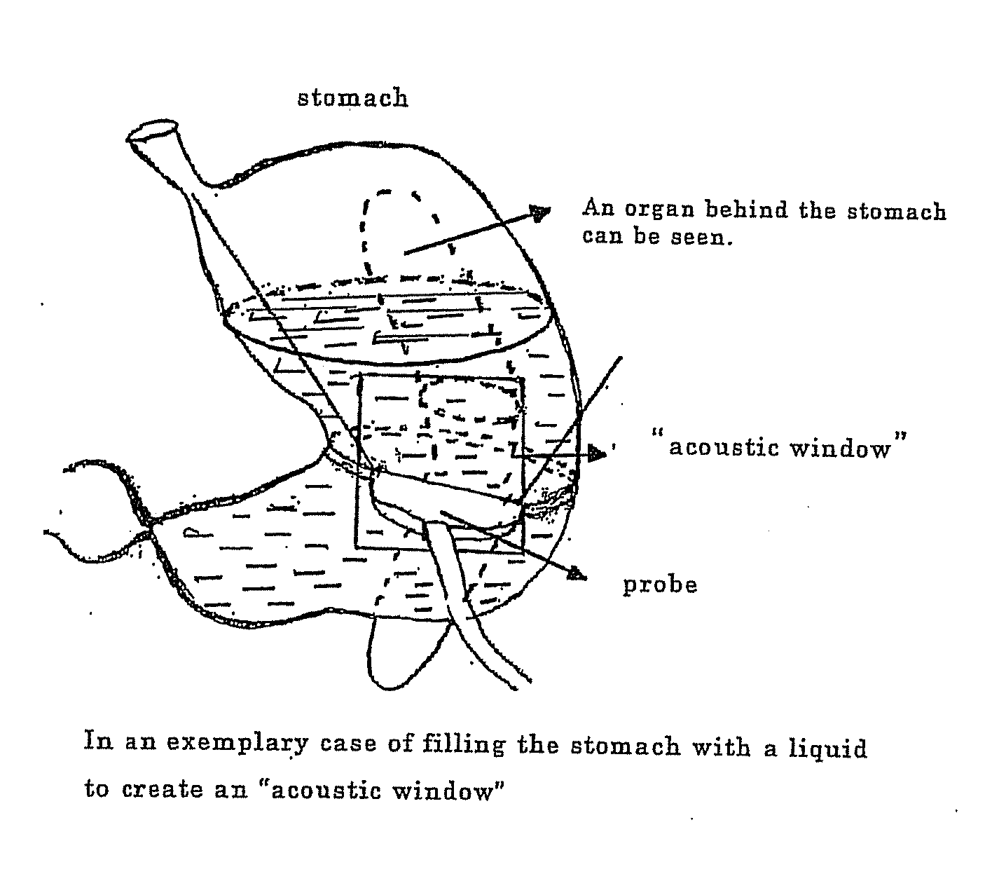
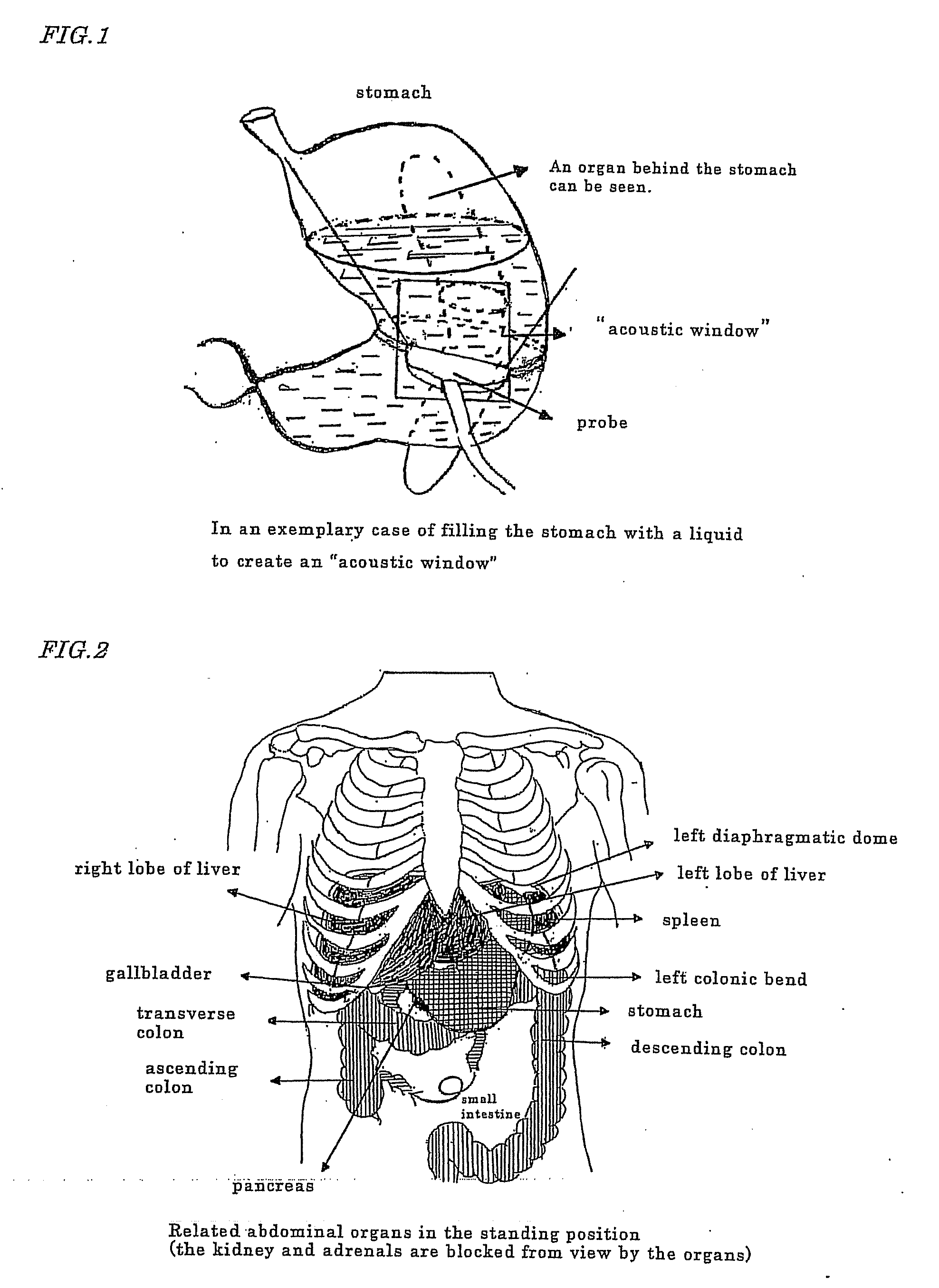
Since ultrasonic waves by nature do not propagate in the air, an “acoustic window” (see FIG. 1) is created in a suitable position on the surface of the human body that will not interfere with the propagation of ultrasound, whereby the organ of interest is viewed through a two-dimensional (2D) section and recognized as a three-dimensional (3D) image by individual observers subjectively, also depending on their experience and the like; however, this always presents the problem with objectivity.
The “acoustic window” cannot be created at a position that passes through an air-containing organ such as the lung or stomach although this position is preferred for observation and it is well known that the space between ribs is also unsuitable as a place to be contacted by the probe.
In short, the position of the “acoustic window” is limited.
Hence, it is difficult to obtain a useful image at the edge of the outward area of the left lobe of the liver, the left side of an adrenal, the superior pole and the area inward of the left kidney, and the upper portion of the spleen (under the dome of the left diaphragm) and a certain lesion sometimes fail to be noticed.
Hence, it is difficult to create an “acoustic window” for viewing the pancreas, especially the tail of pancreas, and in many cases the stomach is filled with water and observation is made through the stomach lumen but water will readily flows out of the stomach.
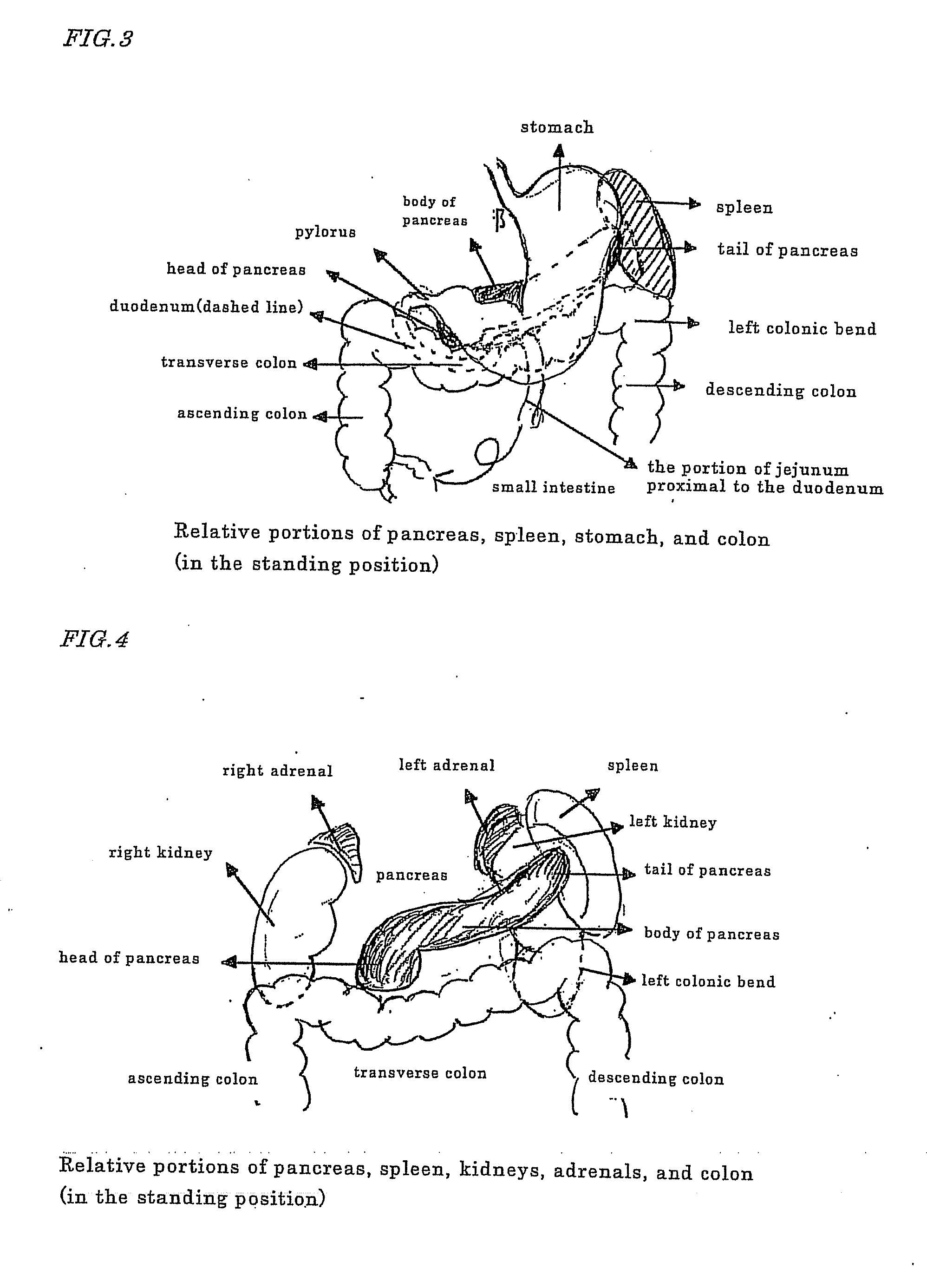
Hence, it is generally held that ultrasonic examination is difficult to perform on the pancreas and other retroperitoneal organs (see FIGS. 2, 3, and 4).
According to Patent document 1, the auxiliary agent is allowed to stay within the digestive tract by virtue of viscosity; however, increased viscosity makes the auxiliary agent difficult to swallow and impairs the convenience of ultrasonic diagnosis.
The difficult-to-digest viscosity enhancer has colloid osmotic pressure and induces diarrhea.
If air bubbles enter into the auxiliary agent as it is swallowed, the chance of their disappearing will decrease with the increasing viscosity and the air bubbles that entered will reflect ultrasound, impairing its propagation.
Patent document 1 further states that viscosity is enhanced in an acidic environment or in an environment where polyvalent ions are present; however, in these environments, the alginate becomes insoluble and forms a gel rather than a uniform solution.
In other words, ultrasound is reflected or refracted and its propagation is impaired, which is unsuitable for the creation of an “acoustic window.”
According to Patent document 2, the auxiliary agent contains at least one viscosity enhancer and insoluble polyvinyl pyrrolidine and as in Patent document 1, the purpose is considered to achieve retention within the digestive tract by virtue of viscosity; however, increased viscosity makes the auxiliary agent difficult to swallow and impairs the convenience of ultrasonic diagnosis.
The difficult-to-digest viscosity enhancer has colloid osmotic pressure and induces diarrhea.
If air bubbles enter into the auxiliary agent as it is swallowed, the chance of their disappearing will decrease with the increasing viscosity and the air bubbles that entered will reflect ultrasound, impairing its propagation.
In addition, the solution containing the insoluble polyvinyl pyrrolidine is not a uniform solution and a gel-like substance interspersed with the insoluble polyvinyl pyrrolidine will reflect or refract ultrasound and impairs its propagation, which is unsuitable for the creation of an “acoustic window.”
In Patent document 4, an aqueous dispersion of edible inorganic particles is used and this itself is an ultrasound reflector, impairing the propagation of ultrasound and making it impossible to create an “acoustic window.” As in Patent document 1, the purpose is considered to achieve retention within the digestive tract by virtue of viscosity; however, increased viscosity makes the testing aid difficult to swallow and impairs the convenience of ultrasonic diagnosis.
The difficult-to-digest viscosity enhancer has colloid osmotic pressure and induces diarrhea.
If air bubbles enter into the testing aid as it is swallowed, the chance of their disappearing will decrease with the increasing viscosity and the air bubbles that entered will reflect ultrasound, impairing its propagation, which is unsuitable for the creation of an “acoustic window.”
As in Patent document 1, the purpose is considered to achieve retention within the digestive tract by virtue of viscosity; however, increased viscosity makes the testing aid difficult to swallow and impairs the convenience of ultrasonic diagnosis.
The difficult-to-digest viscosity enhancer or polymer solution has colloid osmotic pressure and induces diarrhea.
If air bubbles enter into the auxiliary agent as it is swallowed, the chance of their disappearing will decrease with the increasing viscosity and the air bubbles that entered will reflect ultrasound, impairing its propagation.
Furthermore, the use of the silicon-containing compound, which is not water-soluble, results in an interface that reflects ultrasound, impairing its propagation, which is unsuitable for the creation of an “acoustic window.”
In Patent document 6, the composition is adjusted to have a gastric emptying time of at least 20 minutes and high osmotic pressure (300 millimoles / kilogram or more) so that a stomach emptying suppressing effect (already known) will be achieved; however, in order to generate this osmotic pressure, a water-soluble substance is required but the difficult-to-digest polydextrose solution and the like that are mentioned in the document have colloid osmotic pressure together with viscosity and this viscosity makes the composition difficult to swallow, impairs the convenience of ultrasonic diagnosis, and induces diarrhea on account of the colloid osmotic pressure.
If air bubbles enter into the auxiliary agent as it is swallowed, the chance of their disappearing will decrease with the increasing viscosity and the air bubbles that entered will reflect ultrasound, impairing its propagation.
In addition, the glyceride and the like that are described in the document usually form an interface in the aqueous solution to reflect ultrasound and impair its propagation, which is unsuitable for the creation of an “acoustic window.”
(1) their viscosity should not be high (because high viscosity makes the preparation difficult to swallow, impairs the convenience of ultrasonic examination, and the more viscous the liquid is, the smaller the chance of air bubbles to disappear);
(2) an oil-in-water emulsion and the dispersion of oil droplets should be avoided (because they reflect or refract ultrasound);
(3) insoluble particles and gel-like particles should be avoided (because they reflect or refract ultrasound);
(4) a difficult-t-digest water-soluble polymer should be avoided (because it gengerates a colloid osmotic pressure and induces diarrhea.)

Method used


Image

  • Novel method of using triacetin and auxiliary agent for ultrasonic diagnostic examination
  • Novel method of using triacetin and auxiliary agent for ultrasonic diagnostic examination
  • Novel method of using triacetin and auxiliary agent for ultrasonic diagnostic examination

Examples


example 1

Triacetin (0.5%)

[0083]To 200 mL of water under agitation with a magnetic stirrer, 1.5 g of triacetin, 0.24 g of citric acid and 1.74 g of trisodium citrate, both as a pH modifier, and 3 g of glycerin as a sweetener were added, followed by heating at 80° C.

[0084]Subsequently, 0.012 g of ethyl paraoxybenzoate and 0.027 g of butyl paraoxybenzoate that had been preliminarily dissolved in 0.45 g of propylene glycol were added; after cooling, a preliminarily prepared ethanol solution of 20% 1-menthol was added in such an amount that 0.006 g of 1-menthol would be incorporated and water was added to make a volume of 300 mL. As a result, a colorless and clear preparation was completed.

[0085]Thus, the completed preparation contains the following components (A) to (I) in 300 mL of water.

[0086]Since triacetin tastes bitter, additives and a method for eliminating the bitterness may optionally be used.

(A) triacetin: 1.5 g

(B) buffer: citric acid: 0.24 g

(C) buffer: trisodium citrate: 1.74 g

(D) swee...

example 2

Triacetin (0.5%)

[0087]To 200 mL of water under agitation with a magnetic stirrer, 1.5 g of triacetin, 0.24 g of citric acid and 1.74 g of trisodium citrate, both as a pH modifier, and 0.03 g of saccharin sodium as a sweetener were added, followed by heating at 80° C.

[0088]Subsequently, 0.012 g of ethyl paraoxybenzoate and 0.027 g of butyl paraoxybenzoate that had been preliminarily dissolved in 0.45 g of propylene glycol were added; after cooling, 3 g of glycine and a preliminarily prepared ethanol solution of 20% 1-menthol were added in such an amount that 0.006 g of 1-menthol would be incorporated and water was added to make a volume of 300 mL. As a result, a colorless and clear preparation was completed.

[0089]Thus, the completed preparation contains the following components (A) to (J) in 300 mL of water.

(A) triacetin: 1.5 g

(B) buffer: citric acid: 0.24 g

(C) buffer: trisodium citrate: 1.74 g

(D) sweetener: saccharin sodium: 0.03 g

(E) solvent promoter: propylene glycol: 0.45 g

(F) an...

example 3

Triacetin (10%)

[0090]Thirty grams of triacetin and 4.5 g of polyoxyethylene(30) hardened castor oil (Nikko Chemicals Co., Ltd.: NIKKOL HCO-30) were mixed to give the indicated proportions and agitated with a high-performance stirrer / disperser, ULTRA-TARAX (product of IKA JAPAN CO., LTD.) to yield a liquid mixture.

[0091]To 200 mL of water heated at about 70° C., 0.6 g of carmellose sodium (CELOGEN F-SC of DAI-ICHI KOGYO SEIYAKU CO., LTD.) was added under agitation with a magnetic stirrer to form a solution. After cooling to room temperature, 0.24 g of citric acid and 1.74 g of trisodium citrate, both as a pH modifier, and 0.03 g of saccharin sodium as a sweetener were added, followed by heating at 80° C.

[0092]Then, 0.012 g of ethyl paraoxybenzoate and 0.027 g of butyl paraoxybenzoate that had been preliminarily dissolved in 0.45 g of propylene glycol were added; after thorough agitation, the temperature of the liquid was lowered to about 50° C.

[0093]Subsequently, the whole quantity o...


PUM

PropertyMeasurementUnit
Massaaaaaaaaaa
Volumeaaaaaaaaaa
Volumeaaaaaaaaaa

Abstract

A drug in the form of a solid preparation soluble or suspendable in water or an aqueous solution characterized by containing at least one biocompatible, gastric motility suppressing component such as triacetin, which is capable of transmitting ultrasonic wave to a target organ via the solution pooled in the body in a noninvasive examination (for example, an ultrasonic examination of an abdominal organ), identifying the area in which the solution exists, or clarifying the boundary of the solution and an organ in contact therewith, is employed as an auxiliary agent in the examination. In addition, triacetin can be extensively used as a gastric motility suppressor.

Description

TECHNICAL FIELD[0001]The present invention relates to a stomach motility suppressant containing triacetin, a testing aid for ultrasonic imaging diagnosis, and a kit for use in ultrasonic imaging diagnosis.BACKGROUND ART[0002]In order to diagnose abdominal parenchymal organs such as the liver, gallbladder, pancreas, adrenals, kidneys, and spleen, both functional and organic changes in disease must be known accurately and various means are employed to this end; they include not only fundamental tests such as history taking (covering the environment surrounding the patient, his or her background, etiology, and course of development), phonacoscopy, background investigation, physical examination, hematological test, biochemical examination of blood, and urine test but also imaging tests such as X-ray examination (with plain film or contrast medium), endoscope, ultrasonic test (US), endoscopic ultrasonography (EU), CR, MRI, and PET; these means are improving day by day. Among others, imag...

Claims


Application Information

Patent Timeline
IPC IPC(8): A61B8/00B65D71/00
CPCA61K49/221A61K31/22A61P1/00
Inventor KOYAMA, SHOZOTOKUDA, MASAAKIHORI, YOSHIYUKIOHSAKA, KAZUMASA
Owner SHINSHU UNIVERSITY
Learn More