Composition for a Tissue Repair Implant and Methods of Making the Same

a tissue repair and implant technology, applied in the field of tissue repair implants, can solve the problems of limiting each, and achieve the effect of increasing the affinity of growth factors

Active Publication Date: 2010-02-11
LIFENET HEALTH
View PDF44 Cites 94 Cited by
  • Summary
  • Abstract
  • Description
  • Claims
  • Application Information

AI Technical Summary

Benefits of technology

[0008]The tissue repair implant of the present invention may be preshaped, elastic, resilient, and insoluble in water. The implant will maintain its shape in a dry or a wet state. In its dry state, the implant will quickly absorb fluid because it is porous. For example, in its wet state, the implant will maintain its porosity, cohesiveness, and integrity.
[0014]The methods of the present invention may include combining the tissue repair implant with one or more bioactive supplements. The bioactive supplements may be growth factors, differentiation factors, cytokines, or anti-inflammatory agents. The growth or differentiation factors may be a growth factor of the the FGF-family or TGF-family, IGF-1, PDGF, EGF, VEGF, HGF, PTHrP, Ihh, dexamethasone, insulin, transferrin, selenium, ITS, ascorbate, or a combination thereof. The cytokines may be GM-CSF, G-CSF, TNF-α, IL-1β, IL-4, IL-6, IL-8, IL-10, SLP1, MCP1, MIP-1α, MIP-2, IL-18, angiopoietin, KGF, endothelin, IFN-α, or IFN-β. The anti-inflammatory agents may be an IL-1αR antibody, TNF-a receptor antagonist, cyclooxygenase-2 specific inhibitors, MAP kinase inhibitors, NO synthase inhibitors, NF-κB inhibitors, or inhibitors of MMP. The tissue repair implant may also be combined with one or more agents having bioactive supplement binding sites comprising hyaluronan, heparin, heparin sulfate, keratin sulfate, dermatan sulfate, chondroitin sulfate, betaglycan, heparan sulfate proteoglycan, syndecan, biglycan, or decorin. The one or more bioactive supplement binding sites on the agents may increase the affinity of growth factors, differentiation factors, cytokines, or anti-inflammatory agents to said tissue repair implant. Accordingly, the present invention also discloses a tissue repair implant comprising one or more bioactive supplements and one or more agents having bioactive supplement binding sites.

Problems solved by technology

Despite the benefits of autografts and allografts, the limitations of each have necessitated the pursuit of alternative graft materials.

Method used


Image

  • Composition for a Tissue Repair Implant and Methods of Making the Same
  • Composition for a Tissue Repair Implant and Methods of Making the Same
  • Composition for a Tissue Repair Implant and Methods of Making the Same

Examples


example 1

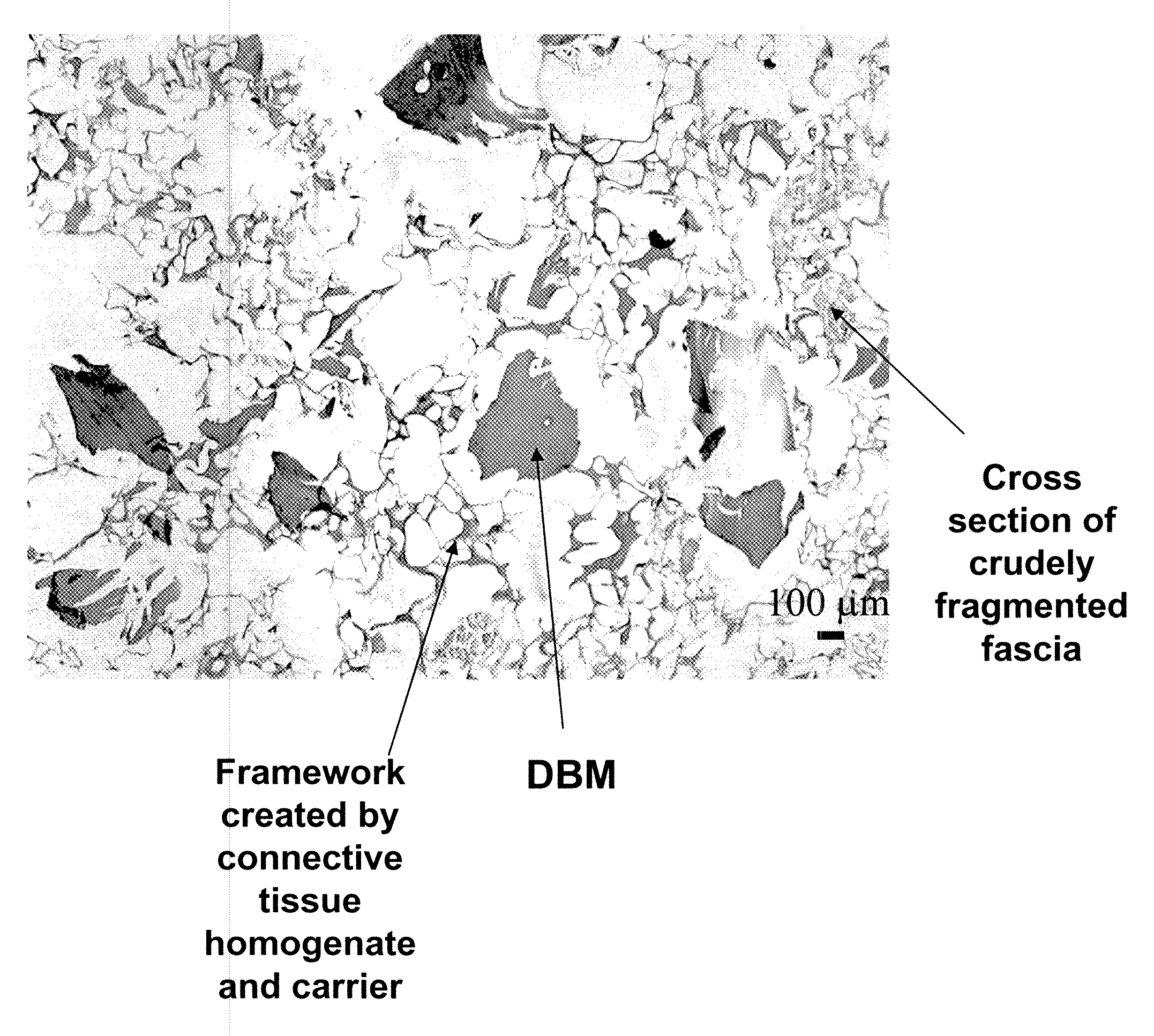
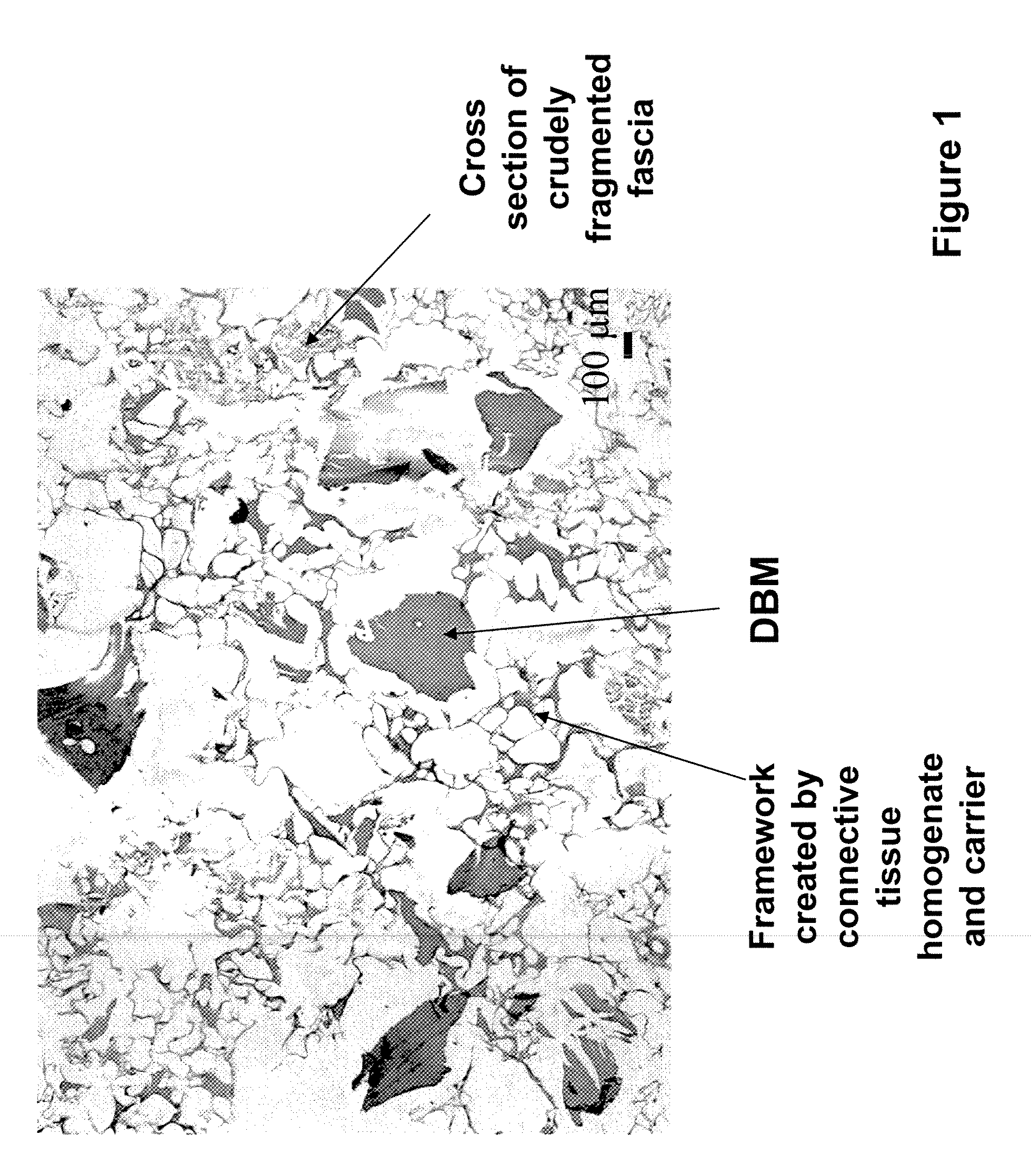
[0082]Preparation of Tissue Repair Implants: Porous Sponge-Like Structure with DMB Particles.

[0083]Fascia lata and bone from a human cadaver were procured and returned to the processing facility under sterile conditions. Donor histories, personal and medical, were obtained following accepted standards of the American Association of Tissue Banks. Microbiological tests were performed following FDA guidelines for testing sterility of products.

[0084]The bone and fascia were cleaned of unwanted tissues and freeze-dried. The freeze-dried fascia was cut into small (about 1.5 cm by 1.5 cm) pieces (e.g., crude fragments). Sterile water in a volume (1 cm3 of isotonic saline corresponds to about 1 g) approximating 20 times the weight of tissue, was added to a container containing the cut fascia. The ingredients were heated to a temperature of 100 degree C. using a hot plate, and maintained at this temperature for about 5 minutes. Optionally, water was added to the heated composition to replace...

example 2

[0089]Preparation of Tissue Repair Implants: Porous Sponge-Like Structure without Bone Fragment.

[0090]The homogenized fascia, sodium alginate solution, crudely fragmented fascia, and CaCl2 solution were prepared as described in Example 1.

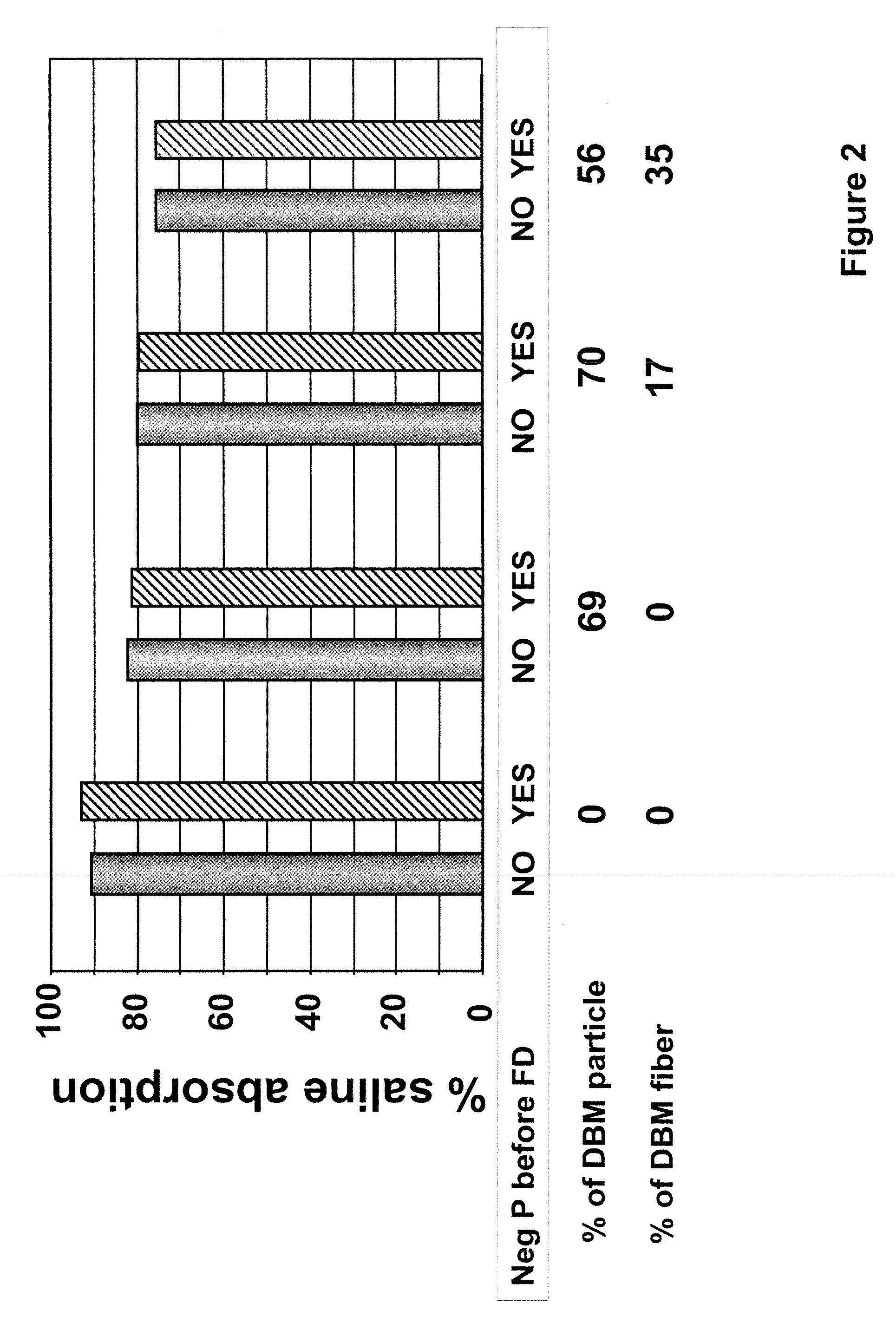
[0091]To prepare the tissue repair implant, the homogenized fascia was mixed with a sodium alginate solution first to produce a connective tissue carrier, which was mixed further with crudely fragmented fascia. After mixing all the components, the mixture was distributed into molds with pre-determined shapes and sizes, freeze-dried, treated with CaCl2, washed, and freeze-dried again. Before the first freeze-drying cycle, some samples were exposed to a negative hydrostatic pressure to allow the expansion of the mixture to a preset thickness. The resultant porous sponge-like structure was then sent out for sterilization by gamma irradiation.

[0092]In one of the tissue repair implants, the porous sponge-like structure contained about 20% alginate, 60% h...

example 3

[0093]Preparation of Tissue Repair Implants: Porous Sponge-Like Structure with DBM Particles and DMB Fiber Bone.

[0094]The homogenized fascia, sodium alginate solution, crudely fragmented fascia, demineralized bone powder, and CaCl2 solution were prepared as described in Example 1.

[0095]Demineralized fiber bone was prepared by cutting cortical bone to produce cut fiber bone having an average length from about 1 mm to about 100 mm. Then the cut fiber bone was cleaned and disinfected, and a total of 463 grams of bone materials were dermineralized to 2.5% residual calcium using 2 cycles of 0.5 N HCl and acid volumes of 4.0 liters / cycle and 3.0 liters / cycle, 1 cycle of ultrapure water of 3.0 liters / cycle, and 2 cycles ultrapure water plus buffer of 3.0 liters / cycle to terminate the demineralization process. The bone fibers were finally washed in 3.0 liters of ultrapure water, stored frozen at −80 degree C. in a sterile container, and freeze-dried for 96 hrs.

[0096]To prepare the tissue re...


PUM


Abstract

The invention is directed to a process for making a tissue repair implant having a porous sponge-like structure to repair bone, cartilage, or soft tissue defects by producing a connective tissue homogenate from one or more connective tissues; mixing the connective tissue homogenate with a carrier solution to produce a connective tissue carrier; optionally mixing one or more natural or synthetic bone fragements with said connective tissue carrier to produce a tissue repair mixture; freezing or freeze-drying the tissue repair mixture to produce a porous sponge-like structure and create a three-dimensional framework to entrap the natural or synthetic bone fragments, treating the frozen or freeze-dried porous sponge-like structure with one or more treatment solutions to produce a stabilized porous sponge-like structure. A crudely fragmented connective tissue from one or more connective tissues is optionally mixed with the tissue repair mixture before freezing or freeze-drying. The tissue repair implant having a porous sponge-like structure is optionally combined with one or more bioactive supplements or one or more agents that have bioactive supplement binding site(s) to increase the affinity of growth factors, differentiation factor, cytokines, or anti-inflammatory agents to the tissue repair implant. The invention is further directed toward applying such tissue repair implant for tissue repair.

Description

FIELD OF THE INVENTION[0001]The present invention is related to a tissue repair implant having a porous sponge-like structure and to methods of preparing such an implant. The present invention is also related to methods of using the implant to repair defective tissues.BACKGROUND OF THE INVENTION[0002]The ability to promote tissue regrowth in vivo can facilitate wound healing and post-surgical recovery of patients who have suffered tissue damage or destruction. The need for such methods and compositions is readily apparent, when considering that in 1999, approximately 500,000 bone graft procedures were performed in the United States alone. Ideal bone graft materials for use in such procedures possess characteristics necessary to new bone growth, namely osteoconductivity and osteoinductivity.[0003]Osteoconductivity refers to a graft's ability to support the attachment of new osteoblasts and osteoprogenitor cells. The osteoconductive components of a graft provide an interconnected stru...

Claims


Application Information

Patent Timeline
Patent Type & Authority Applications(United States)
IPC IPC(8): A61F2/02A61F2/28
CPCA61F2/28A61F2/30756A61L27/3604A61L27/3608A61F2310/00359A61L27/38A61L27/56A61L2430/40A61L27/3683
Inventor CHEN, SILVIA S.CHEN, JINGSONGWOLFINBARGER, JR., LLOYD
Owner LIFENET HEALTH
Why Eureka
Try Eureka