Atomic force microscope as an analyzing tool for biochip

a biochip and microscope technology, applied in the field ofatomic force microscopy, can solve the problems of biomolecules lacking intrinsic properties, biomolecules still suffering from limitations, and conventional microarrays have limitations in flexibility, speed, cost, and sensitivity, and achieve good measurable force related to hybridization/binding events, easy to adapt to other biochip systems, and easy to detect the effect of afm

Inactive Publication Date: 2010-10-14
POSTECH ACAD IND FOUND +1
View PDF6 Cites 14 Cited by
  • Summary
  • Abstract
  • Description
  • Claims
  • Application Information

AI Technical Summary

Benefits of technology

[0009]In one aspect the invention is directed to a force based atomic force microscope as an analyzing tool for genotyping and gene expression profiling without labeling process. The invention also relates to a nanoscale-engineered dendron surface to immobilize DNA, which provides good measurable force related to hybridization / binding events. The inventive method exhibits a sensitivity of ≦103 target DNAs detectable without labeling, a level that is better than the 105 number achievable with a high-density microarray system, and approaching the 103-104 level usually observed for quantitative PCR (qPCR) for genotyping study. The sensitivity of Bio AFM measurement was increased 105 times over conventional microarray for gene expression profiling. Lateral spacing of dendron modified surface can scale in a highly predictable manner. Force based AFM is readily adaptable to other bio-chip systems.

Problems solved by technology

It has been used for monitoring of gene expression, mutation detection, single nucleotide polymorphism analysis, and many other applications.8 However, conventional microarrays have limitations in flexibility, speed, cost, and sensitivity.
In addition, most biochemical assays require secondary detection of a label, because biomolecules lack intrinsic properties that are useful for direct high-sensitivity detection.
But organic dyes still suffer from limitations such as photobleaching and discrete excitation bands that preclude their use in many applications.9
An increasing number of studies have demonstrated the ability of ultrasensitive force measurement methods to study the unbinding kinetics of single molecular receptor / ligand pairs and to probe the mechanical properties of single biopolymeric molecules.10-13 Such studies have provided much insight into the role of force in a range of biological processes, including in cell adhesion, in protein unfolding, and on the dissociation of DNA / RNA oligonucleotide duplexes.14-16 However, many of these developments are currently hindered by the available biomolecule surface attachment methods, in that it is still not trivial to create surfaces and devices with highly defined surface functionality and / or uniformity.

Method used


Image

  • Atomic force microscope as an analyzing tool for biochip
  • Atomic force microscope as an analyzing tool for biochip
  • Atomic force microscope as an analyzing tool for biochip

Examples


example 1

Materials and Methods

[0141]General. The silane coupling agent N-(3-(triethoxysilyl)propyl)-O-polyethyleneoxide urethane (TPU) was purchased from Gelest Inc. All other chemicals are of reagent grade from Sigma-Aldrich. The UV-grade fused-silica plates were purchased from CV1 Laser Co. The polished prime Si (100) wafers (dopant, phosphorus; resistivity, 1.5-2.1 Ω·cm) were purchased from MEMC Electronic Materials Inc. Deionized water (18 MΩ·cm) was obtained by passing distilled water through a Bamstead E-pure 3-Module system.

example 2

Sample Preparation

Example 2.1

[0142]Cleaning the Substrates. Silicon wafers (and fused-silica plates for dendron surface coverage analysis; see the Supporting Information) were sonicated in Piranha solution (concentrated H2SO4 / 30% H2O2=7:3 (v / v)) for 4 h (Caution: Piranha solution can oxidize organic materials explosively. Avoid contact with oxidizable materials.). The substrates were washed and rinsed thoroughly with deionized water after sonication. Subsequently, they were immersed in a mixture of deionized water, concentrated ammonia solution, and 30% hydrogen peroxide (5:1:1 (v / v / v)) contained in a Teflon beaker. The beaker was placed in a water bath and heated at 80° C. for 10 min. The substrates were taken out of the solution and rinsed thoroughly with deionized water. Again, the substrates were placed in a Teflon beaker containing a mixture of deionized water, concentrated hydrochloric acid, and 30% hydrogen peroxide (6:1:1 (v / v / v)). The beaker was heated at 80° C. or 10 min. ...

example 2.2

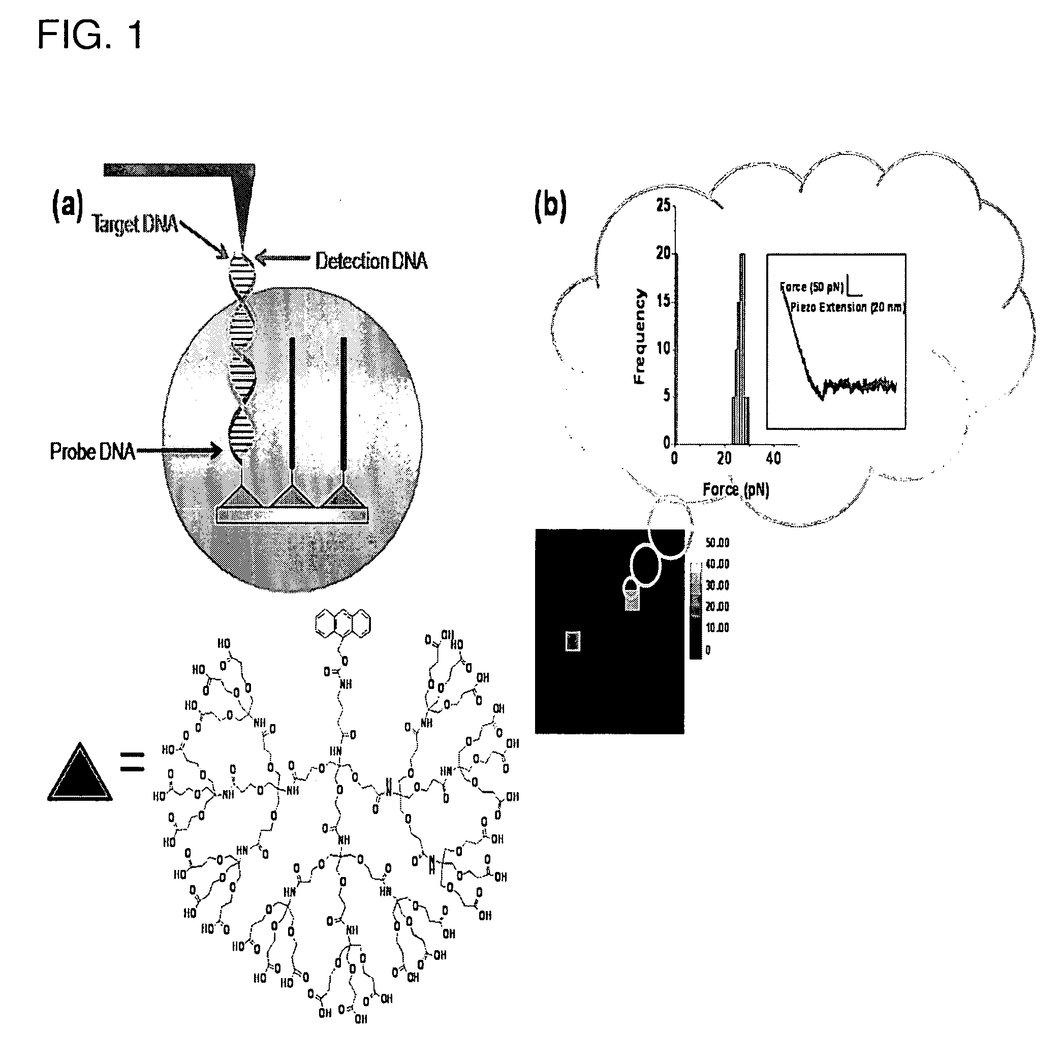
[0143]AFM Probe Pretreatment. Standard V-shaped silicon nitride cantilevers (MLCT-AUNM) with pyramidal tips (Veeco Instruments; k=10 pN / nm) were first oxidized by dipping in 10% nitric acid and heating at 80° C. for 20 min. The cantilevers were taken out of the solution and washed and rinsed thoroughly with a copious amount of deionized water. The clean cantilevers were dried in a vacuum chamber (30-40 mTorr) for about 20 min and used immediately for the next steps.


PUM

PropertyMeasurementUnit
diametersaaaaaaaaaa
diametersaaaaaaaaaa
distanceaaaaaaaaaa

Abstract

The present application discloses a method for detecting a presence of target ligand in a fluid medium which includes the steps of: (i) contacting the fluid medium with a solid substrate that includes an array of dendrons on its surface, wherein each of the dendron includes a central atom, a probe that is attached to the central atom optionally through a linker, and a base portion attached to the central atom and having a plurality of termini that are attached to the surface of the solid support; and (ii) determining the presence of a probe-target ligand complex by measuring binding force between the bound ligand and detection molecule tethered to the tip of an atomic force microscope (“AFM”), which detection molecule has affinity for the ligand, wherein measurement of an increase in force between the probe-target ligand complex and the detection molecule by AFM indicates the presence of the probe-target ligand complex.

Description

CROSS-REFERENCE TO RELATED APPLICATIONS[0001]The present application claims priority to U.S. Provisional Patent Application No. 60 / 944,056, filed Jun. 14, 2007, the contents of which are incorporated by reference herein in their entirety.BACKGROUND OF THE INVENTION[0002]1. Field of the Invention[0003]The present invention relates generally to atomic force microscopy (AFM), and an apparatus and a measuring method of intermolecular interaction between the biomolecules using the same on a chip. The present invention regards the usage of dendron coated Bio-AFM tips in measuring the interaction force between biomolecules on a chip.[0004]2. General Background and State of the Art[0005]Recent advances in bioanalytical sciences and bioengineering have led to the development of DNA chips1,2, miniaturized biosensors3,4, and micro fluidic devices (e.g., microelectromechanical systems or bioMEMS)5-7. In particular, DNA microarray technology is an increasingly important tool since it considerabl...

Claims


Application Information

Patent Timeline
Patent Type & Authority Applications(United States)
IPC IPC(8): C40B30/10C40B60/12
CPCB82Y35/00G01Q60/42G01N33/6845
Inventor PARK, JOON WONHONG, BONG JINKWON, SUNG HONG
Owner POSTECH ACAD IND FOUND
Why Eureka
Try Eureka