Method for obtaining structural and functional information on proteins, based on polarization fluorescence microscopy, and a device implementing said method

a technology of polarization fluorescence and protein structure, applied in the field of polarization microscopy, can solve the problems of limited ability to observe the functional activity of proteins in living cells, the presence and spatial distribution of fluorescence alone do not allow monitoring the functional activity of the studied protein, and all methods have limitations. achieve the effect of rapid acquisition of images and high quality

Inactive Publication Date: 2012-08-30
MIKROBIOLOGICKY USTAV AV CR V V I +1
View PDF5 Cites 2 Cited by
  • Summary
  • Abstract
  • Description
  • Claims
  • Application Information

AI Technical Summary

Benefits of technology

[0009]Since living cells undergo microscopic motions, a precise analysis of images acquired with different polarizations requires that these images be of high quality, and acquired over as short a time span as possible. Rapid acquisition of images with different polarizations also allows monitoring of rapid physiological processes, such as changes in calcium concentrations, or changes in cell membrane voltage.
[0010]A device for multi-photon, preferably two-photon, polarization fluorescence and higher harmonic emission microscopy that allows rapid acquisition of images with different polarizations of excitation light, and carrying out the method described herein, is another subject of this invention. Preferably, this device is in the form of a module attachable to a laser scanning microscope that allows synchronization with external or internal devices, and is suitable, in terms of quality and quantity of available excitation and observation capabilities, for observation of fluorescence of biological samples. The device comprises an excitation laser beam polarization modulator, synchronized with the microscope by a control unit and timing signals. In order to measure polarization of emitted fluorescence, the device can be preferably supplemented by a polarizer or a polarizing beam splitter inserted between the observed sample and the fluorescence detector.

Problems solved by technology

However, presence and spatial distribution of fluorescence alone generally do not allow monitoring functional activity of the studied protein, such as whether a receptor protein is activated, an ion channel open or closed, a transporter protein transporting, etc.
Our ability to observe functional activity of proteins in living cells is very limited.
Although useful, all of these methods have their limitations.
Transcription activation takes minutes to hours, which is too long to observe many systems.
FLIM requires expensive equipment, is not very sensitive, and FLIM data cannot be interpreted in terms of protein structure.
FRET requires two optically active molecules (e.g. fluorescent proteins), which often negatively affects function of the system studied.
Furthermore, the observed transfer of fluorescence energy (FRET signal) is only a fraction of the total fluorescence, and so is often hard to detect on the background fluorescence from the two present fluorescent moieties.

Method used


Image

  • Method for obtaining structural and functional information on proteins, based on polarization fluorescence microscopy, and a device implementing said method
  • Method for obtaining structural and functional information on proteins, based on polarization fluorescence microscopy, and a device implementing said method
  • Method for obtaining structural and functional information on proteins, based on polarization fluorescence microscopy, and a device implementing said method

Examples


example 1

A Device for Obtaining Structural and Functional Information About Proteins, Based on Polarization Fluorescence Microscopy

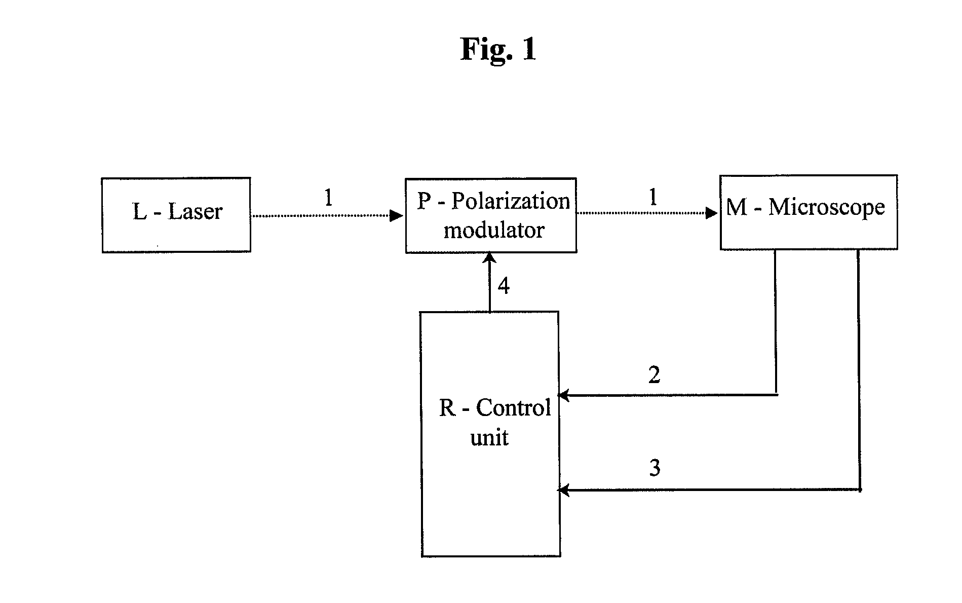
[0053]A device for carrying out polarization fluorescence microscopy, in principle illustrated in FIG. 1, has been created. The device comprises, as major components, a polarization modulator P acting on an excitation beam 1 emitted by a laser L, synchronized with a scanning fluorescence microscope M by a control unit R using timing signals 2 and 4.

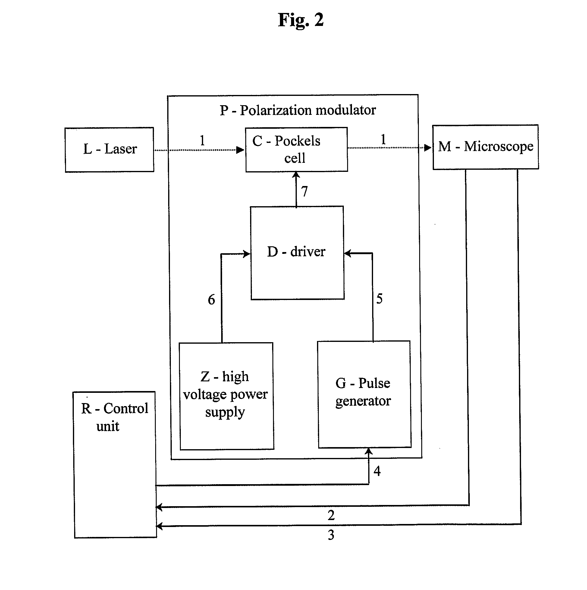
[0054]A device (shown in FIG. 2) has been designed to create a linearly polarized excitation laser beam 1 with direction of polarization modulated in synchrony with scanning of the microscope M. This was achieved by using a polarization modulator P consisting of a Pockels cell C (RTP-3-20-AR800-1000, Leysop Inc., United Kingdom) under the control of a high-voltage driver D (model B2, BME Bergmann GmbH, Germany). Driver D modulates high voltage 6 (0-1.2 kV), generated by a high voltage power supply Z (model HV, BME Berg...

example 2

Obtaining Structural and Functional Information About a Modified GFP Using Polarization Fluorescence Microscopy and the Device of Example 1

[0056]The device of Example 1 was used to acquire an image containing different parts (pixels) acquired with different polarizations of the excitation beam, i.e., a mixed image. The mixed image was deconvolved into separate individual images, each acquired with a different polarization of the excitation beam, in a manner schematically shown in FIG. 4. The result is illustrated in FIG. 5. Steps were taken during processing of the mixed image to account for delays in detector response and other factors. This was done by measuring the temporal response profile of the detectors used, and ascertaining what percentage of the signal (fluorescence) excited during a period of one polarization of the excitation beam is being reported in a pixel acquired during that period, and what percentage is being reported later. During deconvolution of the mixed image...

example 3

Monitoring and Determination of Protein Structure

[0064]The method and device for two-photon polarization microscopy of the invention have been used to determine and monitor structure of G-proteins, structure of a protein sensitive to calcium ion concentration, a protein sensitive to membrane voltage, receptor proteins, and a cytoskeleton attached protein (FIG. 6).


PUM

PropertyMeasurementUnit
timeaaaaaaaaaa
voltageaaaaaaaaaa
polarization fluorescence microscopyaaaaaaaaaa

Abstract

The invention pertains to a method of obtaining structural and functional information on proteins, based on polarization fluorescence microscopy, which comprises subjecting a protein tagged with a fluorophore to two- or multi-photon fluorescence microscopy, whereas the observed protein is irradiated with a laser beam with light of at least two different polarizations, which excites the fluorescence of the fluorophore, and wherein information on localization, intensities and polarizations of the fluorescence excited by the different polarizations of the excitation laser beam is used to identify, localize and quantify anisotropy of absorption and/or fluorescence, which information is then used to infer structural and functional properties of proteins. An example of a device for obtaining structural and functional information on proteins, based on polarization fluorescence microscopy, comprises a modulator (P) for rapid modulation of the excitation beam (1) for eliciting two- or multi-photon fluorescence, and a control unit (R), wherein the function of the modulator (P) and control unit (R) is synchronized with scanning of the microscope (M) such that information on fluorescence intensity acquired by the microscope (M) is attributable to a particular polarization state of the excitation beam (1) by virtue of knowing the temporal profile of the polarization modulation of the excitation beam (1) effected by the modulator (P). The method and device of the invention allow determining and monitoring structure and function of proteins, such as membrane proteins, and thereby observing physiological processes in living cells.

Description

FIELD OF THE INVENTION [0001]The invention described herein concerns polarization microscopy, specifically in two- and multi-photon arrangement. The invention is a method for obtaining structural and functional information about properties of proteins, preferably proteins in live cells, and a device implementing said method. The method and device allow, using two-photon or multi-photon polarization fluorescence microscopy, to determine and monitor structure and function of proteins, for example membrane proteins, and in doing so, observe physiological processes in live cells.BACKGROUND OF THE INVENTION[0002]Proteins in live cells can be studied, after being tagged by an optically observable label, by optical methods. Often, the optically observable tag used is a fluorescent protein. Presence of a fluorescent protein tag then allows observations of presence and spatial distribution of the studied protein in live cells, using a fluorescence microscope. However, presence and spatial di...

Claims


Application Information

Patent Timeline
Patent Type & Authority Applications(United States)
IPC IPC(8): G01N21/64C12M1/34
CPCG01N21/6428G01N21/6458G02B21/0076G01N21/6445G02B21/0068G02B21/16
Inventor LAZAR, JOSEF
Owner MIKROBIOLOGICKY USTAV AV CR V V I
Why Eureka
Try Eureka