Removing Cells from an Organism

a technology of cells and organisms, applied in the field of specific diseases, can solve the problems of severe side effects, inability to eliminate dace-associated diseases, and ineffective approaches, and achieve the effect of preventing the damage of desirable cells, and preventing or delaying cancer metastasis

Inactive Publication Date: 2013-08-01
OTT KARL HEINZ +1
View PDF17 Cites 15 Cited by
  • Summary
  • Abstract
  • Description
  • Claims
  • Application Information

AI Technical Summary

Benefits of technology

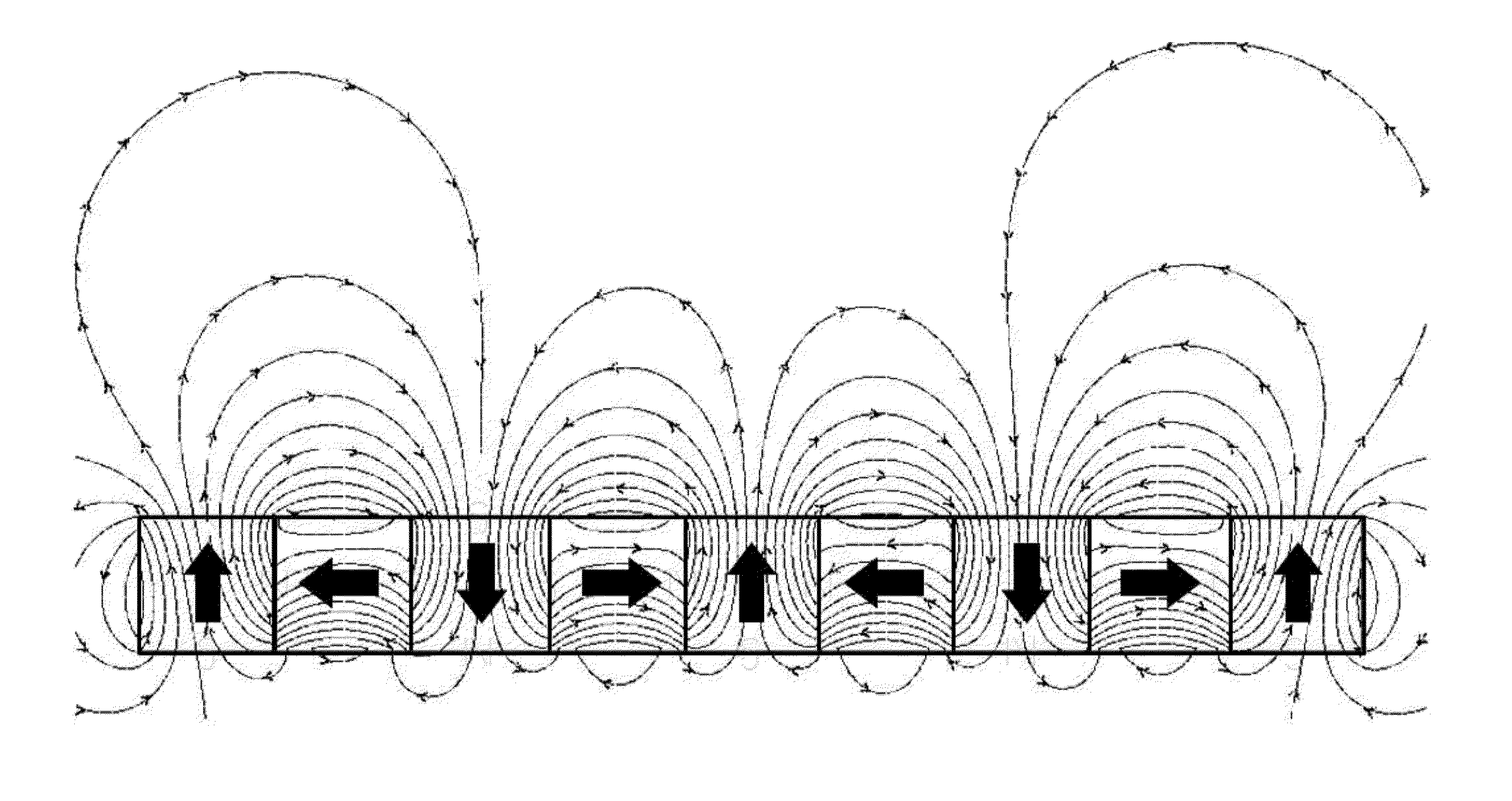
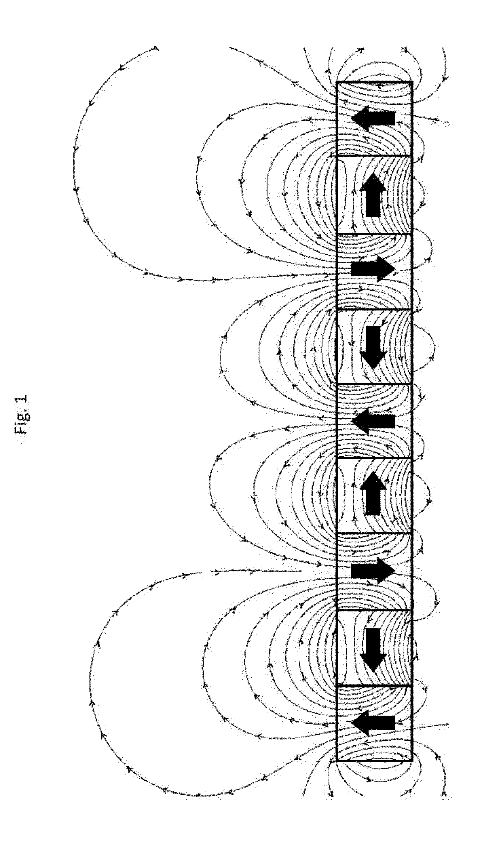
[0052]The invention further relates to methods to enhance the initial association and complex formation of the nanoparticles with the DACE. These methods enhance the kinetics of binding by applying a magnetic force to the superparamagnetic or paramagnetic nanoparticles, resulting in their relative motion with respect to the surrounding fluid that contains the DACEs.
[0055]In one particular embodiment, the DACEs in the circulatory system are removed by introducing fNPs into the circulatory system by injection, the formation of fNP-DACE complexes during circulation in the body, concentration or retention of the complexes in a target site and removal or destruction of the complexes. In one embodiment, the removal comprises venipuncture or a blood draw of the blood volume containing the fNP-DACE complexes at the site of arrest. In another embodiment, the complexes are removed from the lymphatic system by incubation to facilitate the formation of fNP-DACE complexes in the circulatory or lymphatic system and subsequent removal of the resulting fNP-DACE complexes from the lymphatic system, for example by removing of the lymph node in which the particles have been captured.
[0057]The invention draws knowledge about suitable Bioaffinity molecules and Bioaffinity targets from an extensive research into the biology of disease and immune-system associated cell and biological entities. It further builds on the research into disease diagnostic and immunomagnetic methods for identification of superior fNPs. The invention further benefits from experience with in vivo use of nanoparticle as used in some ablation and theragnosic approaches and from many years of experience with nanoparticles in in vivo imaging approaches. The invention adds to the arsenal of local and systemic methods to combat disease. It's novel and minimally invasive approach of the removal of DACEs may surpass other systemic methods as it prevents the damage to desirable cells and makes the treatment accessible to patients even outside of highly specialized and well equipped treatment centers.
[0058]The advantages of the present invention include preventing or delaying cancer metastasis by removing CTCs from circulation. Metastases, seeded from CTCs, are responsible for ˜90% of mortality of cancer patients. Successful removal of CTC circulation would add a significant new option for intervention of cancer progression and supplement other therapeutic options.

Problems solved by technology

Furthermore, it is sometimes desirable to isolate large quantities of specific agents from an organism, e.g., for research or diagnostic purposes.
These approaches, however, are not completely effective due in part to increasingly resistant strains of bacteria or the escape of cancer cells from systemic treatments.
Systemic treatments are often invasive and may cause severe side effects.
Local treatments, such as surgical removal of a tumor, can in most cases not be used to eliminate DACE-associated diseases.
1) poor outcome after surgery and chemotherapy is observed when CTCs remain in the body,
2) the number of CTCs in the blood is highly correlated with disease progression,
3) blood vessels and the lymphatic system act as conduits to transport malignant circulating tumor cells13.
However, these methods typically require extensive equilibration times, cannot process all the blood volume of a patient and are thus only useful for ex-vivo diagnostic, not therapy.
However two major roadblocks exist: 1) such profiling methodology has yet to be validated to yield suitable molecular targets.
2) Even if targets were identified, the process would still require the successful discovery, development and clinical testing of potentially suitable drugs or drug combinations—which is an arduous, time-consuming, expensive and uncertain task.
As such, methods of counting or isolating DACEs ex-vivo do not accomplish the healing of a patient.
These techniques are applicable to solid tumors, but they have generally not been useful for the ablation or elimination of cells circulating in blood or the lymphatic system, or of cells residing in various body cavities such as the bone marrow.
These techniques are not designed for the treatment of specific cells in the blood, but rather provide for the removal of proteins and molecules normally removed by properly functioning body organs.
These techniques do not treat circulating cells, much less specifically targeted cells, during the process.
Various devices have been developed for the separation or enrichment of specific cells from samples of body fluid, yet these devices are limited to operating on only the sample itself and are not capable to treat the entire blood component of a patient.
These devices, however, are designed and limited to utilize only a small fluid sample and therefore are not useful for treating the entire blood volume of a patient.
This device, however, is not selective in its application of the various treatments to specific cell types or blood components of the patient.
Disadvantages of these techniques therefore include the failure to preferentially treat the undesirable cell subpopulation in the irradiated blood stream, as opposed to treating the entire blood volume.
However it does not describe methods for such neutralization, nor does it describe how such methods distinguish between the target and the remaining blood cells.
This requirement severely limits the time during which a sample can make contact with an ablative energy source or a magnetic filter and has turned out to be one of the main reasons for why these devices have as yet failed to become therapeutically useful.
Similarly, the requirement to avoid shearing and other mechanical damage to the vital components of a biofluid severely limits certain design specifications of extracorporeal filtration devices, such as surface contact area, surface modification, pumping action, tube length, channel dimensions, Reynolds number, filter pore size and maximum achievable flow rate: Large pores permit the transmission of more sample but fail to capture small nanoparticles.
The use of large nanoparticles however limits an effective complex formation because of the large diffusion coefficient of the particles and target cells, which makes mixing inefficient and drives incubation times into hours and days.
Such therapeutic approaches, if effective, may supplement current choices in cancer treatments by attacking larger tumors or metastases but are not expected to be applicable to individually circulating DACEs as they lack the necessary ability to distinguish between DACE and natural cells.
However, there were no reports that such particles carried any significant side effects, indicating that properly functionalized nanoparticles (fNPs) may safely be utilized in vivo.
However, a minimally invasive method of direct elimination of a wide variety of DACEs has never been conventionalized.

Method used


Image

  • Removing Cells from an Organism
  • Removing Cells from an Organism
  • Removing Cells from an Organism

Examples


example 1

Production of fNPs

[0235]Superparamagnetic ironoxide nanoparticles containing a NH2 activation (CANdot Series M aq, ca, Hamburg, Germany) were obtained. A phycoerythrin (PE)-labeled human-EpCAM antibody (Miltenyi Bioscience, Germany, 130-091-253) was slightly reduced and linked via a SM(PEG)12 linker (Thermo-Fisher Scientific, Rockford, Ill. #22113) to the nanoparticles69. The nanoparticles are formulated in PBS suspension at a concentration of 10̂-6M to (about 1 mg / ml Fe) and purified by ultrafiltration and sterile filtration. The resulting particles have a size of 50 nm+−10 nm and show the strong fluorescence spectrum, typical for PE (FIG. 6). The particles are labeled fNP-2.

example 2

Validation of Antibodies and Cell Lines

[0236]The following cell lines were used (available from American Type Culture Collection, ATCC, Manassas, Va.):

a.MCF-7(human breast carcinoma)b.HCT-116(human colon carcinoma)c.Caco-2(human colon carcinoma)d.SEM(leukemia)

[0237]The cells were grown in a Petri plate, the growth media was removed, the cells washed twice with PBS buffer and separated by mild trypsination. About 1 million and 3 million cells per ml were obtained. The cells were centrifuged and suspended in 100 μl PBS buffer. A test sample (50 μl) of).

[0238]10 μl of human CD326(EpCAM)-Ab, FITC labeled, (Miltenyi order Nr. 130-096-415) was added to 100 μl of suspended cells. The results (Table 1) demonstrate that the chosen antibody produces strong staining and that the human tumor cell lines are all EpCAM positive. For the purpose of the in vitro validation, cells with low cell aggregation were chosen to facilitate analysis by flow cytometry / FACS.

TABLE 1Results from light and fluores...

example 3

Validation of fNP-DACE Complex Formation

[0239]Cell lines HCT-116 (colon carcinoma cells), BXPC-3 (pancreas carcinoma cells), SU8686 and PANC1 (available from American Type Culture Collection, ATCC, Manassas, Va.) were grown using standard procedures and prepared as in Example 2. The cells were incubated in an eight-well chamber slide in 100 μl medium with 1 μl fNP-1 (h-EpCAM) MicroBeads, Miltenyi Bioscience #130-061-101) or 2 μl fNP-2 (See Example 1) for about 10-20 min at room temperature. Thereafter the cell nuclei were stained with DAPI (blue) for 5 min.

[0240]Under fluorescence microscopy, HCT-116 (colon carcinoma cells) show strong staining with fNP-1 and fNP-2 (FIG. 8). BXPC3 (pancreas carcinoma cells) show strong staining with fNP-1 and fNP-2 as well as possible internalization of the fNPs (FIG. 8). SU8686 and PANC1 (both pancreas carcinoma cell lines) are only weakly stained with both fNP-1 and fNP-2 (FIG. 8). SEM cells (acute lymphoblastic leukemia) were introduced as negati...


PUM


Abstract

A minimally invasive method to eliminate circulating agents from an organism to prevent and treat diseases is disclosed. The method utilizes immunomagnetic methods to concentrate and localize disease-associated agents in a small region of the body. Subsequently, the complexes are removed from the body. Removal of disease-causing or disease-promoting agents (circulating tumor cells, bacteria, viruses and virus-infected cells, certain immune cells) would add a significant new option for intervention of disease progression and supplement other therapeutic options.

Description

CROSS-REFERENCE TO RELATED APPLICATIONS[0001]This application claims priority to U.S. Provisional Application No. 61 / 586,461, filed Jan. 13, 2012, which is herein incorporated by reference in its entirety.BACKGROUND[0002]An organism, in particular, body fluids and body cavities of humans and animals may carry a wide variety of disease-causing, disease-modifying, or otherwise unwanted agents (herein called disease-associated circulating entities, DACEs). Elimination of such agents can help treat diseases associated with such agents.[0003]This invention relates to managing specific diseases, particularly metastatic cancers. The present invention is the specified use of nanostructures. The present invention is a medical treatment for cancer, immunological and infectious diseases.[0004]Disease associated agents, for example disease associated cells, are generally rare in a given body fluid and comprise a small percentage of the total number of endogenous molecules or cells. Therefore, m...

Claims


Application Information

Patent Timeline
Patent Type & Authority Applications(United States)
IPC IPC(8): A61N2/00A61M3/00A61N2/06
CPCA61N2/004A61N2/06A61M3/00B82Y5/00A61K9/5115A61K47/4893A61K9/0009A61K9/5094A61K41/00A61K47/6941
Inventor OTT, KARL-HEINZDAPPRICH, JOHANNES
Owner OTT KARL HEINZ
Why Eureka
Try Eureka