Pharmacology Bioassays for Drug Discovery, Toxicity Evaluation and in vitro Cancer Research Using a 3D Nanocellulose Scaffold and Living Tissue
a nanocellulose and living tissue technology, applied in the field of drug discovery bioassays, can solve the problems of insufficient model development, inability to replicate cell-matrix interactions found in vivo, and the need to provide a 3d architecture, so as to achieve cost-effective screening of drugs and high throughput
- Summary
- Abstract
- Description
- Claims
- Application Information
AI Technical Summary
Benefits of technology
Problems solved by technology
Method used
Image

Examples
example 1
Development of Human Cartilage on Nano-Cellulose Scaffold
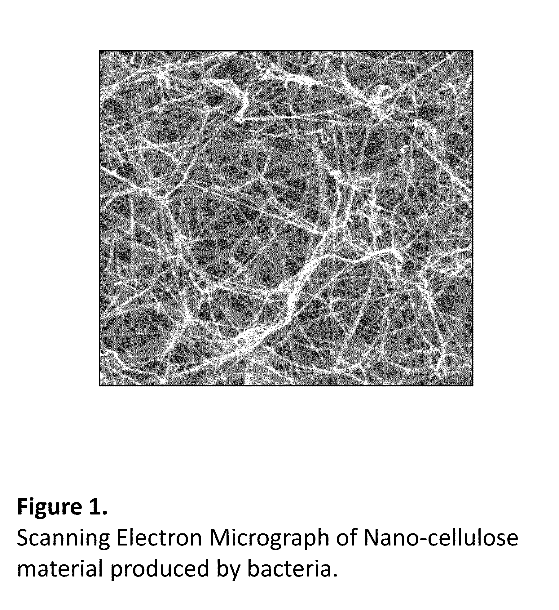
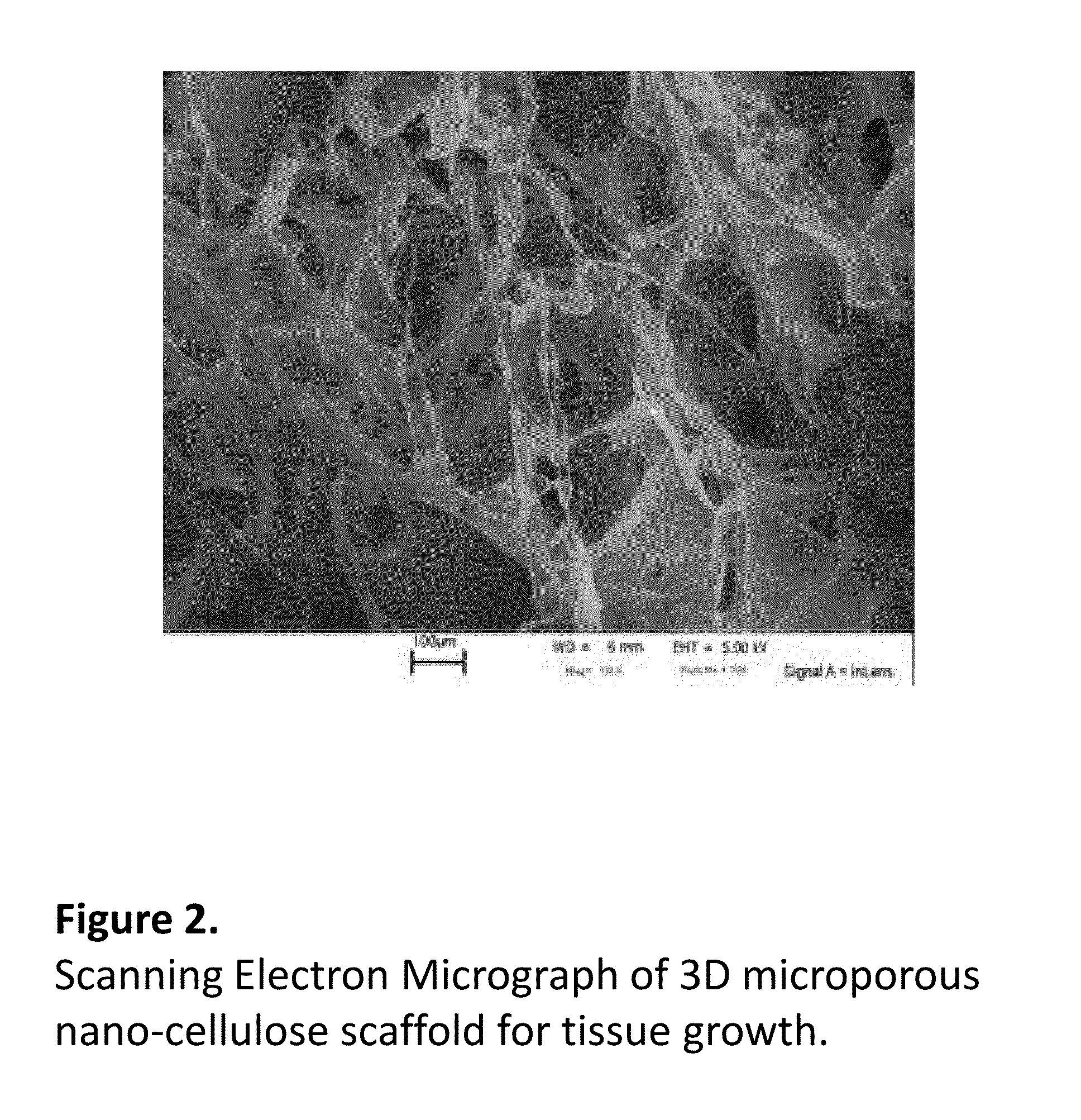
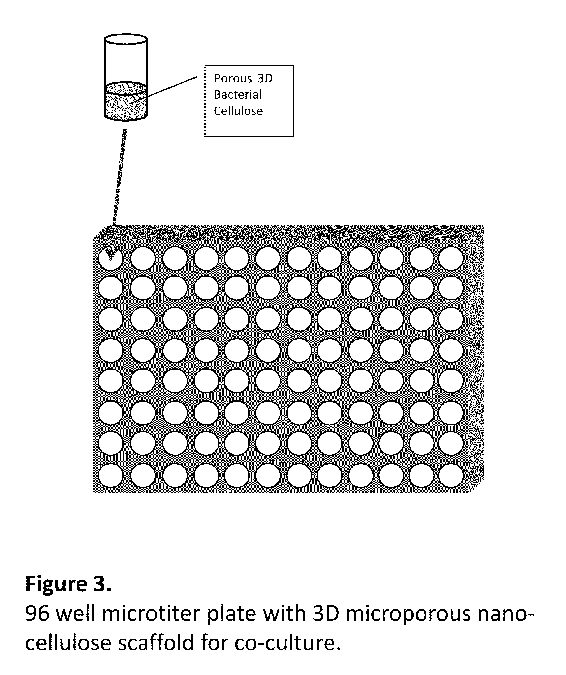
[0046]Nano-cellulose was prepared by fermentation of Gluconoacetobacter Xylinus using corn steep liquor medium. Wax particles size 300 microns were added during the fermentation process and removed by melting and repeated washing. An alternative route of making 3D porous scaffolds is homogenization of bacterial nano-cellulose using Ultratorax followed by freezing of dispersion at −80° C. followed by freeze drying at −50° C. for 24 hours. The resulting porous 3D nano-cellulose scaffold was placed in wells in 96 microtiter plates, sterilized and seeded with human articular chondrocytes. To study formation of cartilage, cells isolated from three patients were expanded and seeded onto 3D porous nano-cellulose scaffolds. Dulbecco's modified eagle's media (DMEM) / F12 (Invitrogen, Grand Island, N.Y.) supplemented with L-ascorbic acid (0.025 mg / mL), gentamicin sulfate (50 mg / L), amphoterricin B (250 mg), L-glutamine (2 mM), and 10% hum...
example 2
Growth of Co-Culture of Endothelial and Smooth Muscle Cells—Model of Arteries and Blood Vessels to Study Arteriosclerosis and Plaque Formation
[0047]3D nano-cellulose scaffolds were designed to mimic (e.g., represent, copy, be similar to, be characterized by, etc.) vascular tissues. Channels were produced by inserting optical fibers with diameter of 500 micron and surrounded by wax particles of diameter 200 microns in bacterial cellulose fermentation process. 3D nano-cellulose scaffold produced this way was purified and sterilized. Scaffold was then placed in the bottom of the titer microplate. Endothelial cells (HSVECs) and smooth muscle cells were isolated from non-diseased human saphenous veins, by-products of coronary bypass surgery. Cells were isolated using an enzymatic technique using a solution of 0.1% collagenase type I in Phosphate Buffered Saline. Endothelial cells were then seeded in channels of nano-cellulose scaffold and smooth muscle cells were seeded in a porous part ...
example 3
Human Mesenchymal Stem Cell Differentiation in 3D Porous Nano-Cellulose
[0048]3D microporous nano-cellulose scaffolds with porosity of 300 microns produced using wax porogens were pretreated with anionic polysaccharides such as carboxymethylcellulose followed by treatment with simulated body fluid to produce biomimetic coating consisting of hydroxyapatite. Such scaffolds were seeded with human mesenchymal stem cells. The differentiation media (growth media supplemented with 0.13 mM ascorbic acid 2-phosphate, 2 mM β-glycerophosphate and 10 nM dexamethasone) was used. Cells were cultivated in an incubator at 37° C., 5% CO2 and 95% relative humidity. The culture medium was changed every third day. The proliferation was studied using MTS assay and results showed that the cells proliferated. Samples at 7, 14 and 21 days were analyzed with Alkaline Phosphatase ELISA Assay Kit assay. Results showed that human mesenchymal stem cells have differentiated into osteoblasts after 21 days cultivat...
PUM
| Property | Measurement | Unit |
|---|---|---|
| Thickness | aaaaa | aaaaa |
| Biodegradability | aaaaa | aaaaa |
| Toxicity | aaaaa | aaaaa |
Abstract
Description
Claims
Application Information
PatSnap Eureka turns technology decisions into work you can execute. Powered by our Innovation Knowledge Graph, it runs expert workflows across engineering, life sciences, materials and intellectual property. Get your review-ready output in minutes.


