Probe for detecting dead cell
a technology for detecting dead cells and probes, applied in the field of probes, can solve the problems that molecules are not suitable for quantitative image analysis, and achieve the effects of accurate detection, high detection accuracy, and quantitative analysis
- Summary
- Abstract
- Description
- Claims
- Application Information
AI Technical Summary
Benefits of technology
Problems solved by technology
Method used
Image
Examples
example 1
Preparation of Wild-Type or Modified mTim4-Fc
[0057]By the following procedure, a fusion protein of a wild-type or modified mouse Tim4 and a human IgG Fc region protein was prepared.
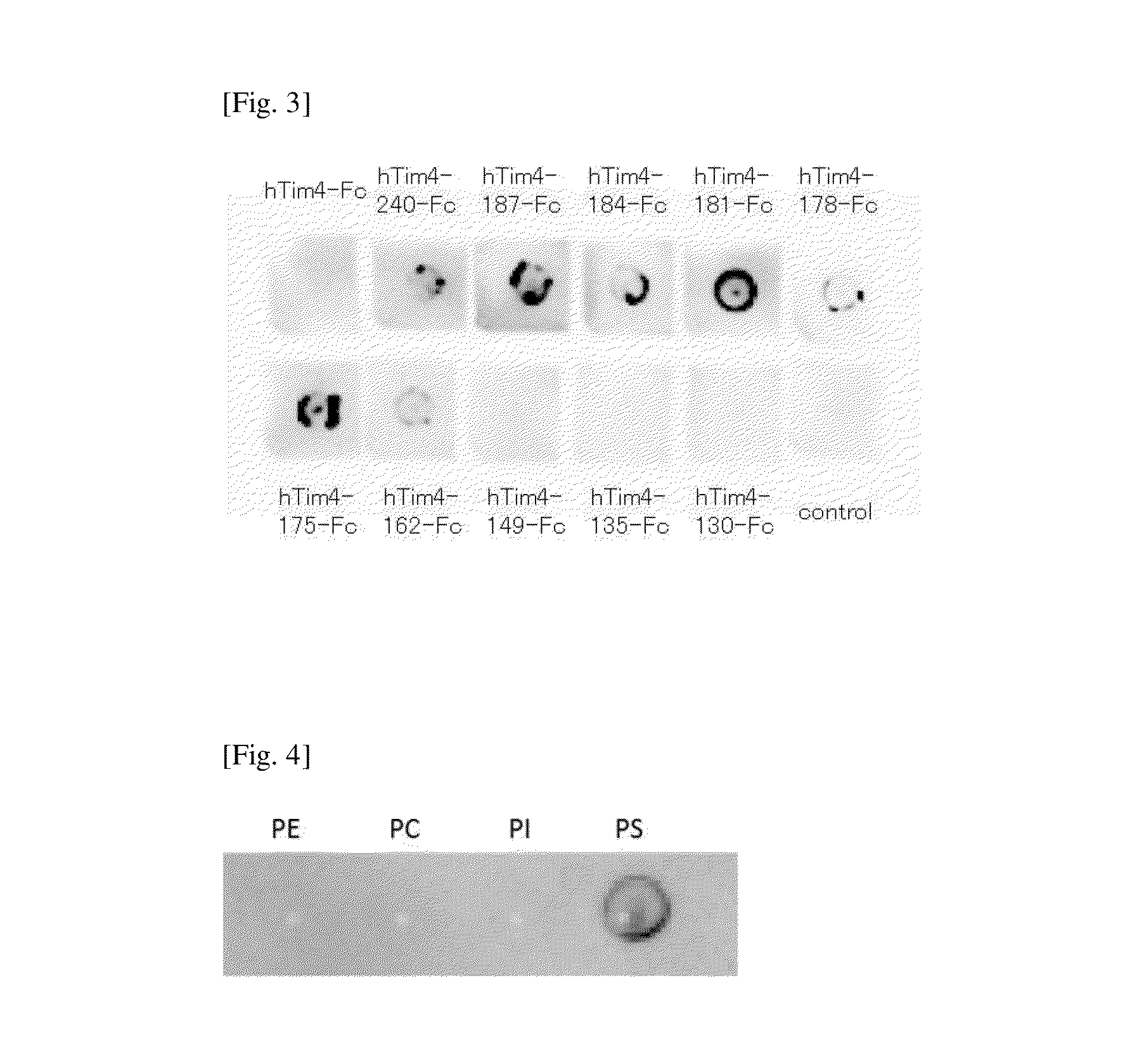
[0058]E. coli (JM109, Takara Bio Inc.) was transformed with a plasmid vector encoding the sequence information of a fusion protein of a wild-type mouse Tim4 and a human IgG Fc region protein (mTim4-Fc), pTim4-Fc (provided by Prof. Shigekazu Nagata; see Nature (2007) 450, 435-439 for the preparation method), so as to amplify the plasmid vector. The plasmid vector was purified using FastPlasmid Mini Kit-250 preps (V Prime), and PCR was carried out using the obtained pTim4-Fc as a template and Expand High Fidelity PCR System (Roche). Using the primers of SEQ ID NOs:11 and 12, a DNA fragment encoding mTim4-ΔM230-Fc (a modified type fusion protein comprising a mTim4 part in which the C-terminal side of the mucin domain of the wild-type mTim4 is deleted so that the amino acid sequence from the N-terminus to the...
reference example 1
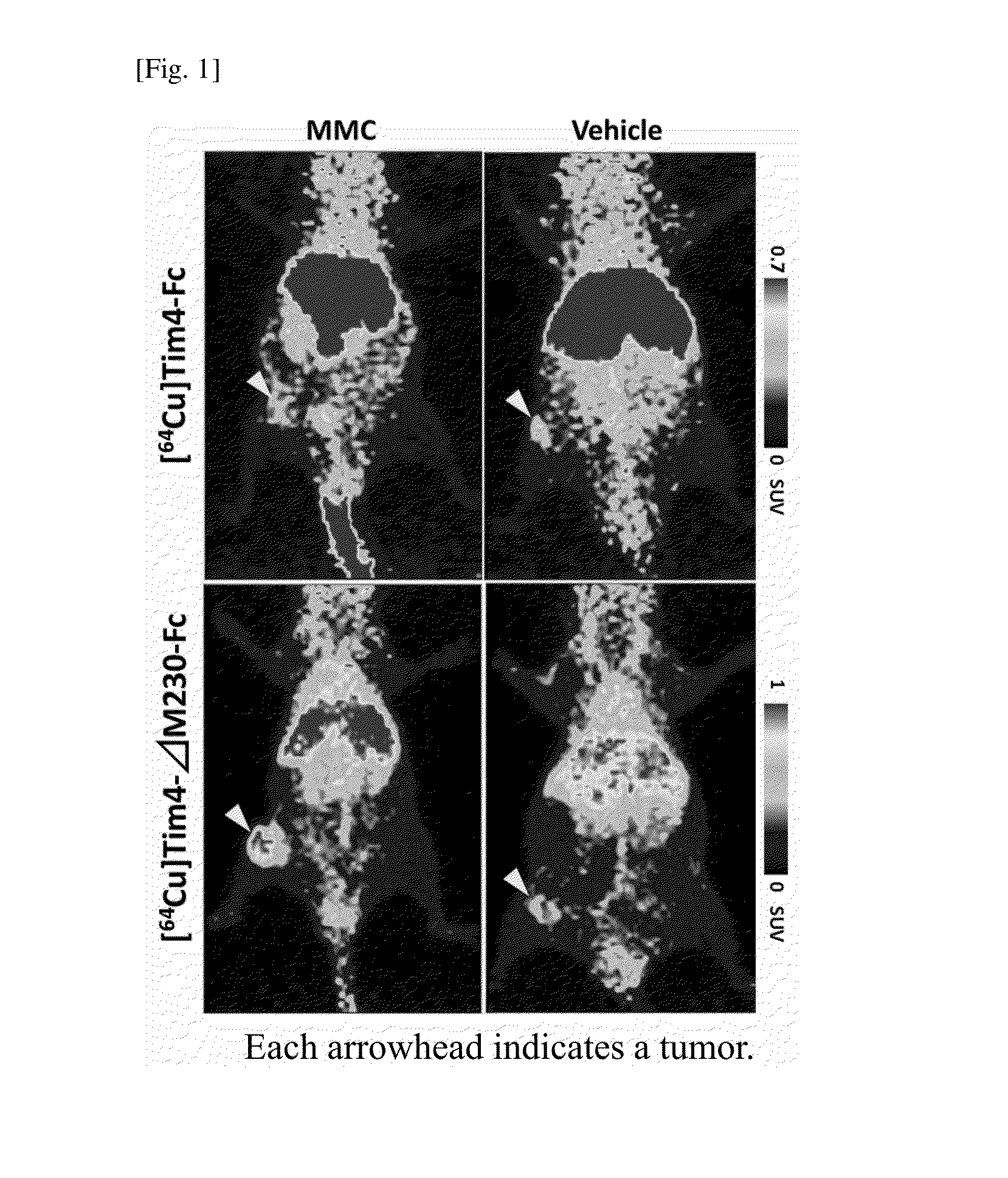
[0060]By the following procedure, the fusion proteins obtained as described above (mTim4-Fc, mTim4-ΔM230-Fc, and mTim4-ΔM184-Fc) were each labeled with RI.
[0061]The stored solution of each fusion protein was subjected to buffer exchange to PBS (D-PBS, Wako Pure Chemical Industries, Ltd.). An aqueous solution of 10 mM p-SCN-Bn-NOTA (NOTA, Macrocyclics) adjusted to pH7.9 to 8.4 with 0.1 N NaOH was added to each fusion protein at an amount of 1000 equivalents, and the resulting mixture was left at 4° C. overnight statically. Thereafter, the reactant was applied to a desalting column (PD-10, Thermo Sci.) equilibrated with PBS, the fusion protein bound to NOTA was separated from unbounded NOTA. The NOTA-binding fusion protein fraction was concentrated using Amicon Ultra, and was subjected to buffer exchange to 100 mM acetate buffer (pH 6.5). Thereafter, 6 MBq of an aqueous solution of [64Cu]CuCl2 per 1 μg of the NOTA-binding fusion protein was added thereto, and the resulting ...
reference example 2
Diagnosis of Apoptosis in Target Tissue
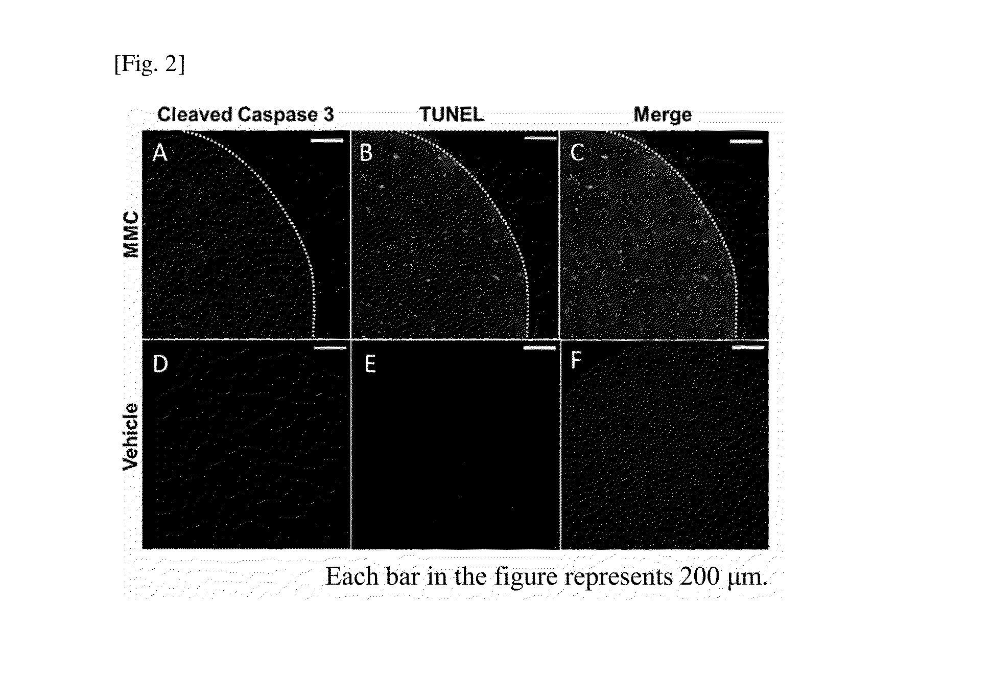
[0067]After the completion of the PET experiment, the tumor was isolated from each mouse, and frozen sections were prepared. The prepared frozen sections were fixed with cold methanol, and stored at −20° C. until use. The sections were used for the experiment within 3 to 5 days after the dissection.
[0068]The frozen sections were subjected to blocking using Protein Block, Serum-Free (Dako), and then subjected to TUNEL staining using DeadEnd Fluorometric TUNEL System (Promega). Thereafter, a 1000-fold diluted primary antibody (Cleaved Caspase-3(Asp175)Antibody, CST) was react with the sections at room temperature for 2 hours, and a secondary antibody (Cy3-conjugated anti-rabbit IgG antibody) was then reacted with the sections at room temperature for 2 hours. Also, nuclear staining was performed using Hoechst 33256 (Dojindo). Thereafter, the samples were observed under a confocal laser microscope.
[0069]The results are shown in FIG. 2. In the froze...
PUM
| Property | Measurement | Unit |
|---|---|---|
| molecular weight | aaaaa | aaaaa |
| molecular weight | aaaaa | aaaaa |
| molecular weight | aaaaa | aaaaa |
Abstract
Description
Claims
Application Information
 - R&D
- Intellectual Property
- Life Sciences
- Materials
- Tech Scout
- Unparalleled Data Quality
- Higher Quality Content
- 60% Fewer Hallucinations
Browse by: Latest US Patents, China's latest patents, Technical Efficacy Thesaurus, Application Domain, Technology Topic, Popular Technical Reports.
© 2025 PatSnap. All rights reserved.Legal|Privacy policy|Modern Slavery Act Transparency Statement|Sitemap|About US| Contact US: help@patsnap.com


