Nanomaterials for the integration of soft into hard tissue

Inactive Publication Date: 2016-04-14
NORTHEASTERN UNIV
View PDF17 Cites 1 Cited by
  • Summary
  • Abstract
  • Description
  • Claims
  • Application Information

AI Technical Summary

Benefits of technology

The invention provides a device called an enthesis regeneration device that can be used to help regenerate an attachment site between a tendon or ligament and bone. The device helps to cushion the insertion of the tendon or ligament to bone and promotes the growth of anenthesis, which helps to improve joint stability and increase the success rate of reconstructive joint surgery. The device is designed to fit snugly in a drilled bone tunnel and allows orthopedic soft tissue to pass through the center of the device. It requires minimal changes to current surgical procedures and promotes the natural graded transition of the tendon bone insertion.

Problems solved by technology

The enthesis lacks a direct blood supply and cannot regenerate once it is injured.

Method used


Image

  • Nanomaterials for the integration of soft into hard tissue
  • Nanomaterials for the integration of soft into hard tissue
  • Nanomaterials for the integration of soft into hard tissue

Examples


example 1

Synthesis of Nanocrystalline Hydroxyapatite

[0037]Following an established procedure, hydroxyapatite (HA) precipitates were synthesized by a wet chemistry process and then treated hydrothermally to produce nanometer-sized HA. See Lopez-Macipe, et al., Wet chemical synthesis of hydroxyapatite particles from nonstoichiometric solutions, J Mater Synth Process, 6, pp. 21-6, 1998; Sato, M., et al., Increased osteoblast functions on undoped and yttrium-doped nanocrystalline hydroxyapatite coatings on titanium, Biomaterials, 27, pp. 2358-69, 2006; and Zhang, L., et al., Biomimetic Helical Rosette Nanotubes and Nanocrystalline Hydroxyapatite Coatings on Titanium for Improving Orthopedic Implants, International Journal of Nanomedicine, 3, pp. 323-34, 2008. With constant stirring, 37.5 mL of a 0.6 M ammonium hydroxide solution was added to 375 mL of deionized water which had been cooled to below 10° C. Approximately 4 mL of ammonium hydroxide was used to adjust the solution pH to about 10. The...

example 2

Nanoparticle Characterization

[0038]A JEOL JEM-101 transmission electron microscope (TEM) was used to characterize the average size and shape of the magnesium oxide (MgO) nanoparticles (purchased from US Research Nanomaterials (20 nm particle size); www.us-nano.com) and HA nanoparticles (synthesized as described in Example 1). The nanoparticle crystal structures were confirmed by x-ray diffraction (XRD), and their chemistry was characterized using Fourier transform infrared spectroscopy (FTIR).

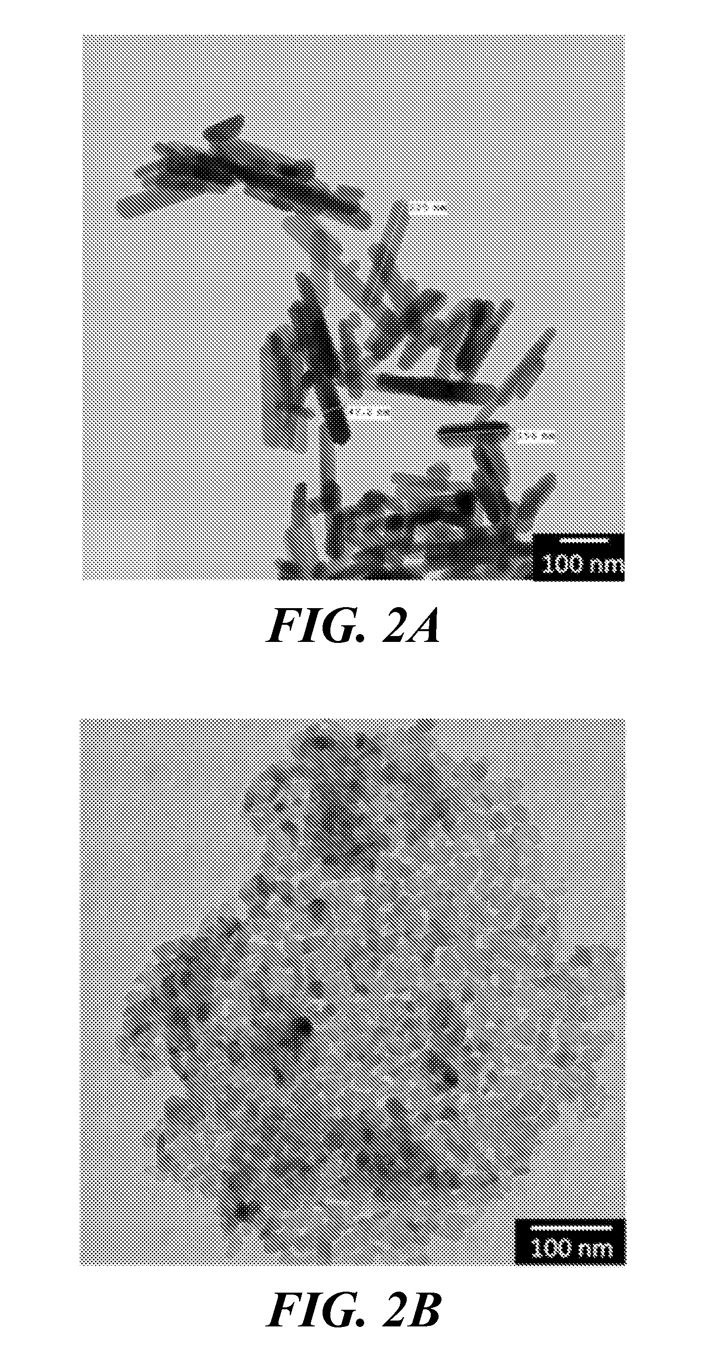
[0039]TEM images revealed that the synthesized HA nanoparticles were rod-shaped with an average length of about 200 nm and an average width of about 40 nm (FIG. 2A). MgO nanoparticles appeared circular under TEM, with an average particle diameter of 20 nm (FIG. 2B). The FTIR spectrum, and therefore the chemical composition, of the synthesized nanoparticulate HA matched a spectrum found in literature (FIG. 3A) (Yang B, et al., Preparation and characterization of bone-like hydroxyapatite / poly(met...

example 3

Preparation of Nanocomposites

[0040]Thin polymer nanocomposite sheets were prepared by a casting method. Poly(L-lactic) acid (PLLA) (Polysciences, MW=50,000 Da), HA nanoparticles produced as described in Example 1, and MgO nanoparticles (US Research Nanomaterials, particle size 20 nm) were placed into 20 mL scintillation vials in the amounts indicated in Table 1. Then, 10 mL of chloroform was added to give 3 wt % dry ingredients in chloroform. Each vial was sealed tightly and sonicated at 40 kHz for 1 hour in a water bath set at 55° C., being careful not to exceed the 60° C. boiling temperature of chloroform. After sonication, the suspension appeared homogeneous. The polymer suspensions were poured into 60-mm diameter Pyrex petri dishes and heated at 55° C. for ˜40 minutes to evaporate excess solvent. Samples were then allowed to sit overnight, producing polymeric sheets ˜0.2 mm in thickness.

TABLE 1Amount (in grams) of dry ingredients used toproduce PLLA nanocompositesPLLA (g)Nano-HA...


PUM

PropertyMeasurementUnit
Thicknessaaaaaaaaaa
Diameteraaaaaaaaaa
Diameteraaaaaaaaaa

Abstract

Nanocomposite materials are provided for attaching soft tissue to hard tissue of a mammalian subject. The materials include a biodegradable polymer network suffused with mineral nanoparticles. The nanocomposite materials have a surface structure that promotes the infiltration, adhesion and proliferation of cells such as osteoblasts and fibroblasts, and are useful to reconstruct enthesis tissue, such as a tendon bone insertion. Devices containing the nanocomposites and methods for implantation of the devices at a tendon-bone interface or ligament-bone interface are provided for reconstructive surgery.

Description

CROSS REFERENCE TO RELATED APPLICATIONS[0001]This application claims the priority of U.S. Provisional Application No. 61 / 827,164 filed 24 May 2013 and entitled “Nanomaterials for the Integration of Soft into Hard Tissue”, the whole of which is hereby incorporated by reference.STATEMENT REGARDING FEDERALLY SPONSORED RESEARCH OR DEVELOPMENT[0002]The invention was developed with financial support from Grant No. 0965843 from the National Science Foundation. The U.S. Government has certain rights in the invention.BACKGROUND[0003]Biomaterials for the regeneration of bone tissue have been investigated for use in treatment of a growing population of people with damaged and degrading bone. While successful regeneration of bone defects has been demonstrated, there is considerable room for improvement. Ideally, biomaterials for tissue regeneration should mechanically match their implant location and possess suitable chemical and topographical properties for promoting cellular adhesion, prolife...

Claims


Application Information

Patent Timeline
IPC IPC(8): A61F2/08A61L27/54A61L27/44A61L27/58A61L27/46
CPCA61F2/0811A61L27/58A61L27/46A61L27/446A61L27/54A61L2430/10A61L2300/102A61L2300/222A61L2300/64A61L2300/414A61L2300/112A61K38/39A61K35/28A61K35/32A61K35/33A61L27/3804A61L27/3817A61L27/3821A61L27/3834A61L27/50A61L2400/12C08L67/04C08L89/06
Inventor HICKEY, DANIEL J.WEBSTERERCAN, BATUR
Owner NORTHEASTERN UNIV
Why Eureka
Try Eureka