
Methods for Development and Use of Minimally Polarized Function Cell Micro-Aggregate Units in Tissue Applications Using LGR4, LGR5 and LGR6 Expressing Epithelial Stem Cells

a function cell and micro-aggregate technology, applied in the field of constructs of micro-aggregate multicellular grafts, can solve the problems of complex full thickness injuries to human and mammalian tissues, complex injuries involving multiple tissue elements, and inability to produce viable and self-sustaining epithelial compartments, etc., to achieve the effect of stabilizing primary tissues and reducing the viability of micro-organisms during transport and processing

Active Publication Date: 2016-06-02
GRANDER ACQUISITION LLC
View PDF0 Cites 2 Cited by
  • Summary
  • Abstract
  • Description
  • Claims
  • Application Information

AI Technical Summary

Benefits of technology

This patent describes a new formulation for a cell sustaining medium used to reduce the viability of micro-organisms during transport and processing of tissues. The formulation includes a mixture of epithelial cells and keratinocytes, as well as certain antibiotics and antimycotics. This formulation is effective at stabilizing the primary tissues and reducing the risk of infection during wound healing. Additionally, this patent describes the use of this formulation in combination with a scaffolding substrate for regenerating skin, muscle, and bone in mammals. The stem cells used in this composition have the ability to generate native anti-microbial peptides, which further reduce the risk of infection and promote faster healing. A final aspect of this patent is the fact that the use of LGR stem cells, particularly when combined with a scaffolding substrate, can enhance and accelerate tissue generating, regenerating, enhancing, and / or healing in mammals.

Problems solved by technology

At this time, there is limited research on how transplantation and application of isolated LGR4, LGR5 and LGR6 expressing epithelial stem cells alter wound healing gene expression.
It is known that LGR4, LGR5 and LGR6 expressing epithelial stem cell populations are often destroyed following severe full-thickness damage to the skin, leaving tissues incapable of producing a viable and self-sustaining epithelial compartment.
Complex full thickness injuries to human and mammalian tissues and / or complex injuries involving multiple tissue elements (skin, muscle, fat, blood vessels, nerves and bone) are difficult in nature to heal.
Such injuries and subsequent resulting wounds are also difficult to treat through current wound care methods, surgical interventions with current approved technologies utilizing cells, tissues, devices, biologics, drugs and / or growth factors.
Such deficiency in the cellular niche, ECM (extracellular matrix) scaffolding and related interactive interfaces subsequently results in failure to re-generate or generate the essential multi-dimensional architecture required for cellular migration, differentiation, and tissue polarization.
Without these cell-to-cell and cell-to-matrix interactions, remaining cellular entities within the wound bed, no matter their proliferative or lineage potential, are forced to provide primarily a barrier utility rather than develop a more complex, multi-tissue construct capable of recognizable “function.” Consequently, the wound—whether involving skin, muscle, fat, tendon, bone—becomes subsequently scarred, disorganized and dysfunctional.
However, in the absence of the above-identified inter-dependent, necessary components, tissue engineering applications, cell therapy applications, regenerative medicine applications, tissue healing applications and tissue transplant therapy applications do not possess the natural cellular micro-aggregate architecture needed to competently assemble functional polarized tissues.
These products, although beneficial, are often expensive and do not provide the patient with a true multi-compartment tissue construct.
For example, cultured epithelial autograft (CEA) remains unable to restore both epithelial and dermal compartments seen in native skin.
This failure, in turn, leads to monolayer fragility, epithelial instability, barrier breakdown, and scar.
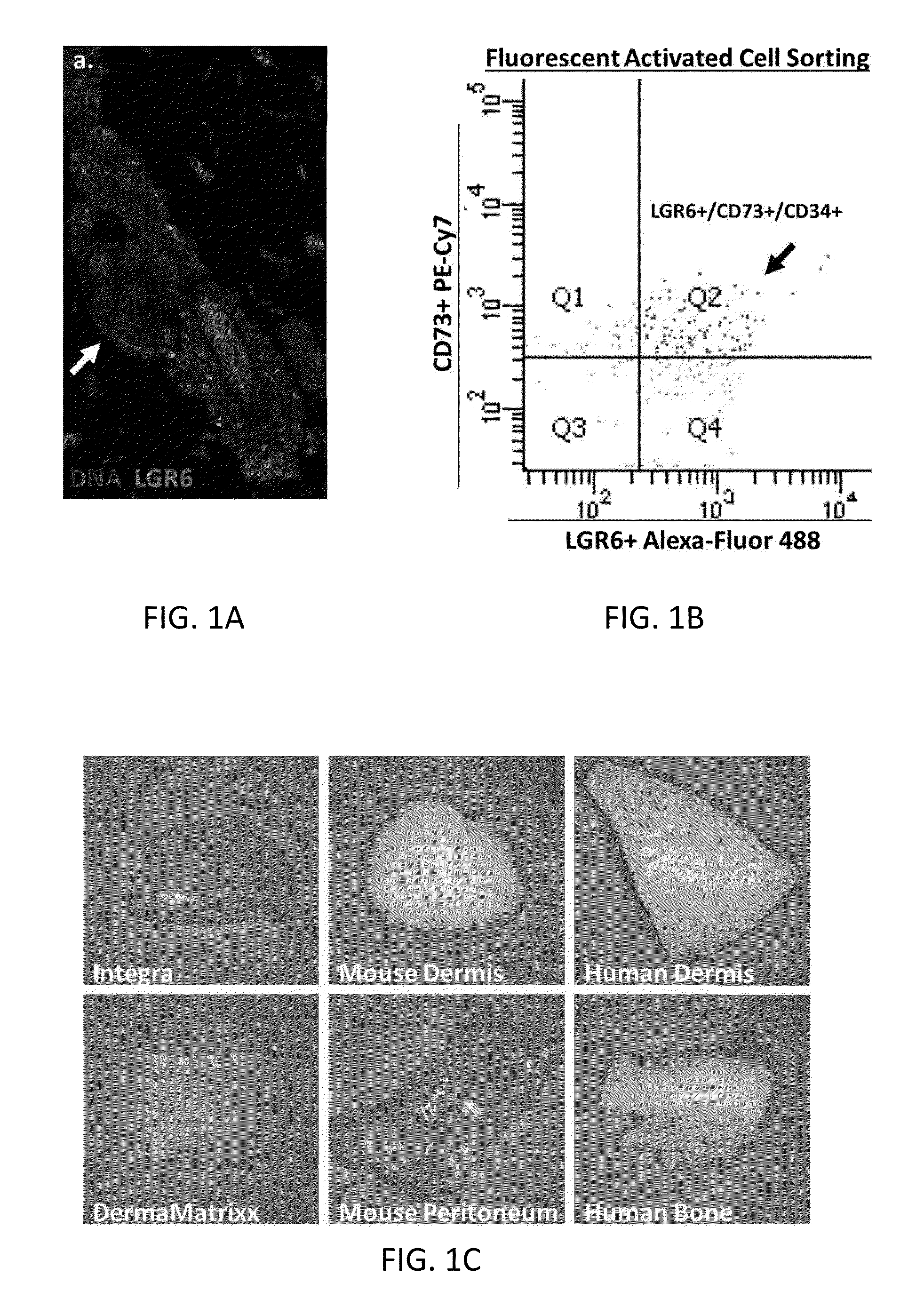
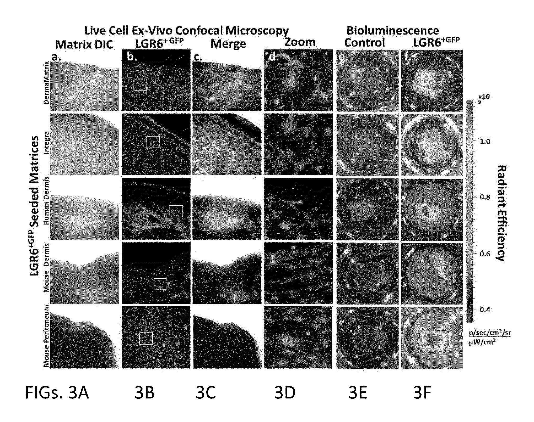
Alternatively, the more robust acellular matrices such as Alloderm® from LifeCell Corporation, Integra® from Integra LifeSciences Corporation and DermaMatrix a product from Musculoskeletal Transplant Foundation, although excellent reconstructive options, lack those properly placed lineage specific stem cell populations which are necessary to develop functional native tissues.

Method used


Image

  • Methods for Development and Use of Minimally Polarized Function Cell Micro-Aggregate Units in Tissue Applications Using LGR4, LGR5 and LGR6 Expressing Epithelial Stem Cells
  • Methods for Development and Use of Minimally Polarized Function Cell Micro-Aggregate Units in Tissue Applications Using LGR4, LGR5 and LGR6 Expressing Epithelial Stem Cells
  • Methods for Development and Use of Minimally Polarized Function Cell Micro-Aggregate Units in Tissue Applications Using LGR4, LGR5 and LGR6 Expressing Epithelial Stem Cells

Examples


example 2

[0148]Example 2 is directed to processing of hypodermis and subdermal fat cellular components. Example 2 recites the following steps:[0149]i. Spraying 70% ethanol (EtOH) on the outer side of the tissue container and placing the tissue container into laminar air flow cabinet;[0150]ii. Sending a sample of the tissue or transfer medium for microbiological testing;[0151]iii. Placing the previously washed adipose and hypodermal tissue in 150 mm sterile petri dish;[0152]iv. Washing the tissue two times with PRM;[0153]v. Trimming the tissue into small (3 mm) pieces with sterile surgical instrument and place into sterile culture holding dish containing pulse media while the dissection is completed;[0154]vi. Aspirating media from holding dish and removing the specimen with sterile scoop or forceps followed by placing the specimen into 50 ml conical tube containing MSC Enzymatic Digestive Media, a pre-mixed digestive enzyme solution (collagenase and dispase-based), which is placed into a 37° ...

example 3

[0164]Example 3 is directed to addition of hypodermis and subdermal fat components to the example of a construct according to an embodiment of the invention. The illustrative component addition example involves:[0165]i. Placing a sterile Nunc Skin Graft Cell Culture Dish or automated dish already containing the assembled and washed scaffold into a laminar flow hood and washing the scaffold again two times with pulse media prior to adding cells;[0166]ii. Inserting a label on each culture vessel with tracking number;[0167]iii. Transferring around 5×10⁵ to 1×10⁶ mixed SVF cells per dish system and 1×10⁵ adipocytes per dish;[0168]iv. Adding a complete culture medium with or without autologous PRP as dictated by the particular requirements of a situation, to the loading reservoir;[0169]v. Transferring the dishes into an incubator onto slow rocker for 1 hour followed by removal therefrom and resting flat for 48 hours in separate sentinel incubator;[0170]vi. Washing the culture medium afte...

example 4

[0175]Example 4 concerns enrichment of the minimally polarized, epithelial stem cell singularity units.

[0176]Following Example 1, the MPFUs is placed in pulse rescue media in a 15 ml conical tube and spin / centrifuged into a soft pellet. The material is then subject to the following process of partial digestion:[0177]i. Obtaining a previously aliquoted frozen 10 ml digestion buffer (collagenase and dispase-based), which has been brought to room temperature prior adding to MPFUs;[0178]ii. Adding the digestion solution to the soft pellet of MPFUs and gently mixing, by flicking, the tube to allow MPFUs to distribute throughout the solution;[0179]iii. Placing the tube into 37° C. water bath or dry incubator for 10 minutes;[0180]iv. Removing the tube from the bath / incubator, gently flicking tube and examining the content for string;[0181]v. Having observed string, centrifuging the content into a soft pellet;[0182]vi. Washing the cell pellet in 5-10 mL complete Defined Keratinocyte SFM med...


PUM


Abstract

Provided herein are constructs of micro-aggregate multicellular, minimally polarized grafts containing Leucine-rich repeat-containing G-protein coupled Receptor (LGR) expressing cells for wound therapy applications, tissue engineering, cell therapy applications, regenerative medicine applications, medical / therapeutic applications, tissue healing applications, immune therapy applications, and tissue transplant therapy applications which preferably are associated with a delivery vector / substrate / support / scaffold for direct application.

Description

PRIORITY CLAIM[0001]This application claims priority of U.S. Provisional Application Ser. No. 62 / 086,526 filed Dec. 2, 2014.FIELD OF INVENTION[0002]The present invention relates to constructs of micro-aggregate multicellular grafts containing Leucine-rich repeat-containing G-protein coupled Receptor (LGR) expressing cells for wound therapy applications, tissue engineering, cell therapy applications, regenerative medicine applications, medical / therapeutic applications, tissue healing applications, immune therapy applications, and tissue transplant therapy applications. More particularly, the invention provides a deliverable micro-aggregate multi-cellular LGR construct on a delivery vector / substrate / support / scaffold for direct application.BACKGROUND[0003]Over the years, clinicians and researchers have searched for antimicrobial agents that not only reduce microorganism wound burden but also possess less cytotoxic side effects. From burns to both acute and chronic wounds, there is the ...

Claims


Application Information

Patent Timeline
IPC IPC(8): A61L27/38A61L27/54A01N1/02C12N5/071C12N5/0775
CPCA61L27/3813C12N5/0625C12N5/0667A61L2430/00A61L27/3886A61L27/54A61L2300/412A01N1/021A01N1/0221C12N2513/00A61P17/02A61P43/00A61P5/00
Inventor LOUGH, DENVER M.
Owner GRANDER ACQUISITION LLC
Learn More