Tissue repair scaffold and preparation method and purpose thereof

a tissue repair and scaffold technology, applied in the field of tissue repair scaffold and preparation, can solve the problems of difficult material to form favorable adhesion with the tissue, product rough surface, relatively hard texture, etc., and achieve the effect of high porosity, unique structure and specific surface area

Inactive Publication Date: 2016-06-23
MEDPRIN REGENERATIVE MEDICAL TECH
View PDF3 Cites 11 Cited by
  • Summary
  • Abstract
  • Description
  • Claims
  • Application Information

AI Technical Summary

Benefits of technology

[0026]The tissue repair scaffold of the present disclosure has unique structure, higher porosity and specific surface area; the growth of the cells into the scaffold is promoted, and the repair rate of the newly generated tissue is accelerated.
[0027]After implantation, the scaffold form proper attachment to the tissue anatomical structure through physical force; the body fluid in the tissue around is rapidly absorbed, a connection with tissue is further formed through the biologically induced adhesive effect; along with the scrawling and ingrowth of the collagen fibers, the scaffold material integrates with the tissue around to achieve repair.
[0028]The composite fibers formed with different material differ in the controlled degradation rate. The space left through the degradation of the material which degrades faster is advantageous for the fibroblasts to grow in, while the material which degrades slowly may prevent the defect formation due to the fast degradation of the material at the early stage, to ensure the sufficient mechanical support before complete repair.
[0029]The tissue repair scaffold of the present disclosure prepared with biomimetic technology has large specific surface area as a feature of nanofibers; the tissue repair scaffold is advantageous for the adhesion and proliferation of the cells, and has favorable compliance and attachment to the tissue. The requirements for a tissue repair scaffold, in consideration of biological and mechanical properties, are fulfilled. In addition, the surface topological structure of the scaffold is advantageous for directing the differentiation of the cells. Due to the light and soft texture of the tissue repair scaffold of the present disclosure, the comfortable feeling of the patients is improved, the trauma of the patients is relieved, and the recovery of the patient is facilitated.
[0030]In an embodiment of the present disclosure, the composite fibers formed with different materials are firstly prepared through biomimetic technology. The composite fibers contain both hydrophobic material and hydrophilic material. In the solution of hydrophilic material, the composite fiber layer swells, and the dimensional structure thereof expands, therefore the hydrophilic material may penetrate into the composite fiber layer. Then the swollen structure is fixed through techniques such as freeze-drying, and the novel unique porous dimensional structure and surface structure are achieved, the porous 3D reticulate structure where multiple structures simultaneously exist is formed. The hydrophobic material degrades slowly; therefore it maintains favorable mechanical support function at the early stage of implantation. The combination of hydrophobic material and hydrophilic material generates favorable compatibility and attachment to the tissue around, therefore advantageous repair effect is achieved.

Problems solved by technology

Particularly, since the hydrophobic synthetic material prepared by common methods lacks porous structures similar with those observed in extracellular matrix, therefore the material is incompatible with the characteristics of the tissue anatomical structure; due to the poor tissue compliance, it is difficult for the material to form favorable adhesion with the tissue anatomical structures.
However such products usually have problems as follows: 1) the product has rough surface, relatively hard texture, and poor tissue compliance.
When the product is used, the poor tissue compliance leads to friction between the product and the tissue, which makes patient uncomfortable.
It may produce foreign body sensation and pain, and lead to common complications such as erosion and infection; 2) due to the poor biocompatibility, such material triggers severe immunological rejection, and leads to extensive sequelae of surgery; when being in direct contact with viscera and organs, the material is prone to produce damage, and it may cause severe adhesion, triggers serious foreign body and immune responses, therefore a secondary surgery is required to take the material out, which brings patient pain and even life-threatening.
An electrospinning membrane in current study often has the defect that the growth of the cells into the membrane is difficult or slow.
The properties of the existing fibrous membrane for tissue repair formed by weaving or electrospinning technology are far from ideal.

Method used


Image

  • Tissue repair scaffold and preparation method and purpose thereof
  • Tissue repair scaffold and preparation method and purpose thereof
  • Tissue repair scaffold and preparation method and purpose thereof

Examples


first embodiment

[0045]In this embodiment, the tissue repair scaffold comprises a surface of at least one layer of composite fiber layer(s), said composite fiber layer comprises composite fibers formed with an adhesion factor and a hydrophobic synthetic material. The tissue repair scaffold may be formed with only one layer of composite fiber layer, in this instance the tissue repair scaffold is substantially a “composite fiber membrane”. The “composite fiber membrane”, when used as a layer of the tissue repair scaffold, is referred to as a “composite fiber layer”. Therefore it should be understood that the “composite fiber membrane” and the “composite fiber layer” in the present disclosure are synonyms only differ in nomenclature; they possess the same structure and composition, both comprise composite fibers formed with adhesion factors and hydrophobic synthetic materials.

[0046]

[0047]The composite fiber layer of the present disclosure comprises composite fibers formed with an adhesion factor and a ...

second embodiment

[0067]In this embodiment, the tissue repair scaffold has a porous 3D reticulate structure formed by a composite fiber layer and a hydrophilic material; said composite fiber layer contains composite fibers formed with adhesion factors and hydrophobic synthetic materials. Wherein the composite fiber layer is the same with that described in the first embodiment above.

[0068]

[0069]In this embodiment, the tissue repair scaffold has a porous 3D reticulate structure formed by a composite fiber layer and a hydrophilic material. The porous 3D structure is formed on and / or inside the surface of the composite fiber layer.

[0070]The porous 3D reticulate structure can be formed through the steps of: immersing the prepared composite fiber layer (membrane) into a solution of hydrophilic material; taking out the composite fiber layer; and freeze-drying the composite fiber layer.

[0071]The hydrophilic material used for forming the porous 3D reticulate structure may be proteins with certain degree of hy...

example 1

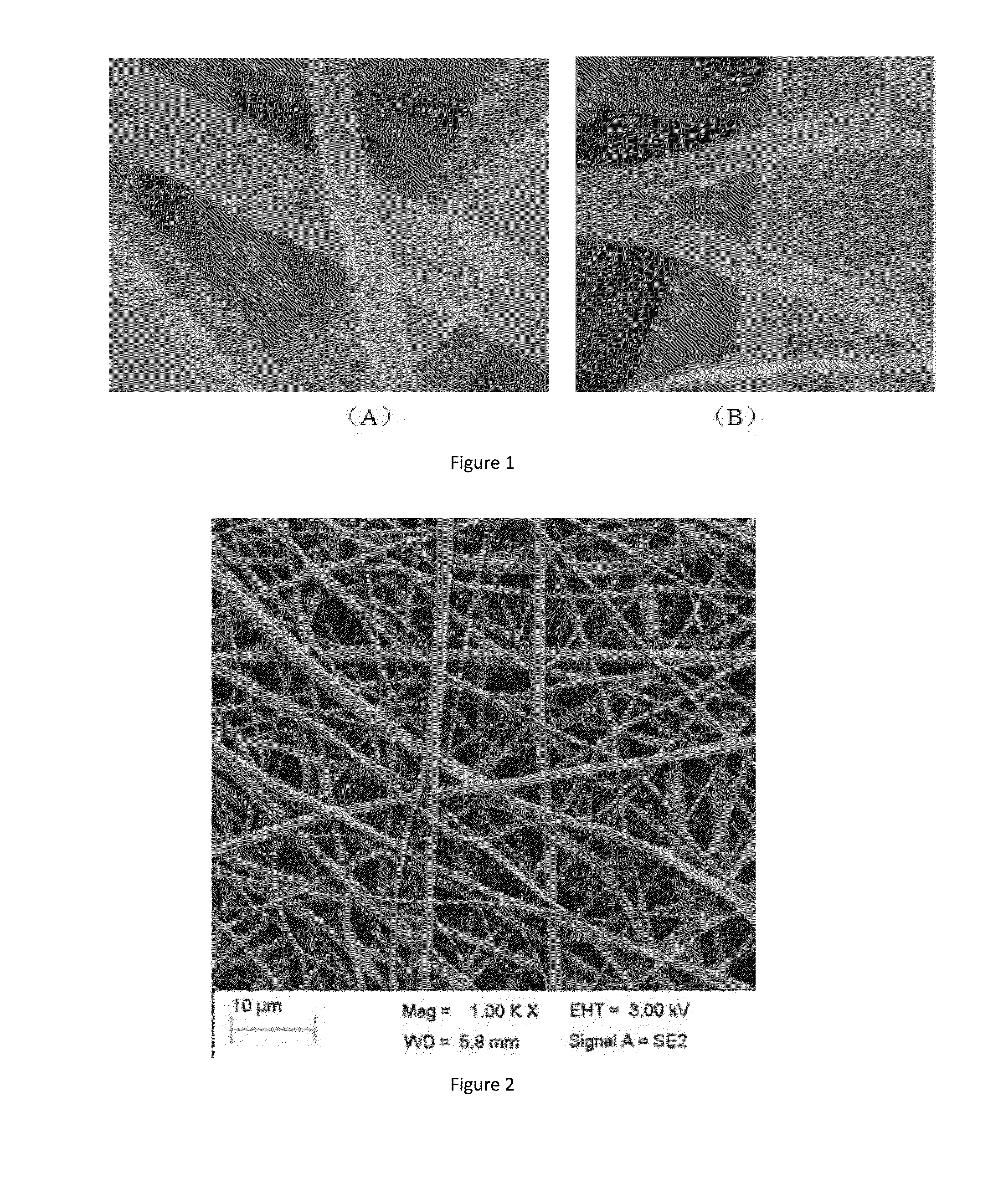
[0083]PCL and gelatin were dissolved in trifluoroethanol in a mass ratio of 10:1 to form a solution. The solution entered into a centrifugal spinning machine via a propel pump. The propelling rate of the propel pump was 150 g / min; the velocity of centrifugal coiling was 6000 m / min. The solution was rapidly spun into fibers at a spinning temperature of 50° C. Once the procedure was completed, the membrane was taken from the receiver, and the solvent was removed by vacuum drying, so that a composite fiber membrane was obtained.

[0084]An SEM image of the composite fibers is shown in FIG. 1-A. After being treated with water at a temperature of 50° C., which may dissolve gelatin but may not dissolve PCL, the composite fiber has an SEM image as shown in FIG. 1-B. It can be clearly observed that the composite fibers have a composite structure formed by PCL and gelatin.


PUM

PropertyMeasurementUnit
contact angleaaaaaaaaaa
water absorption ratioaaaaaaaaaa

Abstract

The present invention provides a tissue repair scaffold and a preparation method and purpose thereof. The tissue repair scaffold comprises at least one composite fiber layer, and the composite fiber layer comprises a composite fiber formed by an adhesion factor and a hydrophobic synthetic material. The composite fiber layer may further form a porous three-dimensional mesh structure with a hydrophilic material. The tissue repair scaffold is applied to brain meninge repair, spinal meninge repair, a tissue engineering scaffold material, artificial skin, a wound assistant, a biological membrane, a wound cladding material, a hemostatic material, a postoperative adhesion prevention material, or a cosmetic material.

Description

TECHNICAL FIELD[0001]The present disclosure relates to a medical device and preparation thereof, particularly relates to a tissue repair scaffold and preparation method and uses thereof.BACKGROUND ART[0002]Tissue repair scaffolds are quite common in clinical work, e.g. tendon repair patches, ligament repair patches, hernia repair patches, dura mater repair patches, female pelvic floor dysfunction repair patches; repair of blood vessels, sling for repair; repairs of skin, fistulae, nerve conduits and bone coloboma.[0003]The existing material itself and the pore structure have some deficiencies. It has great difference from the composition units of human tissue. Particularly, since the hydrophobic synthetic material prepared by common methods lacks porous structures similar with those observed in extracellular matrix, therefore the material is incompatible with the characteristics of the tissue anatomical structure; due to the poor tissue compliance, it is difficult for the material t...

Claims


Application Information

Patent Timeline
Patent Type & Authority Applications(United States)
IPC IPC(8): A61L27/48A61L27/22A61L27/60A61L27/18A61L27/54A61L27/24A61L27/26A61L27/56A61L27/20
CPCA61L27/48A61L27/56A61L27/222A61L27/60A61L27/20A61L2430/38A61L27/24A61L27/26A61L27/18A61L2430/34A61L27/54A61L2430/32A61L27/44A61L31/125A61L31/146A61L31/16
Inventor DENG, KUNXUEYANG, YAYAXU, TAOYUAN, YUYU
Owner MEDPRIN REGENERATIVE MEDICAL TECH
Why Eureka
Try Eureka