
Particle conjugated prosthetic patches and methods of making and using thereof

Active Publication Date: 2018-03-01
YALE UNIV
View PDF4 Cites 1 Cited by
  • Summary
  • Abstract
  • Description
  • Claims
  • Application Information

AI Technical Summary

Benefits of technology

This patent describes a new type of prosthetic patch that can be used to treat various diseases and conditions. The patch contains small particles made of biodegradable polymers that release therapeutic agents over time. The particles are covalently bound to the patch, which can be applied to various locations in the body. The patch can be used to prevent, suppress, or treat symptoms of diseases such as restenosis, neointimal hyperplasia, scarring, and over-proliferation of cells. The patch can be implanted using standard techniques. Overall, this patent provides a new and effective way to deliver therapeutic agents to target tissues in the body.

Problems solved by technology

Historically, the first choice of patch material for blood vessels was autogenous saphenous veins, but a separate harvest procedure on patients, risk of infection, and early reports of vein patch blowout led to surgeons using “off-the-shelf” prosthetic materials.
However, long-term clinical results have identified several issues that may be related to these materials used as patches including restenosis, pseudoaneurysm formation, infection, fibrosis, calcification and thrombosis (Li, et al., Ann Vasc Surg., 25(4): 561-568 (2011)).
Although open venous surgery is performed less frequently compared to open arterial surgery, patches are also used in venous surgery (Ohwada, et al., Hepatogastroenterology, 46, 855-858 (1999)), as venous procedures are frequently complicated by aggressive neointimal hyperplasia and restenosis, possibly due to the lower shear stress in the venous system.
A main cause of restenosis is neointimal hyperplasia, i.e. over-proliferation and migration of vascular muscle cells in the tunica intima, in the area at or near the patch, resulting in the thickening of blood vessel walls and luminal narrowing in vessels.
However, oral administration of rapamycin induces systemic toxicity, leading to severe adverse effects including fever, anemia, and capillary leak syndrome (Kaplan et al, Arch Dermatol, 135(5):553-557 (1999)).
However, rapamycin-loaded nanoparticles are inconsistent in inhibiting restenosis.
Another complication of vascular anastomotic techniques is the potential for development of anastomotic aneurysm.
Several factors in current formulations hinder the successful delivery of pharmaceuticals from prosthetic patches.

Method used


Image

  • Particle conjugated prosthetic patches and methods of making and using thereof
  • Particle conjugated prosthetic patches and methods of making and using thereof
  • Particle conjugated prosthetic patches and methods of making and using thereof

Examples


example 1

on and Characterization of Nanoparticle-Patch Composites

[0248]Materials and Methods

[0249]Synthesis of Nanoparticles (NPs)

[0250]NPs were prepared using an emulsion method. Carboxylated PLGA (inherent viscosity 0.55-0.75 dL / g) (100 mg) and rapamycin (5 mg) were dissolved in chloroform, and then added drop-wise to 5% polyvinyl alcohol (PVA). The mixture was sonicated three times and then added to 0.2% PVA solution. The solvent was evaporated for 2 hours while stirring and the PLGA particles were centrifuged before lyophilization (Bal, et al., Sci Rep.; 7: 40142 (2017)).

[0251]The size and polydispersity Index (PDI) of the nanoparticles were measured using a Zetasizer (Malvern, Westborough, Mass.). Light scattering was measured by back-scattering at a detection angle of 173 and a wavelength of 532 nm; the hydrodynamic radius (RH) was calculated using the Stokes-Einstein equation:

RH=kBT6πηD0

[0252]where kB=Boltzmann constant, T=absolute temperature, η=solvent viscosity, D0=diffusion coeffi...

example 2

cle-Mediated Release of Active Agents from Pericardial Patches is Sustained for 15 Days and Inhibits the Proliferation of Smooth Muscle Cells In Vitro

[0260]Materials and Methods

[0261]Assessment of Active Agent Release from Patches

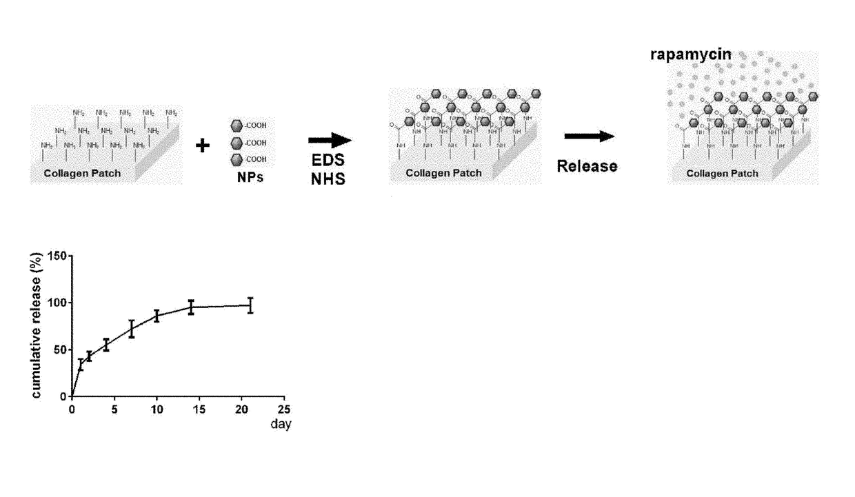
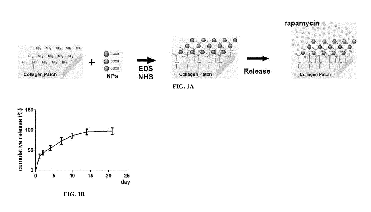
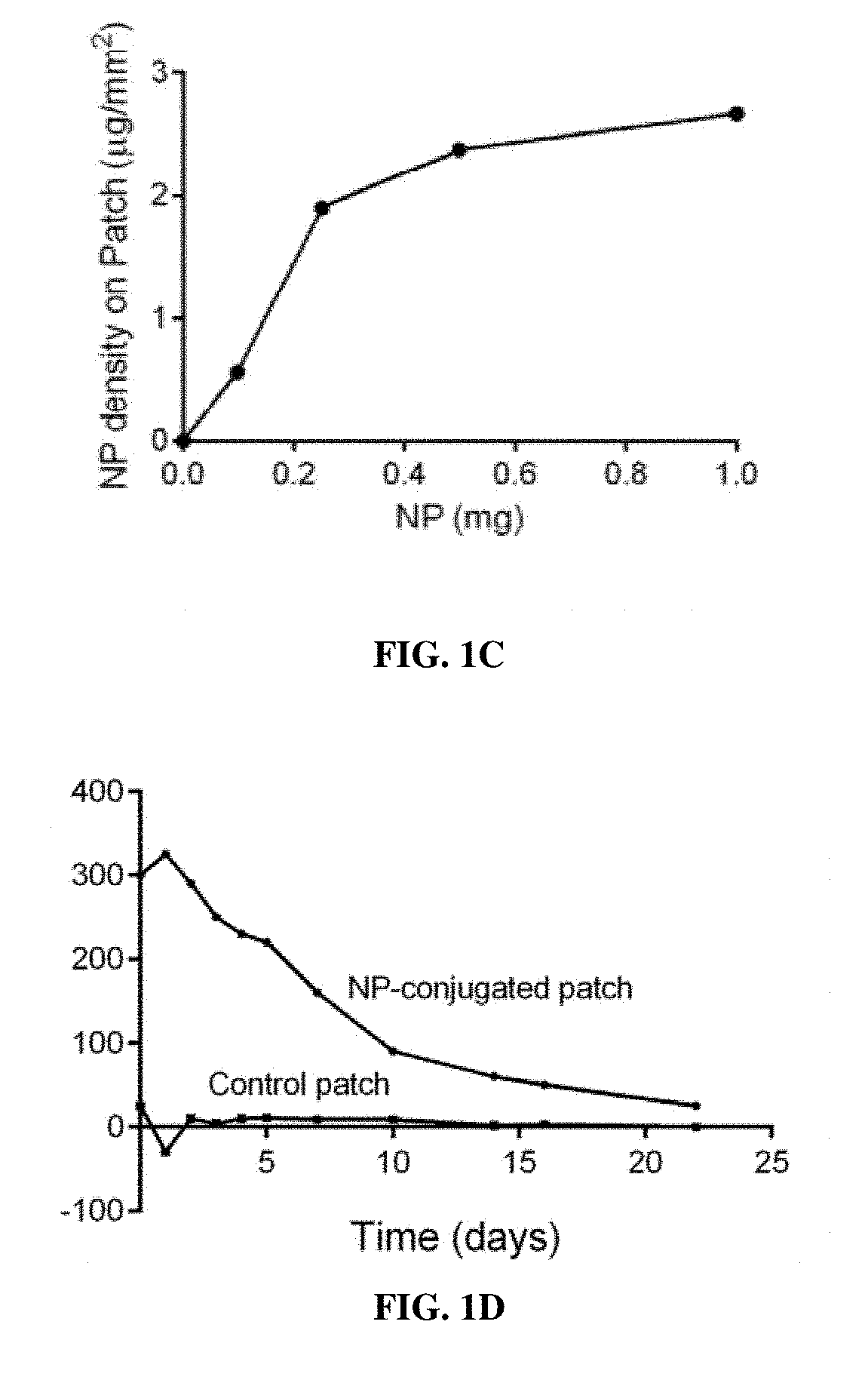
[0262]Patches conjugated with NP containing rapamycin (NP-rapamycin) were incubated in phosphate buffered saline (PBS) at 37° C. The supernatant of each sample was collected and analyzed for absorption at 400 nm at each time point using a SpectraMax plate reader (Molecular Devices, Sunnyvale, Calif.). Patches conjugated with NP containing rhodamine (NP-rhodamine) were incubated in PBS at 37° C.; control patches were incubated in PBS containing free rhodamine. At various time points patches were washed in PBS and their fluorescence intensity was measured.

[0263]Culture of Human Smooth Muscle Cells

[0264]Human smooth muscle cells (SMCs), passages 6-8, were cultured in endothelial basal medium 2, supplemented with endothelial cell growth media-2 MV SingleQuot Ki...

example 3

cle-Patch Composite Delivery System does not Introduce Toxicity to Lungs In Vivo

[0270]Materials and Methods

[0271]Rats were anesthetized with isoflurane inhalation, and tissues were fixed by transcardial perfusion of phosphate buffered saline (PBS) followed by 10% formalin. Tissue was removed and fixed overnight in 10% formalin followed by a 24-hour immersion in 70 percent alcohol. Tissue was then embedded in paraffin and sectioned (5 μm thickness). Tissue sections were de-paraffinized and stained with hematoxylin and eosin.

[0272]Results

[0273]FIG. 3A demonstrates that there is no decrease in proliferation in the lungs of rats that had NP-rapamycin patches implanted compared to control or NP-control patches. FIG. 3B demonstrates that there is also no increase in apoptosis in the lungs of rats that had NP-rapamycin patches compared to control or NP-control patches. Further, rapamycin was not present in detectable quantities in serum on days 1, 3 and 7 after implantation of NP-rapamycin...


PUM

PropertyMeasurementUnit
Timeaaaaaaaaaa
Timeaaaaaaaaaa
Massaaaaaaaaaa

Abstract

Composite prosthetic patches with covalently bound particles for controlled drug release, and methods of making and using thereof, have been developed. The particles may encapsulate one or more therapeutic, prophylactic or diagnostic agent(s). Generally, the prosthetic patches are decellularized extracellular matrix such as bovine or porcine pericardium or synthetic polymeric materials. The size of the particles ranges from between 1 nm and 1000 μm, preferably from between 10 nm and 500 nm. In some embodiments, the agent is a therapeutic agent for treatment of neointimal hyperplasia. In other embodiments, the agent is a therapeutic agent for suppressing or resolving inflammation. In yet other embodiments, the agent is a therapeutic for mitigating scarring.

Description

CROSS-REFERENCE TO RELATED APPLICATIONS[0001]This application claims the benefit of and priority to U.S. Ser. No. 62 / 380,685 filed Aug. 29, 2016 and which is incorporated by reference in its entirety.STATEMENT REGARDING FEDERALLY SPONSORED RESEARCH[0002]This invention was made with government support under HL095498 and HL128406 awarded by National Institutes of Health and under I01-BX002336 awarded by The United States of America as represented by the Department of Veterans Affairs. The government has certain rights in the invention.FIELD OF THE INVENTION[0003]The present invention relates to the field of prosthetic implants and drug delivery, and more particularly, to localized sustained delivery via particle-conjugated prosthetic implants and grafts.BACKGROUND OF THE INVENTION[0004]Prosthetic patches are widely used for repairs of vasculature, congenital diaphragmatic hernia, orthopedic and fascial defects, as well as in reconstructive surgery and other types of tissue repair. His...

Claims


Application Information

Patent Timeline
IPC IPC(8): A61L27/36A61L27/24A61L27/26A61L27/30
CPCA61L27/3641A61L27/24A61L27/3604A61L27/26A61L2420/06A61L27/306A61L2400/12A61L2400/18A61L27/3687A61L27/34A61L27/3633A61L27/54A61L2300/62A61K31/436A61K31/4439C08L67/04
Inventor FAHMY, TAREK M.DARDIK, ALAN
Owner YALE UNIV
Learn More