

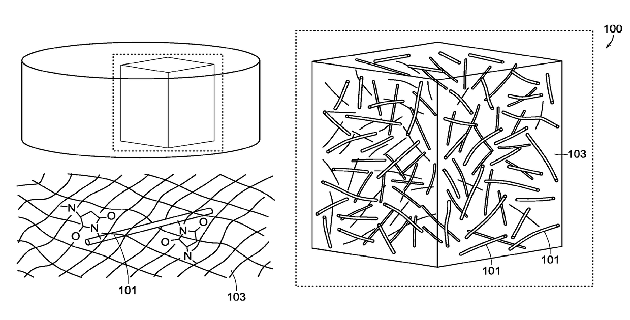
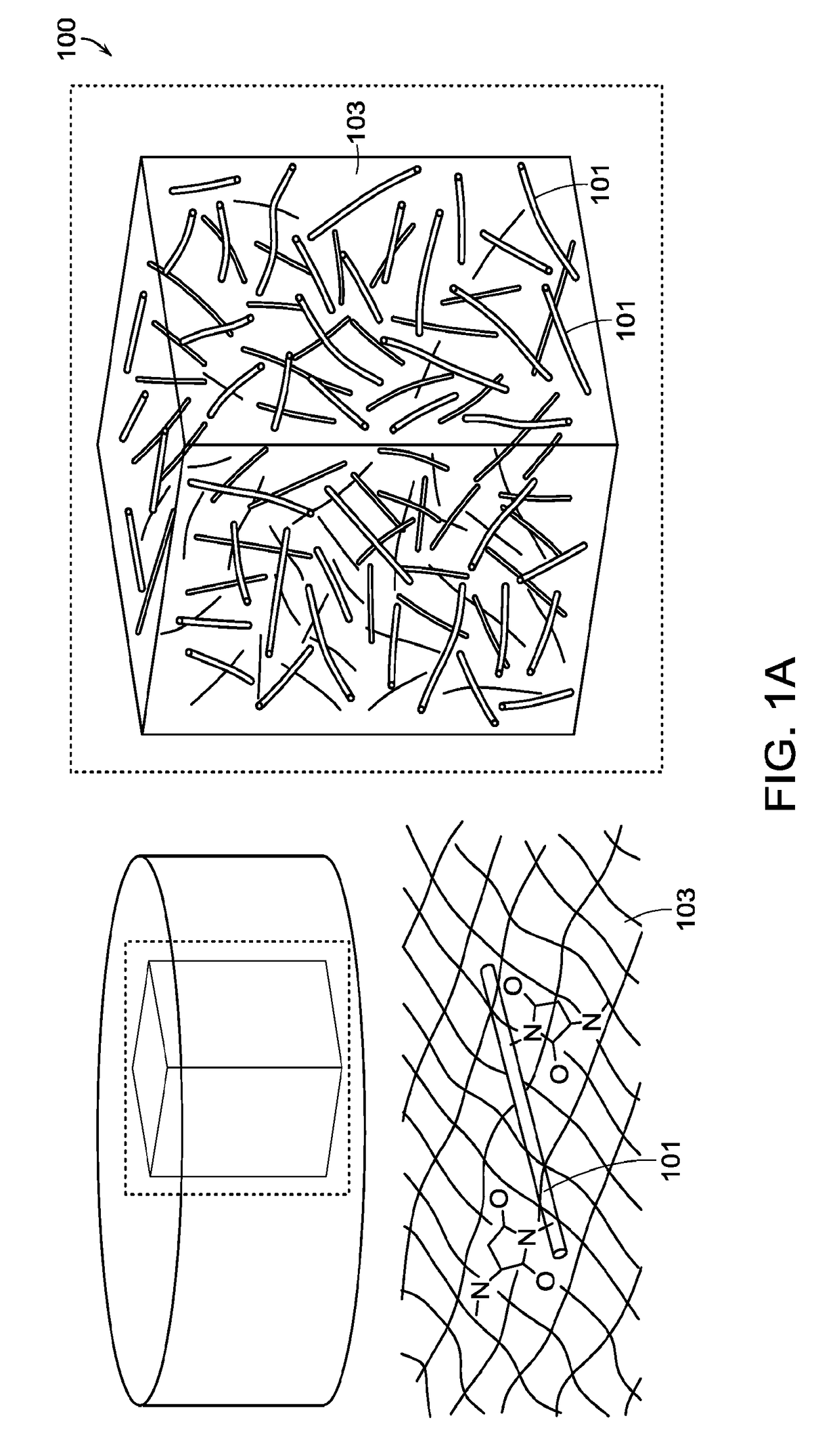
Fiber-hydrogel composite surgical meshes for tissue repair
a fiber-hydrogel and composite technology, applied in the field of composite materials and methods, can solve the problems of donor site defects, soft tissue defects resulting from trauma, oncologic resection, congenital malformations that are difficult to treat by conventional means, etc., and achieve the effect of improving the quality of reconstruction and improving the properties
- Summary
- Abstract
- Description
- Claims
- Application Information
AI Technical Summary
Benefits of technology
Problems solved by technology
Method used
Image

Examples
example 1
on of a Composite Surgical Scaffold Device
[0246]Nanofibers were produced by electrospinning PCL (polycaprolactone, 80 k from Sigma Aldrich). The nanofibers were spun into a random mesh. The spinning parameters were a 10% wt solution of PCL in 90% / 1% w / w DCM-DMF, at a flow rate of 0.6 ml / h through a 27 gauge blunt needle 15 cm from the target metal plate. The needle voltage was +10 kV, with the target plate was negatively biased with a voltage of −3 kV. One mL of solution was spun per round for each of the nanofiber sheet.
[0247]The fibers were then functionalized with a multistep process. Briefly, the fibers were plasma-treated to have reactive groups on the fiber surface, to which acrylic acid was conjugated by UV photoinitiation. The acrylate groups were then reacted with EDC and diazimine to form primary amines. These amines could then be reacted with SMCC to attach maleimide groups, which could readily react with the thiol groups in the hydrogel.
[0248]A composite surgical mesh wa...
example 2
on Test of the Nanofiber-Hydrogel Composite
[0278]For compression testing, the fiber-hydrogel samples were formed as cylinders 8.5 mm in diameter and ˜4 mm in height, allowed to set overnight in molds at 37° C. The elastic moduli were determined via compression testing with a Bose EnduraTEC ELF 3200 (Eden Prairie, Minn.). The sample underwent uniaxial compression between two parallel plates, compressed to 50% Strain. The elastic moduli were determined by measuring the slope of the initial linear region. Two sample groups were tested, with the same hydrogel formulations, with and without fibers. The hydrogel-only sample was formed with 4.5 mg / mL of thiolated hyaluronic acid (Gylcosan Glycosil) and 10 mg / mL PEG-DA (polyethyl-glycol diacrylate, molecular weight 3350). The fiber-hydrogel composite group had the same hydrogel concentrations, but additionally has 6.75 mg / mL PCL nanofibers that have a surface functionalized with maleimide groups that can readily react with the thiolated hya...
example 3
rials Interaction
[0281]To test for the cellular response to the composite hydrogel, the migratory potential of adipose-derived stem cells (ASCs) was tested in varying formulations of hydrogels, with and without fibers.
[0282]ASCs were transfected to express GFP, then formed into spheroid clusters by seeding the cells overnight in alginate molds made by Microtis sues molds. The cells were seeded as spheroids to better evaluate cell motility, as the spheroids are a distinct point source from which migrating cells can be easily measured. The spheroids were mixed in to the hydrogel before being pipetted into a 96-well plate and being allowed to set. The cells were then imaged over the next several days to observe their migration. The cells were able to migrate progressively further as the concentrations of hyaluronic acid and PEG-DA were lowered, due to the respectively increasing pore sizes. At the same hydrogel densities (4.5 mg / mL hyaluronic acid and 2.5 mg / mL PEG-DA), cells were bett...
PUM
| Property | Measurement | Unit |
|---|---|---|
| mean diameter | aaaaa | aaaaa |
| pore diameter | aaaaa | aaaaa |
| diameter | aaaaa | aaaaa |
Abstract
Description
Claims
Application Information