

Radio-wave responsive doped nanoparticles for image-guided therapeutics
a nanoparticle and radio wave technology, applied in the direction of emulsion delivery, pharmaceutical delivery mechanism, heating surgery instruments, etc., can solve the problem of limited lesion size that can be treated to 4 cm or less
- Summary
- Abstract
- Description
- Claims
- Application Information
AI Technical Summary
Benefits of technology
Problems solved by technology
Method used
Image

Examples
example — 1
Example—1 Preparation of Iron Doped Calcium Phosphate Nanoparticles (nCP: Fe) for Dual T1-T2 Magnetic Contrast Guided Radiofrequency Ablation of Tumor—Synthesis and Characterization
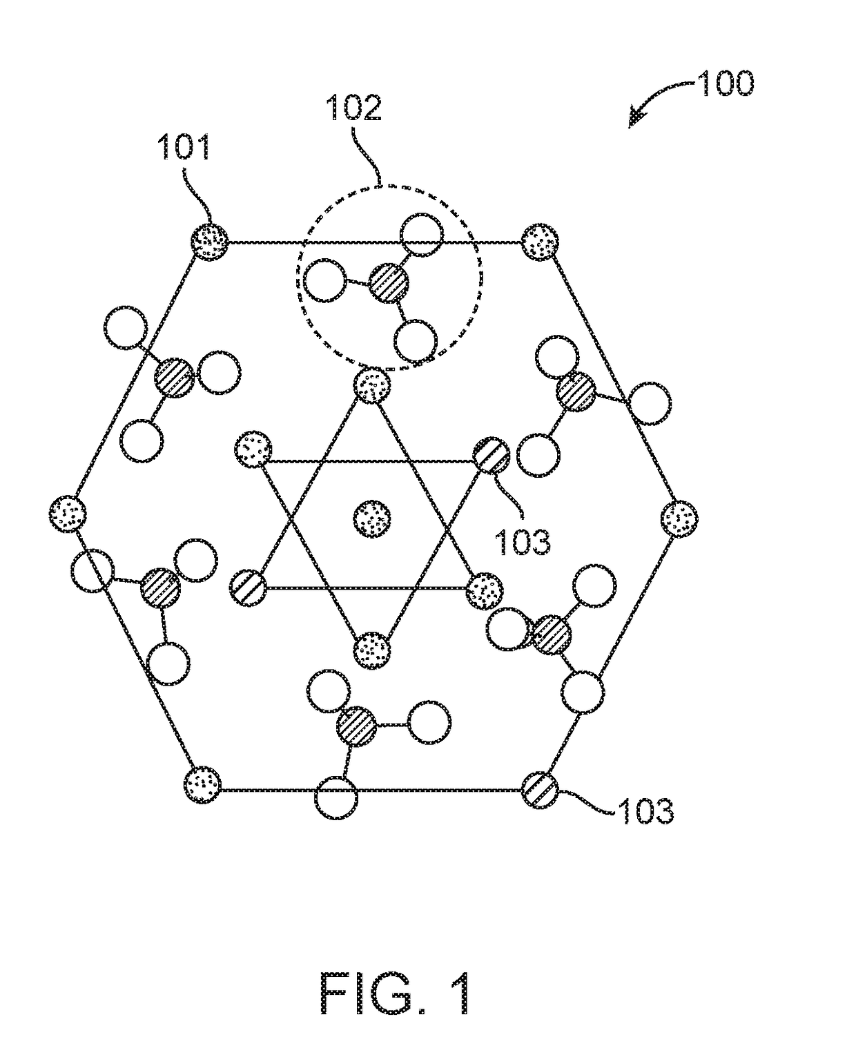
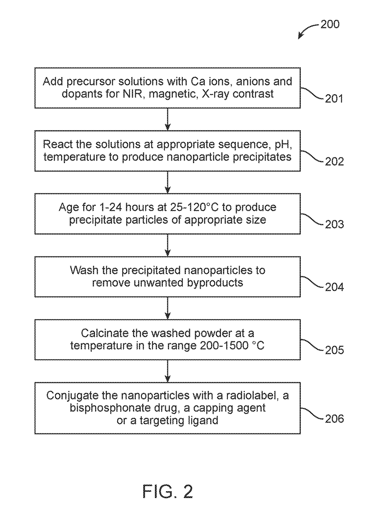
[0084]20 mL of 0.5 M calcium chloride (CaCl2, Sigma, USA) was mixed with 20 mL of 0.2 M trisodium citrate (Na3C6H5O7, Fisher Scientific, India) and 0.1 M FeCl3 (Sigma, USA). Volume of 0.1 M FeCl3 added was varied as per the required percentage of doping. 5 mL of 0.3 M diammonium hydrogen phosphate ((NH4)2HPO4, S.D Fine Chemicals, India) mixed with 0.2 mL of 3 N ammonium hydroxide (NH4OH, Fisher Scientific, India) was added drop wise to the above mixture of CaCl2, Na3C6H5O7 and FeCl3 under constant stirring to obtain Fe doped calcium phosphate nanoparticles, Fe-nCX (X=phosphate). The precipitate was washed 4 times in hot distilled water by centrifugation at 8500 rpm for 15 minutes and redispersed in PBS. FIG. 4A shows the schematic of Fe-nCX, wherein Fe3+ replaces Ca2+ in the calcium phosphate crystal latt...
example 2
tion of In Vivo Dual Mode T1 and T2 Contrast
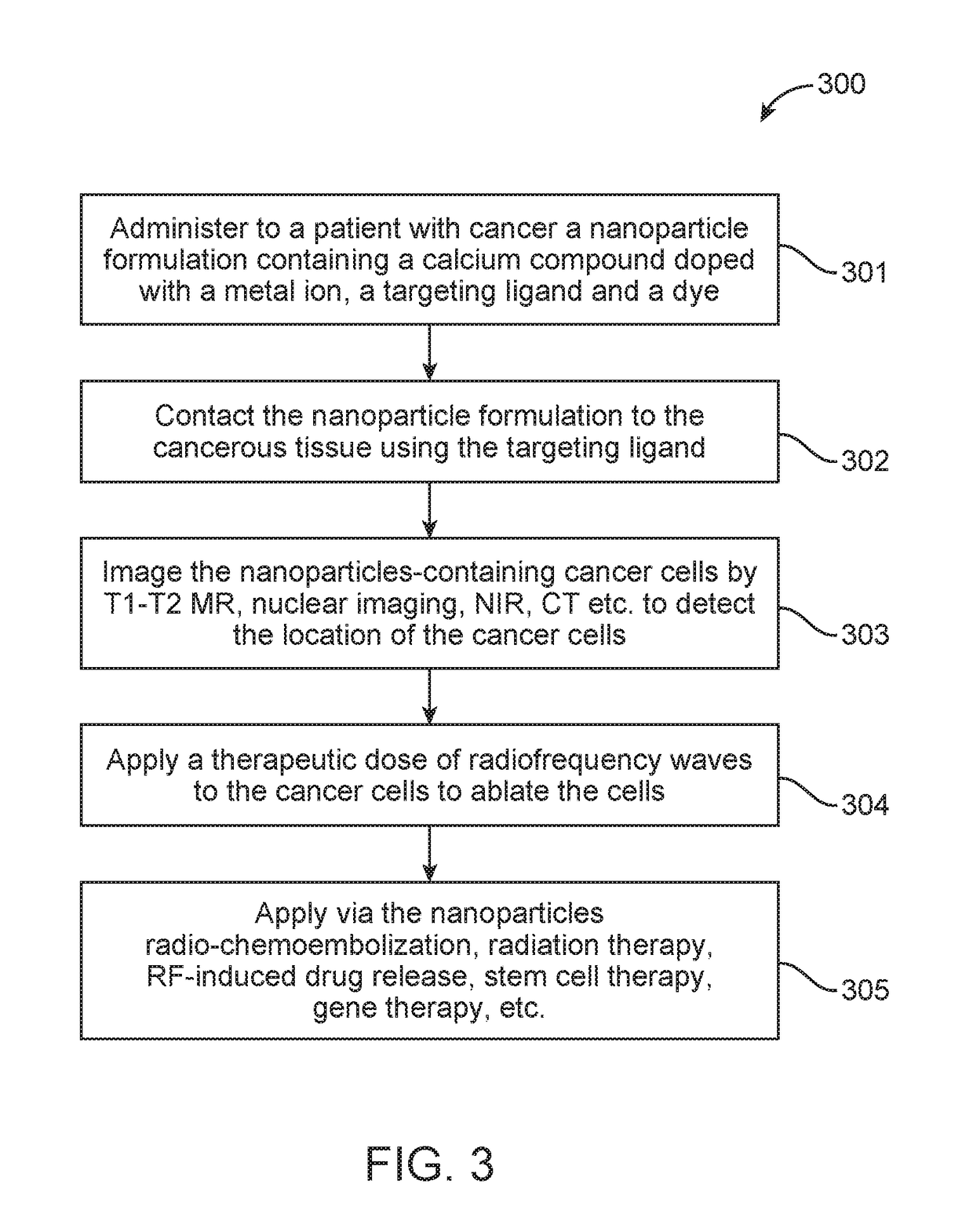
[0088]1 hour after Fe-nCX(X=phosphate) injection, an enhancement of both T1 and T2 contrast was observed especially in the liver and heart region (shown in white dotted box: FIG. 8A, 8B). Axial T2 weighted images of liver sections before (FIG. 8C) and after (FIG. 8D) Fe-nCX injection clearly showed enhancement in T2 contrast intensity after sample injection that was also reflected in T2 mapping data (FIG. 8E) obtained from selected ROI (white circles in FIG. 8B, 8C). Reduction in T2 contrast in liver was associated with reduction of T2 relaxation time from 40 ms to 25 ms. To evaluate the biodistribution of Fe-nCX, MRI of sample injected animal was carried out over a period of 96 hours. T2 weighted whole body coronal (FIG. 9A) and axial (FIG. 9B) MRI showed an increase in T2 contrast in liver (shown in white dotted box) within the first one hour which gradually reduced to initial contrast by 96 hours. This variation was also reflected in th...
example 3
Radiofrequency Response of Fe-nCX (X=Phosphate)
[0090]RF response of the nanoparticles was measured in a custom made non-invasive 13.5 MHz RF instrument. Different concentrations of Fe-nCX varying from 10-500 μg / mL were taken in a small glass petri dish and 100 W RF power was applied for 1 minute. The temperature of the solution was measured before and after RF irradiation. In the frequency range applied for RF ablation (350-550 kHz), there was an increase in dielectric loss factor, tan delta value, from 2.4 for undoped nCP to 3.54 for Fe-nCX (FIG. 11), that indicates lossy character of doped sample under RF exposure. We compared the RF mediated heating of nCP and Fe-nCX at varying concentration at 100 W RF power for 1 minute exposure in a non-invasive RF machine. In the concentration range of 50-500 μg / mL, there was significant increase in temperature of up to 22° C. for Fe-nCX samples compared to only <10° C. for undoped nCP (FIG. 12). We presumed that this rise in temperature obse...
PUM
| Property | Measurement | Unit |
|---|---|---|
| size | aaaaa | aaaaa |
| size | aaaaa | aaaaa |
| temperature | aaaaa | aaaaa |
Abstract
Description
Claims
Application Information